Insights into Alpha-Hemolysin (Hla) Evolution and
Expression among
Staphylococcus aureus
Clones with
Hospital and Community Origin
Ana Tavares1, Jesper B. Nielsen2, Kit Boye2, Susanne Rohde2, Ana C. Paulo3, Henrik Westh2,4, Kristian Schønning2,4, Hermı´nia de Lencastre1,5, Maria Miragaia1,6*
1Laboratory of Molecular Genetics, Instituto de Tecnologia Quı´mica e Biolo´gica (ITQB), Oeiras, Portugal,2Dept. of Clinical Microbiology 445, Copenhagen University Hospital, Hvidovre, Denmark,3Molecular Microbiology of Human Pathogens, ITQB, Oeiras, Portugal,4Faculty of Health and Medical Sciences, University of Copenhagen, Copenhagen, Denmark,5Laboratory of Microbiology and Infectious Diseases, The Rockefeller University, New York, New York, United States of America,6Laboratory of Bacterial Evolution and Molecular Epidemiology, ITQB, Oeiras, Portugal
Abstract
Background:Alpha-hemolysin (Hla) is a major virulence factor in the pathogenesis ofStaphylococcus aureusinfection, being active against a wide range of host cells. Althoughhla is ubiquitous in S. aureus, its genetic diversity and variation in expression in different genetic backgrounds is not known. We evaluated nucleotide sequence variation and gene expression profiles ofhlaamong representatives of hospital (HA) and community-associated (CA)S. aureusclones.
Methods:51 methicillin-resistantS. aureusand 22 methicillin-susceptibleS. aureuswere characterized by PFGE,spatyping, MLST and SCCmectyping. The internal regions ofhlaand the hlapromoter were sequenced and gene expression was assessed by RT-PCR.
Results:Alpha-hemolysin encoding- and promoter sequences were diverse, with 12 and 23 different alleles, respectively. Based on phylogenetic analysis, we suggest thathlamay have evolved together with theS. aureusgenetic background, except for ST22, ST121, ST59 and ST93. Conversely, the promoter region showed lack of co-evolution with the genetic backgrounds. Four non-synonymous amino acid changes were identified close to important regions ofhlaactivity. Amino acid changes in the RNAIII binding site were not associated tohlaexpression. Although expression rates ofhla were in general strain-specific, we observed CA clones showed significantly higherhlaexpression (p = 0.003) when compared with HA clones.
Conclusion: We propose that the hla gene has evolved together with the genetic background. Overall, CA genetic backgrounds showed higher levels ofhlaexpression than HA, and a high strain-to-strain variation of gene expression was detected in closely related strains.
Citation:Tavares A, Nielsen JB, Boye K, Rohde S, Paulo AC, et al. (2014) Insights into Alpha-Hemolysin (Hla) Evolution and Expression amongStaphylococcus aureusClones with Hospital and Community Origin. PLoS ONE 9(7): e98634. doi:10.1371/journal.pone.0098634
Editor:J. Ross Fitzgerald, University of Edinburgh, United Kingdom
ReceivedFebruary 7, 2014;AcceptedMay 6, 2014;PublishedJuly 17, 2014
Copyright:ß2014 Tavares et al. This is an open-access article distributed under the terms of the Creative Commons Attribution License, which permits unrestricted use, distribution, and reproduction in any medium, provided the original author and source are credited.
Funding:This work was funded by project Ref. P-99911 from Fundac¸a˜o Calouste Gulbenkian (http://www.gulbenkian.pt/Institucional/pt/Homepage) and Project PTDC/BIA-MIC/3195/2012 from Fundac¸a˜o para a Cieˆncia e Tecnologia (http://www.fct.pt/) awarded to HdL; Project PTDC/BIA-EVF/117507/2010 from Fundac¸a˜o para a Cieˆncia e Tecnologia (http://www.fct.pt/) awarded to MM; and through grant Ref. Pest-OE/EQB/LAO004/2011 from Fundac¸a˜o para a Cieˆncia e Tecnologia (FCT), Portugal. A. Tavares was supported by grant SFRH/BD/44220/2008 from FCT. The funders had no role in study design, data collection and analysis, decision to publish, or preparation of the manuscript.
Competing Interests:Co-author Herminia de Lencastre is a PLOS ONE Editorial Board member. This does not alter the authors’ adherence to PLOS ONE Editorial policies and criteria.
* Email: miragaia@itqb.unl.pt
Introduction
Staphylococcus aureus is a human opportunistic pathogen responsible for a wide range of infections that can vary in its clinical presentation and severity. Methicillin-resistant S. aureus
(MRSA) emerged in 1960 in the United Kingdom and has been a major problem in hospitals (HA-MRSA) worldwide during the last 40 years; however since the late 1990s, MRSA has been emerging as a leading cause of severe infection also in the community, in individuals without recent health-care contact (CA-MRSA) [1,2]. CA-MRSA present distinct genetic backgrounds from their hospital counterparts, are more susceptible to antibiotics other
than beta-lactams, carry the smallest staphylococcal cassette chromosome mec types (SCCmec IV or V), and have higher virulence capacity [1,2,3]. The underlying reasons behind the enhanced virulence of CA-MRSA appear to be multiple including a different capacity to overcome host cell response [4], different distribution of mobile genetic elements carrying virulence deter-minants [5], allelic variation in virulence deterdeter-minants located in the core genome and in mobile genetic elements [6], and different levels of expression and protein production of virulence determi-nants (alpha-hemolysin, collagen adhesin, staphylokinase,
lase, lipase, enterotoxins C3 and Q, V8 protease and cysteine protease) [7,8,9].
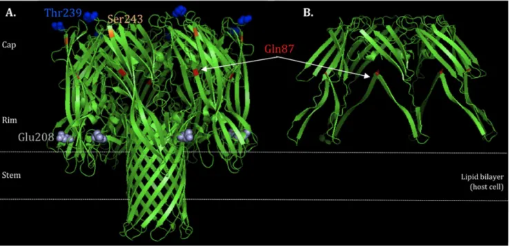
The alpha-hemolysin or a-toxin (Hla), is one of the major virulence determinants implicated in the pathogenesis of S. aureus, associated to severe skin and soft tissue infections (SSTI), necrotizing pneumonia and even sepsis [10]. Hla is the most prominentS. aureuscytotoxin that can act against a wide range of host cells including erythrocytes, epithelial cells, endothelial cells, T cells, monocytes and macrophages [10,11,12]. The gene encoding Hla is located in the core genome and is expressed as a water-soluble monomer (33.2 kD) that assembles to form a membrane-bound heptameric b-barrel pore (232.4 kD) on susceptible cells leading to cell death and lysis [11]. The overall structure is mushroom-like, divided into three domains: 1) Cap domain: largely hydrophobic, defining the entry of the pore; 2) Rim domain: underside of the Cap, in close proximity to membrane bilayer; 3) Stem domain: part of the transmembrane channel, forming the membrane-perforating b-barrel pore (Figure 1) [10,11]. Hla expression is mainly controlled by the global toxin accessory gene regulator (agr), via the regulatory effector molecule RNAIII [13]. While agrprovides the first and most important mechanism of up-regulation ofhla, expression can also be modulated by other regulators, such as SaeR, SarZ, ArlS [14,15,16] (up-regulators) and Rot, SarT [17](down-regulators).
Although polymorphisms in thehlapromoter region have been described [18], the range of genetic diversity and evolution of this toxin has never been assessed in a large representativeS. aureus
collection. Furthermore, although differences in hla expression have been described between community- and hospital-associated MRSA, these studies have been performed with a limited number of CA-MRSA epidemic clones [9], or almost exclusively with representatives of the USA300 clone [19,20,21]. To better understand the evolutionary history ofhlaand its importance as a virulence factor for CA-MRSA, in this study we compared the
hla nucleotide sequence and expression among the major
epidemic and minor CA and HA clones, including both MRSA and MSSA strains.
Materials and Methods
Ethics Statement
Isolates were obtained from routine diagnostic and were analyzed anonymously and only the isolates, not humans, were studied. All data was collected according to the European Parliament and Council decision for the epidemiological surveil-lance and control of communicable disease in the European community. Ethical approval and informed consent were for that reason not required.
Bacterial collection
A total of 73S. aureus, including 51 MRSA and 22 MSSA were analyzed in this study. Strains were collected in 13 different countries (Belgium, Bulgaria, Czech Republic, Denmark, Greece, Netherlands, Portugal, Romania, Spain, Sweden, United King-dom, USA and Brazil), between 1961 and 2009 from both community (n = 46) and hospital (n = 27). The strains comprised a total of 52spatypes and 23 sequence types (STs) (see Table S1).
Strains were defined as belonging to CA or HA clones if they contained the same or related genetic backgrounds as the reference CA-MRSA and HA-MRSA epidemic control strains, based on ST,spatype and SCCmec(in case of MRSA).
Media and bacterial growth conditions
Before RT-PCR analysis, strains were grown overnight at 37uC on tryptic soy agar plates (TSA). Bacterial growth experiments were performed by growing bacteria in tryptic Soy Broth (TSB) at 37uC with shake and measuring OD (600 nm) each hour in the follow up automatic incubator Bioscreen C (Oy Growth Curves AB, Helsinki, Finland). Plates of 100-well honeycomb (Oy Growth Curves AB, Helsinki, Finland) were filled with 300ml/well of
Figure 1. HLA protein structure.A) wildtype (highlighted the non-synonymous mutations Gln87, Glu208, Thr239 and Ser243) and B) truncated protein due to a stop codon at Gln87. Structure generated by the program PyMOL v.1.6.
doi:10.1371/journal.pone.0098634.g001
Hla Evolution and Expression inS. aureusfrom Community and Hospital
overnight culture diluted to OD600= 0.05 in TSB growth medium. Three individual growth experiments (SetC, SetD and SetE) were performed for each strain and named accordingly e.g. HLZ6C, HLZ6D and HLZ6E (see Figure S2.I to III).
Nucleotide sequence ofhlaand promoter region Chromosomal DNA was extracted from overnight cultures, using the boiling method (100uC for 10 min followed by centrifuged at 13.000 g for 5 minutes). Two sets of primers were designed to span the most polymorphic regions within thehlagene andhlapromoter (considered as the region located2600 bp from
hla starting codon), after alignment of sequences available on NCBI forS. aureus. One set of primers (Forward: hla-F_CGAA-AGGTACCATTGCTGGT; Reverse: hla-R_CCAATCGATTT-TATATCTTTC) amplified an internal fragment of thehlagene (nt 1170419–1170982, CP000730.1) and the other set (Forward: F_CACTATATTAAAAATACATAC; Reverse: hlaPro-R_GTTGTTACTGAGCTGAC) amplified an internal fragment of the hlapromoter region (nt 1171289–1171773, CP000730.1) (Figure S3). PCR products were sequenced (Macrogen Europe, Amsterdam, The Netherlands) and sequences were analyzed using SeqMan (DNAstar, Lasergene v9, Madison, WI, USA). To each unique hla promoter (P) and gene sequence (hla) - allotype - a single Arabic number was attributed (e.g. P1, P2;hla1,hla2). Gene and promoter sequences were deposited in GenBank (accession numbers KM019547–KM019606; KM019607–KM019674).
Phylogenetic analysis
Phylogenetic relatedness was analyzed using the MEGA5 v5.05 software (http://www.megasoftware.net/) for gene, promoter region and concatenated sequences obtained from 1) gene with promoter region and 2) seven MLST alleles from the 23 representative STs within the collection. Phylogenetic trees were constructed using the Neighbor-Joining clustering method, and 1000 bootstrap replicates, which assigns confidence values for the groupings in the tree.
Moreover, nucleotide diversity (ND) between the two clusters was calculated based on the estimation of the average evolutionary divergence over sequence pairs within the two groups, where the number of base substitutions per site from averaging over all sequence pairs within each group are compared using the maximum composite likelihood model [22].
Detection of recombination
Alignments from the hla gene, hla promoter and internal fragments of each of the seven MLST gene were screened for the occurrence of putative recombination events using Recombination Detection Program version 4 (RDP4) (http://web.cbio.uct.ac.za/) with the default settings (with highest acceptable probability value of 0.05). Identification of recombinant sequences recombination breakpoints and major parent was determined using simulta-neously nine recombination detection methods (RDP, BOOT-SCAN, GENECONV, MAXCHI, CHIMAERA, SIBOOT-SCAN, PhylPro, LARD and 3SEQ. The ‘‘minor parent’’ is considered a sequence closely related to that from which sequences in the proposed recombinant region may have been derived (the presumed donor). The ‘‘major parent’’ was considered as a sequence closely related to that from which the greater part of the recombinant’s sequence may have been derived.
RT-PCR analysis
Culture growth was stopped at late exponential phase, when alpha-toxin is described to have maximal activity [23],
corre-sponding to the time-points 1) 3 hours 30 min in one group (65 strains) and 2) 4 hours 30 min in another (8 strains). Total RNA was extracted from three biological replicates. Cells were mechanically disrupted with FastPrep-24 Instrument (MP Bio-medicals, Solon, OH, USA) and RNA was protected using RNA Protect (Qiagen, Valencia, USA). RNA was extracted automati-cally using the QIAsymphony platforms (Qiagen, Valencia, USA) with QIAsymphony RNA kit (Qiagen, Valencia, USA).
The RT-PCR assay was performed on a 7500 Real-Time PCR System (Applied Biosystems, Foster City, CA) using the following primers and TaqMan probes: Hla RT_F: TAATGAATCCTG-TCGCTAATGCC; HlaRT_R: CACCTGTTTTTACTGTAG-TATTGCTTCC; Hla RT Probe: 6FAM-AAACCGGTACTA-CAGATAT-MGBNFQ. The RT-PCR reaction was performed using the EZ RT-PCR Core Reagents (Applied Biosystems, Foster City, USA), in which RNA is reverse transcribed and amplified in a single reaction. The following PCR protocol was used: 50uC for 2 min, 60uC for 30 min, 95uC for 5 min, followed by 42 cycles of 95uC for 20 sec and 62uC for 1 min. The 16S gene was used as internal or reference control. The primers used for 16S RNA amplification were those previously described [24].
RT-PCR data analysis
The relativehlagene expression was calculated based on the Ct (RT-PCR output) of the gene of interest (Cthla) as compared to the Ctof the internal control (Ct16S) as follows: Delta Ct= Ct hla-Ct16S. The lower the Delta Ctthe higher is the amount ofhla mRNA and the more the gene is expressed. The reproducibility of the assay was evaluated by the calculation of the arithmetic mean of the relative expression of the three biological replicates (Mean Delta Ct1–3= Average (Delta Ct1; Delta Ct2; Delta Ct3). The reproducibility of RT-PCR reaction was evaluated by the calculation of the standard deviation (STDEV) of Delta Ct obtained for each biological replica (Delta Ct1; Delta Ct2; Delta Ct3). Values were considered valid when at least two Ctreadings exist with STDEV,2.
Protein structure visualization (pyMOL)
The protein structure was modeled using PyMOL v.1.6 (http:// www.pymol.org/) if a nucleotide mutation gave rise to a stop codon.
Statistical analysis
The statistical analysis was performed using the Graphpad Prism 6 (http://www.graphpad.com/scientific-software/prism/), with the two-tailed Student’s t-test to determine whether the differences of mean expression rates (MSSA versus MSSA; HA backgroundsversusCA backgrounds) were statistically significant (p#0.05).
Regression tree analysis was used to explore which variables could be related with thehlaexpression [25]. Trees explain the variation of a single response variable (in this study thehlamRNA expression) by repeatedly splitting the data into more homoge-neous groups, using combinations of explanatory variables (in our case, the ST,spatype, MRSA, MSSA and the type of SCCmec).
Results
Analysis of polymorphisms in thehlagene andhla
promoter
The sequence analysis of the internal region ofhlaand thehla
promoter region among the 73 strains identified a total of 12hla
and 23 promoter region different sequences (allotypes) (Table 1). We obtained no amplification products forhlaandhlapromoter Hla Evolution and Expression inS. aureusfrom Community and Hospital
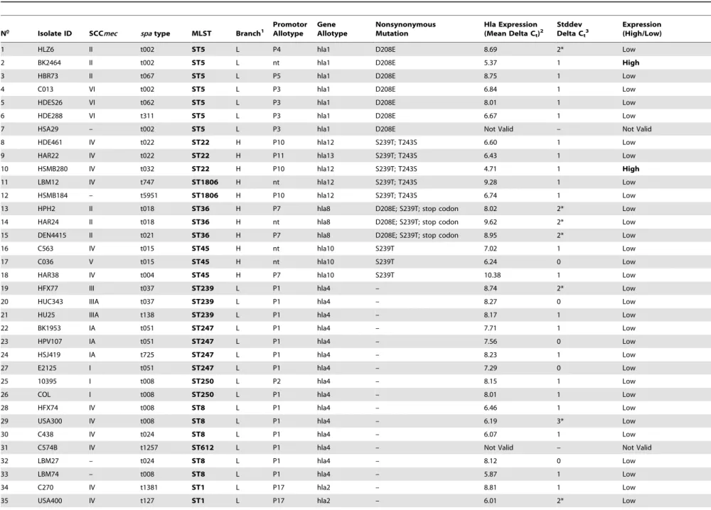
Table 1.Summary of molecular characterization, sequence variation and relative expression rates ofS. aureusstrains collection.
N Isolate ID SCCmec spatype MLST Branch1
Promotor Allotype
Gene Allotype
Nonsynonymous Mutation
Hla Expression (Mean Delta Ct)2
Stddev Delta Ct3
Expression (High/Low)
1 HLZ6 II t002 ST5 L P4 hla1 D208E 8.69 2* Low
2 BK2464 II t002 ST5 L nt hla1 D208E 5.37 1 High
3 HBR73 II t067 ST5 L P5 hla1 D208E 8.75 1 Low
4 C013 VI t002 ST5 L P3 hla1 D208E 6.84 1 Low
5 HDES26 VI t062 ST5 L P3 hla1 D208E 8.01 1 Low
6 HDE288 VI t311 ST5 L P3 hla1 D208E 6.67 1 Low
7 HSA29 – t002 ST5 L P3 hla1 D208E Not Valid – Not Valid
8 HDE461 IV t022 ST22 H P10 hla12 S239T; T243S 6.60 1 Low
9 HAR22 IV t022 ST22 H P11 hla13 S239T; T243S 6.43 1 Low
10 HSMB280 IV t032 ST22 H P10 hla12 S239T; T243S 4.71 1 High
11 LBM12 IV t747 ST1806 H nt hla12 S239T; T243S 9.28 1 Low
12 HSMB184 – t5951 ST1806 H P10 hla12 S239T; T243S 6.74 1 Low
13 HPH2 II t018 ST36 H P7 hla8 D208E; S239T; stop codon 8.02 2* Low
14 HAR24 II t018 ST36 H nt hla8 D208E; S239T; stop codon 9.62 2* Low
15 DEN4415 II t021 ST36 H P7 hla8 D208E; S239T; stop codon 8.95 2* Low
16 C563 IV t015 ST45 H nt hla10 S239T 7.02 1 Low
17 C036 V t015 ST45 H nt hla10 S239T 6.24 0 Low
18 HAR38 IV t004 ST45 H P7 hla10 S239T 10.38 1 Low
19 HFX77 III t037 ST239 L P1 hla4 – 8.74 2* Low
20 HUC343 IIIA t037 ST239 L P1 hla4 – 8.27 0 Low
21 HU25 IIIA t138 ST239 L P1 hla4 – 8.17 1 Low
22 BK1953 IA t051 ST247 L P1 hla4 – 7.71 1 Low
23 HPV107 IA t051 ST247 L P1 hla4 – 7.56 0 Low
24 HSJ419 IA t725 ST247 L P1 hla4 – 8.23 1 Low
27 E2125 I t051 ST247 L P1 hla4 – 7.29 0 Low
25 10395 I t008 ST250 L P2 hla4 – 8.15 1 Low
26 COL I t008 ST250 L P1 hla4 – 8.01 1 Low
28 HFX74 IV t008 ST8 L P1 hla4 – 6.46 1 Low
29 USA300 IV t008 ST8 L P1 hla4 – 6.19 3* Low
30 C438 IV t024 ST8 L P1 hla4 – 6.07 1 Low
31 C574B IV t1257 ST612 L P1 hla4 – Not Valid – Not Valid
32 LBM27 – t024 ST8 L P1 hla4 – 8.12 0 Low
33 LBM74 – t008 ST8 L P1 hla4 – 5.87 1 Low
34 C270 IV t1381 ST1 L P17 hla2 – 8.81 1 Low
35 USA400 IV t127 ST1 L P17 hla2 – 6.01 2* Low
Hla
Evolution
and
Expression
in
S.
aureus
from
Community
and
Hospital
PLOS
ONE
|
www.ploson
e.org
4
July
2014
|
Volume
9
|
Issue
7
|
Table 1.Cont.
N Isolate ID SCCmec spatype MLST Branch1
Promotor Allotype
Gene Allotype
Nonsynonymous Mutation
Hla Expression (Mean Delta Ct)2
Stddev Delta Ct3
Expression (High/Low)
36 LBM36 – t127 ST1 L P18 hla2 – 11.09 1 Low
37 C577 IV t216 ST59 L P20 hla5 – 5.35 0 High
38 C583 IV t437 ST59 L P19 hla5 – 5.31 1 High
39 C434 V t437 ST59 L P19 hla5 – 9.14 1 Low
40 C018 IV t1819 ST93 L nt hla7 – 5.16 1 High
41 C491 IV t202 ST93 L P21 hla7 – 5.45 0 High
42 LBM54 IV t011 ST398 H P12 hla11 – 4.46 2* High
43 C482 IV t011 ST398 H P13 hla11 – 3.25 1 High
44 C496 VII t108 ST398 H nt hla11 – 2.85 1 High
45 LBM40 – t034 ST398 H P12 hla11 – 5.37 1 High
46 C017 IV t019 ST30 H nt hla9 D208E; S239T 4.53 0 High
47 C385 IV t019 ST30 H P7 hla9 D208E; S239T 7.25 1 Low
48 C479 IV t019 ST30 H nt hla9 D208E; S239T 8.10 1 Low
71 HUC585 – t342 ST30 H P7 hla9 D208E; S239T 5.14 1 High
69 HFF204 – t318 ST30 H P9 hla9 D208E; S239T 6.23 1 Low
70 HFA30 – t012 ST30 H P8 hla8 D208E; S239T; stop codon 7.94 1 Low
49 HSJO7 IV t148 ST72 L P14 hla1 D208E 6.56 1 Low
50 USA700 IV t148 ST72 L P14 hla1 D208E 5.76 0 Low
51 COO3 IV t791 ST72 L P15 hla1 D208E 6.28 1 Low
52 SAMS1024 IV t1346 ST1810 L P14 hla1 D208E 4.78 1 High
53 HUC594 – t148 ST72 L P14 hla1 D208E 8.36 1 Low
54 HFA28 – t126 ST72 L P14 hla1 D208E 4.56 2* High
55 C238 – t3682 ST72 L P14 hla1 D208E 4.64 1 High
56 C168 IV t044 ST80 L P16 hla1 D208E 8.20 0 Low
57 C485 IV t044 ST80 L P16 hla1 D208E 5.72 1 High
58 C014 IV t131 ST80 L P16 hla1 D208E 4.87 0 High
59 LBM25 – t1509 ST15 L P2 hla1 D208E 6.69 0 Low
60 C157 – t084 ST15 L P2 hla1 D208E 4.86 1 High
61 C230 – t346 ST15 L P2 hla1 D208E 9.03 2* Low
62 HBA33 – t258 ST25 L P6 hla1 D208E 5.73 1 High
63 C095 – t2909 ST25 L P6 hla1 D208E 4.16 1 High
64 C141 – t081 ST25 L P6 hla1 D208E 4.50 2* High
65 HBA34 IV t308 ST121 L nt hla6 – 5.62 1 High
66 HUC574 – t435 ST121 L P1 hla6 – 5.19 1 High
67 HUC587 – t159 ST121 L P2 hla6 – 5.09 1 High
Hla
Evolution
and
Expression
in
S.
aureus
from
Community
and
Hospital
PLOS
ONE
|
www.ploson
e.org
5
July
2014
|
Volume
9
|
Issue
7
|
region in one and 13 strains, respectively, which probably result from misparing of the primers used.
From the 12hla(hla1–12), we observed that only a single
hla-allotype was found among representatives of a specific ST, except for ST22 (hla12;hla13) and ST30 (hla8;hla9) where two different alleles were identified. On the other hand, the most frequent alleles, hla1 (33.3%, n = 24) and hla4 (20.8%, n = 15), were identified in more than one ST.
Regarding the nucleotide changes identified in the hla, some correspond to non-synonymous mutations (E208, T239 and S243) and, in one particular case, to a stop codon (Table 1 and 2). The substitutions observed did not correspond to any difference in the charge or polarity of the amino acid (aa). However, changes in molecular weight were observed: i) changes from aa D208 to aa E208 (D208E) and from aa S239 to T239 (S239T) gave rise to a higher molecular weight aa; and ii) change from aa T243 to S243 (T243S) resulted in a lower molecular weight aa; of note all changes occurred in the Rim domain of the protein. In a particular case, the aa change gave rise to a stop codon located in the CAP domain, in strains of ST36. Protein structure modeling showed that a protein of about one third of its real size is produced, truncated at the Gln87 (Figure 1, A and B). The truncation is in the outside part of the domain, suggesting that this will affect the capacity of the Hla to form cell wall pores, and ultimately to induce hemolysis.
A high number of sequence variations were identified in thehla
promoter region, (n = 23) (P1–23) (Table 1 and 2). Although we found that some STs were associated to a specific promoter allotype, and some promoters were identified in a single ST, we also identified cases where single STs were associated to different promoters (8 out of 23) and examples in which a single promoter allotype was associated to different STs (5 out of 23). This is the case of the most frequent promoter (P1) that was found in about one third of the strains analyzed (25.4%, n = 16), including several different STs.
A particular highly polymorphic region corresponding to nt
222 to224 from the start codon, was found in the majority (16 out of 23) of the promoter allotypes (exceptions P1, P6, P13, P14, P15, P18 and P23). These polymorphisms are located in the vicinity of RNAIII binding site [26]; however, we could not find a direct correlation between a particular nucleotide sequence and a specific expression pattern (high or low expression). For example, the sequence TTT, observed in two strains belonging to ST398 that have a high level expression, was also observed in strains with low expression belonging to other genetic backgrounds (ST8, ST239, ST247, ST250, ST36, ST45 and ST22).
Alpha-hemolysin evolutionary history
In order to better understand the evolution ofhlagene within theS. aureuspopulation, we constructed phylogenetic trees from thehlaand hlapromoter sequences, separately or concatenated (Figure 2, A) and compared it with the tree constructed from the concatenated sequences of the seven housekeeping genes used in MLST, including all the STs represented in the strain collection described here (Figure 2, B).
The phylogenetic tree constructed for thehlagene showed two distinct major clusters with different evolutionary clocks that differed in their nucleotide diversity (ND, see Materials and Methods): cluster (L) with lower diversity (ND = 0.005), and cluster H with higher diversity (ND = 0.019). Cluster L included more than 70% of strains (71.2%, n = 52), and five sub-clusters; Cluster H contained about 29% of the strains (28.8%, n = 21), and comprised four minor sub-clusters including hla8–hla12 alleles,
Table 1. Cont. N Isolate ID SCC mec spa type MLST Branch 1 Promotor Allotype Gene Allotype Nonsynonymous Mutation Hla Expression (Mean Delta Ct ) 2 Stddev Delta Ct 3 Expression (High/Low) 68 HUC578 – t284 ST121 L P 1 h la6 – 7.10 2* Low 72 LBM23 – t100 ST9 L P 22 hla1 D208E 5.48 2* High 73 HFX84 – t267 ST97 L P 23 hla3 – 9.03 1 Low 1H: High polymorphism; L: Low polymorphism; 2Mean Delta Ct1–3 = A verage (Delta Ct1 ; D elta Ct2 ; Delta Ct3 ), Delta Ct =C t hla 2 Ct 16S; Not valid: only o ne Ct reading; 3*low reproducibility between three CT values (Stddv # 2). n t: non typable; Stddv: standard deviation. doi:10.1371/journal.p one.0098634.t001
Hla Evolution and Expression inS. aureusfrom Community and Hospital
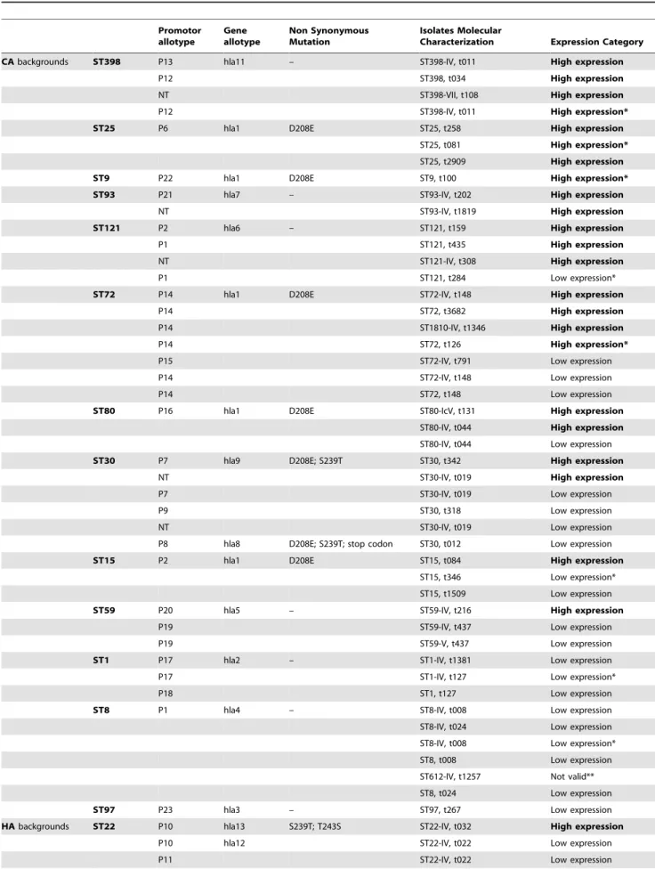
Table 2.Strains data distribution based on promoter allotypes.
Promotor allotype
Gene allotype
Non Synonymous Mutation
Isolates Molecular
Characterization Expression Category
CAbackgrounds ST398 P13 hla11 – ST398-IV, t011 High expression
P12 ST398, t034 High expression
NT ST398-VII, t108 High expression
P12 ST398-IV, t011 High expression*
ST25 P6 hla1 D208E ST25, t258 High expression
ST25, t081 High expression*
ST25, t2909 High expression
ST9 P22 hla1 D208E ST9, t100 High expression*
ST93 P21 hla7 – ST93-IV, t202 High expression
NT ST93-IV, t1819 High expression
ST121 P2 hla6 – ST121, t159 High expression
P1 ST121, t435 High expression
NT ST121-IV, t308 High expression
P1 ST121, t284 Low expression*
ST72 P14 hla1 D208E ST72-IV, t148 High expression
P14 ST72, t3682 High expression
P14 ST1810-IV, t1346 High expression
P14 ST72, t126 High expression*
P15 ST72-IV, t791 Low expression
P14 ST72-IV, t148 Low expression
P14 ST72, t148 Low expression
ST80 P16 hla1 D208E ST80-IcV, t131 High expression
ST80-IV, t044 High expression
ST80-IV, t044 Low expression
ST30 P7 hla9 D208E; S239T ST30, t342 High expression
NT ST30-IV, t019 High expression
P7 ST30-IV, t019 Low expression
P9 ST30, t318 Low expression
NT ST30-IV, t019 Low expression
P8 hla8 D208E; S239T; stop codon ST30, t012 Low expression
ST15 P2 hla1 D208E ST15, t084 High expression
ST15, t346 Low expression*
ST15, t1509 Low expression
ST59 P20 hla5 – ST59-IV, t216 High expression
P19 ST59-IV, t437 Low expression
P19 ST59-V, t437 Low expression
ST1 P17 hla2 – ST1-IV, t1381 Low expression
P17 ST1-IV, t127 Low expression*
P18 ST1, t127 Low expression
ST8 P1 hla4 – ST8-IV, t008 Low expression
ST8-IV, t024 Low expression
ST8-IV, t008 Low expression*
ST8, t008 Low expression
ST612-IV, t1257 Not valid**
ST8, t024 Low expression
ST97 P23 hla3 – ST97, t267 Low expression
HAbackgrounds ST22 P10 hla13 S239T; T243S ST22-IV, t032 High expression
P10 hla12 ST22-IV, t022 Low expression
P11 ST22-IV, t022 Low expression
Hla Evolution and Expression inS. aureusfrom Community and Hospital
which were found in strains of ST30, ST36, ST45, ST398 and ST22.
As opposed to the phylogenetic tree constructed fromhlagene, the one constructed from the promoter region did not show two distinct evolutionary branches (Figure S1). Moreover, dissimilar subgroup clustering was noticed in the tree constructed from the promoter gene sequence. For example, ST45, ST30 and ST36 backgrounds were clustered together in the promoter sequence-based tree whereas in the hla sequence-based tree ST45 was placed separately from ST30 and ST36 cluster (branch H). The same type of observations can be drawn for most of STs. Overall the promoter region showed to be more diverse than thehlagene sequence among the different backgrounds.
On the other hand, when we compared the phylogenetic tree constructed with thehlagene with that constructed from MLST concatenated genes, the same type of division into two distinct main clusters was observed (Figure 2). Moreover, the majority of STs were equally distributed between the two clusters in the two trees. The only exceptions were ST22, ST121, ST59 and ST93 that in the two trees have exchanged their positions from one cluster to the other (Figure 2, B-blue arrows).
Detection of recombination inhlagene, hla promoter and MLST genes
To understand if recombination could explain the incongruence found between the trees constructed from hla and MLST
concatenated genes, we screened thehlagene,hlapromoter and each MLST gene for recombination events using the RDP4 software.
The SiScan and 3Seq methods detected one recombination event in the hla gene. This event corresponded to a fragment ending in positions 385–410 of the hlaalignment, however the beginning breakpoint was not possible to determine. In the collection analyzed this event was detected in five isolates belonging to ST22 or related STs (HSMB280, HDE461, HAR22 and LBM12 (TLV ST22) and HSMB184 (TLV ST22)) and four isolates of ST398 (LBM54, LBM40, C496, C482_ST398). The ST30 HFF204 strain was identified as the minor parent (97.8% identity with ST22 strains and 299.3% identity with ST398 strains) and ST121 strain HUC587 was identified as the major parent (with 100% identity to ST398 strains and 93.5–95.2% identity with ST22 strains) of the recombining fragment. A trace signal of recombination of this same event was also identified among ST45 isolates; however this signal was not statistically significant. Interestingly all the recombination events were detected in strains belonging to the high genetic diversity cluster in the tree constructed fromhlagene. In thehlapromoter region no recombination events were detected.
We have performed the same type of analysis using the internal sequences of each of the seven housekeeping used in MLST scheme, including the alleles present in all STs identified in this Table 2.Cont.
Promotor allotype
Gene allotype
Non Synonymous Mutation
Isolates Molecular
Characterization Expression Category
P10 ST1806, t5951 Low expression
NT ST1806-IV, t747 Low expression
ST5 NT hla1 D208E ST5-II, t002 High expression
P3 ST5-VI, t002 Low expression
P3 ST5-VI, t062 Low expression
P3 ST5-VI, t311 Low expression
P4 ST5-II, t002 Low expression*
P3 ST5, t002, Not valid**
P5 ST5-II, t067 Low expression
ST36 P7 hla8 D208E; S239T; stop codon ST36-II, t018 Low expression*
P7 ST36-II, t021 Low expression*
NT ST36-II, t01 Low expression*
ST45 NT hla10 S239T ST45-IV, t015 Low expression
NT ST45-V, t015 Low expression
P7 ST45-IV, t004 Low expression
ST239 P1 hla4 – ST239-IIIA, t037 Low expression
ST239-III, t037 Low expression*
– ST239-IIIA, t138 Low expression
ST247 P1 hla4 – ST247-I, t051 Low expression
ST247-IA, 051 Low expression
ST247-IA, t051 Low expression ST247-IA, t725 Low expression
ST250 P1 hla4 – ST250-I, t008 Low expression
P2 ST250-I, t008 Low expression
(*)(**) relative expression values not valid (SDV#2 or only one CTreading).
doi:10.1371/journal.pone.0098634.t002
Hla Evolution and Expression inS. aureusfrom Community and Hospital
Hla Evolution and Expression inS. aureusfrom Community and Hospital
study, however no recombination events were detected in any of the genes.
Altogether the data gathered suggest that for the majority of strains hla gene evolved together with the genetic background. The different clustering of ST22 and ST121 strains, in the trees constructed from MLST concatenated genes and hlagene, may derive from recombination events occurring in the hla gene. Similarly these type of events might explain the genetic diversity observed in cluster H in thehlatree in strains belonging to ST22, ST398, ST45, ST30 and ST36 (H cluster ofhlatree).
Expression of alpha-hemolysin
The expression of alpha-hemolysin in the 73 strains was assessed by RT-PCR, in three biological replicates. Fifteen of the 73 strains (20.5%) were excluded from the final analysis, either because a single valid determination for Delta Ct (N = 2) was obtained or because CTobtained from the different biological replicates were not reproducible (N = 13).
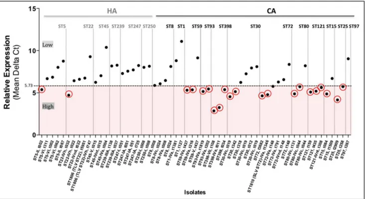
The analysis of the regression tree split the response variable into two distinct groups, according to thespatype of the strains. There was a group of strains with mean Delta Ct1–3#5.73, that was classified as a high expression group and a second group with a mean Delta Ct1–3.5.73 classified as a low expression group (Table 1, Table 2 and Figure 3). Overall the regression tree explained 60% of the variance in the data. This is mostly because there were strains expressing a low or high mean Delta Ctthat were classified in the same spatype; those were the cases ofspa
types t002, t019, t044 and t437.
Furthermore, we explored in each of thespatypes what other explanatory variables (ST, MRSA, MSSA and type of SCCmec) could differentiate the inclusion of some strains in the low or high expression group, but we found no associations with the variables we measured in the study.
We observed that thehlaexpression level varied within strains of the same ST (Figure 3; Table 1 and 2). In fact, in some cases the same ST comprised strains with both high and low levels of expression (ST5, ST15, ST22, ST30, ST59, ST72 and ST80). Moreover, we found that the expression rates did not differ significantly (P = 0.665) between MRSA and MSSA strains. However, we did find a correlation between the hlaexpression and the origin of the genetic backgrounds. Actually, strains of CA genetic backgrounds showed, in general, higher mean expression rates than strains of HA backgrounds (p = 0.003) (Figure 4). Among the 21 strains (36.2%, 21 out of 58) with high expression level, only two (9.5%) belonged to HA backgrounds (ST22-IVh, t032 and ST5-II, t002) whereas the majority (90.5%, n = 19) were represented by CA backgrounds (Table 1 and Table 2). Moreover, two additional CA strains, ST72-IVa-t148 and ST8-MSSA-t008, showed expression rates near the cutoff value (5.73), with 5.76 and 5.87, respectively. These were considered as belonging to the low-level expression group.
The three strains with the highest expression rate were ST398-VII-t108 (2.85), ST398-IVa-t011 (3.25) and ST25-MSSA-t2909 (4.16) and strains with the lowest rate were ST1806 (TLV ST22)-IVh-t747 (9.28), ST45-IVa-t004 (10.38) and ST1-MSSA-t127 (11.09).
We observed that some promoters and gene alleles (P6, P12/ P13, P21; andhla7,hla9,hla11) were exclusively associated to a
high expression level profile, while others (P3/P4/P5, P7, P8/P9, P11, P15, P17/P18, P23; andhla4,hla8,hla10) were exclusively associated to a low expression level (Table 1 and 2). But we also found promoter and gene allotypes that were associated to both high and low expression levels.
Discussion
Although Hla is one of the most importantS. aureusvirulence factors [10], to the best of our knowledge, this is the first study in which the variation in hla nucleotide sequence and gene expression was assessed in such a large and representative collection.
We found that the nucleotide sequence of hla was highly diverse. The high degree of diversity found within hla is in accordance to results obtained for other exotoxins, which are generally highly polymorphic [27]. Four non-synonymous substi-tutions (Q87 stop codon, D208E, S239T and T243S) were identified, that are located in two structural protein domains which are essential for Hla oligomerization and pore formation (Rim and Cap) [11,28,29]. The impact of these amino acid (aa) changes on
hlaactivity is uncertain. If by one hand, the aa changes described implicate differences in the molecular weight of the aa, that can have influence in the three dimensional structure stability and activity of the protein; on the other hand these aa changes did not match any of the aa previously described to be essential for Hla pore formation.
Furthermore, Walker and Bayley showed that multiple muta-tions in this same region (residues spanning Hla235–250) did not alter Hla activity in terms of binding, oligomerization or lysis. Thus, it would not be expected that S239T or T243S had significant biological impact in terms of toxin function. The unique mutation with an identified role in Hla function is the stop codon found in the ST36 and ST30 strains that was previously described by DeLeo and co-authors [30] to hinder toxin production and to originate a less virulent strain in a murine infection model. The true effect of the non-synonymous substitutions identified in our study in the activity of the protein would have to be tested by the construction of site directed mutagenesis mutants and by performing binding, oligomerization, hemolysis and in vivo
models assays.
The construction of phylogenetic trees from thehladefined the existence of two clusters with different levels of genetic diversity suggesting thathlais evolving at different rates in different genetic backgrounds. Interestingly, the most diverse cluster included the clonal types which are presently more disseminated or that emerged recently (like ST398). This might be related to the fact that these clones still need to evolve to evade the human immune system and not enough time as elapsed for the most adapted allele to have been selected [31]. On the other hand the recombination events detected in the hlagene in this study were all in strains belonging to the high genetic diversity cluster, suggesting that this mechanism might have been important in the most recent hla
evolution and diversification.
Interestingly, the phylogenetic tree constructed from the hla
gene was similar to that constructed from MLST genes, in the sense that both trees distributed the different STs similarly in two main clusters. This observation suggests thathlagene has evolved
Figure 2. Phylogenetic trees ofhlagene (A) and concatenated sequences of MLST alleles (B) from 23 STs representatives of the strains collection.The tree was constructed using MEGA 5 with Neighbour-joining method and bootstrap values provided as percents over 1000 replications. Branch length values are indicated and the percentage of replicate trees (bootstrap test) are shown next to the branches. The dashed line indicates the separation of the two evolutionary branches.
doi:10.1371/journal.pone.0098634.g002
Hla Evolution and Expression inS. aureusfrom Community and Hospital
together with theS. aureusgenetic background. A similar type of correlation with the genetic background was previously described for adhesins, either located in the core genome (clfA,clfB,fnbA,
map,sdrC, andspa) or accessory genome (ebpS,fnbB,sdrD, and
sdrE) [32]. Although this was the case for the great majority of
STs, we observed that four STs (ST22, ST121, ST59, ST93) were located in different clusters in thehlaand MLST trees. Our results suggest that recombination occurring at the hla level, might explain the different clustering of strains belonging to ST22 and ST121. No recombination events were, however, detected in
Figure 3. HA and CA strains relative expression distribution. Mean of expression rates from three biological replicates. Dashed line corresponding to the mean Ct value 5.73 results from the regression tree analysis which split strains in two distinct groups, atspatype level: a) high expression group - corresponding to strains with Mean Delta Ct#5.73 and b) low expression group- corresponding to strains with Mean Delta Ct.
5.73). Highlighted in red are the high expressing strains. doi:10.1371/journal.pone.0098634.g003
Figure 4. Distribution of the relativehlaexpression.Mean of relative expression of three independent readings. Expression comparison between a) MRSA and MSSA and b) HA and CA backgrounds using the Two-tailed Student’s t-test. Statistically significance (p#0.05) (**). doi:10.1371/journal.pone.0098634.g004
Hla Evolution and Expression inS. aureusfrom Community and Hospital
MLST genes orhlasequences of strains belonging to ST59 and ST93, suggesting that their displacement in the two trees could derive from different phenomena, like random mutation.
It was previously suggested that CA-MRSA expressed morehla
than HA-MRSA [9]. Results from our study allowed us to extend this conclusion to virtually all epidemic CA, but also in two particular cases of HA genetic backgrounds. The CA strains belonging to ST398, ST25, ST121 and ST93 showed uniformly high relative expression rates and strains belonging to ST36, ST45, ST239, ST247 and ST250 showed uniformly low expression rates. To understand if in fact these patterns of expression are characteristic of these clones, more strains within each clone should be studied forhlaexpression. Nevertheless, we could not correlate the hla expression rate with any particular polymorphism within the promoter or any aa substitution in the
hla gene. The results suggest that hla regulation is probably a result of combination of factors which are redundant, rather than associated to a single genetic event. In fact, it has been demonstrated by several authors that alpha-hemolysin is part of a complex regulatory network, that includes the main two-component systems (TCS) – Agr – that in turn is controlled by a diverse pool of regulatory networks that coordinately interact in response to external stimulus and cell signals, namely others TCS (SaeRS, ArlRS and SrrAB), alternative sigma factors (sB
), and transcription factors (e.g. SarS, SarT, Rot, SarA, SarZ) [33,34].
We showed that hla evolved together with the genetic background. Moreover, the most epidemic CA-MRSA genetic backgrounds express morehlathan the most epidemic HA-MRSA genetic backgrounds. However, the finding of frequent strain-to-strain variation in the expression level ofhlawithin strains of the same clonal types suggests thathlapolymorphisms cannot be used as genetic markers of virulence and investigators should remain cautious when inferring conclusions for the entire MRSA population from studies performed with a limited number of strains.
Supporting Information
Figure S1 Phylogenetic trees of thehlagene, promoter gene and concatenated sequences of both. The tree was constructed using MEGA 5 with Neighbour-joining method and bootstrap values provided as percents over 1000 replications. Branch length values are indicated and the percentage of replicate trees (bootstrap test) are shown next to the branches. The dashed line indicates the separation of the two evolutionary branches (L and H).
(TIF)
Figure S2 I. Growth curves for triplicates of each S. aureus
strain – Set C.II.Growth curves for triplicates of eachS. aureus
strain – Set D.III.Growth curves for triplicates of eachS. aureus
strain – Set E. (TIFF)
Figure S3 Internal sequences ofhlapromoter (highlighted blue) andhlagene (highlighted orange) used for analysis in this study. Primers used are highlighted. The sequence shown corresponds to the promoter and hla regions of USA300 strain from our collection blasted against USA300_TCH1516.
(TIF)
Table S1 Molecular characterization of the 73 MRSA and MSSA strains included in this study [35–50].
(DOC)
Author Contributions
Conceived and designed the experiments: HdL MM. Performed the experiments: AT. Analyzed the data: AT MM ACP JBN KS. Contributed reagents/materials/analysis tools: HdL MM HW. Wrote the paper: AT MM. Manuscript revision: HdL HW KS JBN KB SR ACP.
References
1. Deurenberg RH, Stobberingh EE (2009) The molecular evolution of hospital-and community-associated methicillin-resistantStaphylococcus aureus. Curr Mol Med 9: 100–115.
2. David MZ, Daum RS (2010) Community-associated methicillin-resistant Staphylococcus aureus: epidemiology and clinical consequences of an emerging epidemic. Clin Microbiol Rev 23: 616–687.
3. Otto M (2013) Community-associated MRSA: What makes them special? Int J Med Microbiol.
4. Kobayashi SD, Voyich JM, Burlak C, DeLeo FR (2005) Neutrophils in the innate immune response. Arch Immunol Ther Exp (Warsz) 53: 505–517. 5. Baba T, Takeuchi F, Kuroda M, Yuzawa H, Aoki K, et al. (2002) Genome and
virulence determinants of high virulence community-acquired MRSA. Lancet 359: 1819–1827.
6. Diep BA, Gill SR, Chang RF, Phan TH, Chen JH, et al. (2006) Complete genome sequence of USA300, an epidemic clone of community-acquired meticillin-resistantStaphylococcus aureus. Lancet 367: 731–739.
7. Burlak C, Hammer CH, Robinson MA, Whitney AR, McGavin MJ, et al. (2007) Global analysis of community-associated methicillin-resistant Staphylococcus aureusexoproteins reveals molecules produced in vitro and during infection. Cell Microbiol 9: 1172–1190.
8. Loughman JA, Fritz SA, Storch GA, Hunstad DA (2009) Virulence gene expression in human community-acquired Staphylococcus aureus infection. J Infect Dis 199: 294–301.
9. Li M, Cheung GY, Hu J, Wang D, Joo HS, et al. (2010) Comparative analysis of virulence and toxin expression of global community-associated methicillin-resistantStaphylococcus aureusstrains. J Infect Dis 202: 1866–1876. 10. Berube BJ, Bubeck Wardenburg J (2013)Staphylococcus aureusalpha-Toxin:
Nearly a Century of Intrigue. Toxins (Basel) 5: 1140–1166.
11. Song L, Hobaugh MR, Shustak C, Cheley S, Bayley H, et al. (1996) Structure of staphylococcal alpha-hemolysin, a heptameric transmembrane pore. Science 274: 1859–1866.
12. Valeva A, Palmer M, Bhakdi S (1997) Staphylococcal alpha-toxin: formation of the heptameric pore is partially cooperative and proceeds through multiple intermediate stages. Biochemistry 36: 13298–13304.
13. Novick RP, Ross HF, Projan SJ, Kornblum J, Kreiswirth B, et al. (1993) Synthesis of staphylococcal virulence factors is controlled by a regulatory RNA molecule. EMBO J 12: 3967–3975.
14. Ballal A, Ray B, Manna AC (2009)sarZ, asarAfamily gene, is transcriptionally activated by MgrA and is involved in the regulation of genes encoding exoproteins inStaphylococcus aureus. J Bacteriol 191: 1656–1665.
15. Liang X, Yu C, Sun J, Liu H, Landwehr C, et al. (2006) Inactivation of a two-component signal transduction system, SaeRS, eliminates adherence and attenuates virulence ofStaphylococcus aureus. Infect Immun 74: 4655–4665. 16. Liang X, Zheng L, Landwehr C, Lunsford D, Holmes D, et al. (2005) Global
regulation of gene expression by ArlRS, a two-component signal transduction regulatory system ofStaphylococcus aureus. J Bacteriol 187: 5486–5492. 17. Schmidt KA, Manna AC, Gill S, Cheung AL (2001) SarT, a repressor of
alpha-hemolysin inStaphylococcus aureus. Infect Immun 69: 4749–4758.
18. Liang X, Hall JW, Yang J, Yan M, Doll K, et al. (2011) Identification of single nucleotide polymorphisms associated with hyperproduction of alpha-toxin in Staphylococcus aureus. PLoS One 6: e18428.
19. Bubeck Wardenburg J, Bae T, Otto M, Deleo FR, Schneewind O (2007) Poring over pores: alpha-hemolysin and Panton-Valentine leukocidin inStaphylococcus aureuspneumonia. Nat Med 13: 1405–1406.
20. Bubeck Wardenburg J, Patel RJ, Schneewind O (2007) Surface proteins and exotoxins are required for the pathogenesis ofStaphylococcus aureuspneumonia. Infect Immun 75: 1040–1044.
21. Inoshima I, Inoshima N, Wilke GA, Powers ME, Frank KM, et al. (2011) A Staphylococcus aureuspore-forming toxin subverts the activity of ADAM10 to cause lethal infection in mice. Nat Med 17: 1310–1314.
22. Tamura K, Nei M, Kumar S (2004) Prospects for inferring very large phylogenies by using the neighbor-joining method. Proc Natl Acad Sci U S A 101: 11030–11035.
23. Vandenesch F, Kornblum J, Novick RP (1991) A temporal signal, independent ofagr, is required for hla but notspatranscription inStaphylococcus aureus. J Bacteriol 173: 6313–6320.
24. Zielinska AK, Beenken KE, Joo HS, Mrak LN, Griffin LM, et al. (2011) Defining the strain-dependent impact of the Staphylococcal accessory regulator
Hla Evolution and Expression inS. aureusfrom Community and Hospital
(sarA) on the alpha-toxin phenotype ofStaphylococcus aureus. J Bacteriol 193: 2948–2958.
25. De’ath G, Fabricius KE (2000) Classification and regression trees: a powerful yet simple thechnique for ecological data analysis. Ecology. Ecology 81: 3178–3192. 26. Morfeldt E, Taylor D, von Gabain A, Arvidson S (1995) Activation of alpha-toxin translation inStaphylococcus aureusby the trans-encoded antisense RNA, RNAIII. EMBO J 14: 4569–4577.
27. Wilson GJ, Seo KS, Cartwright RA, Connelley T, Chuang-Smith ON, et al. (2011) A novel core genome-encoded superantigen contributes to lethality of community-associated MRSA necrotizing pneumonia. PLoS Pathog 7: e1002271.
28. Montoya M, Gouaux E (2003) Beta-barrel membrane protein folding and structure viewed through the lens of alpha-hemolysin. Biochim Biophys Acta 1609: 19–27.
29. Walker B, Bayley H (1995) Key residues for membrane binding, oligomeriza-tion, and pore forming activity of staphylococcal alpha-hemolysin identified by cysteine scanning mutagenesis and targeted chemical modification. J Biol Chem 270: 23065–23071.
30. DeLeo FR, Kennedy AD, Chen L, Bubeck Wardenburg J, Kobayashi SD, et al. (2011) Molecular differentiation of historic phage-type 80/81 and contemporary epidemicStaphylococcus aureus. Proc Natl Acad Sci U S A 108: 18091–18096. 31. Castillo-Ramirez S, Harris SR, Holden MT, He M, Parkhill J, et al. (2011) The impact of recombination on dN/dS within recently emerged bacterial clones. PLoS Pathog 7: e1002129.
32. Kuhn G, Francioli P, Blanc DS (2006) Evidence for clonal evolution among highly polymorphic genes in methicillin-resistant Staphylococcus aureus. J Bacteriol 188: 169–178.
33. Novick RP (2003) Autoinduction and signal transduction in the regulation of staphylococcal virulence. Mol Microbiol 48: 1429–1449.
34. Thoendel M, Kavanaugh JS, Flack CE, Horswill AR (2011) Peptide signaling in the staphylococci. Chem Rev 111: 117–151.
35. Tavares A, Miragaia M, Rolo J, Coelho C, de Lencastre H (2013) High prevalence of hospital-associated methicillin-resistantStaphylococcus aureusin the community in Portugal: evidence for the blurring of community-hospital boundaries. Eur J Clin Microbiol Infect Dis 32: 1269–1283.
36. Oliveira DC, Tomasz A, de Lencastre H (2001) The evolution of pandemic clones of methicillin-resistant Staphylococcus aureus: identification of two ancestral genetic backgrounds and the associatedmecelements. Microb Drug Resist 7: 349–361.
37. Roberts RB, de Lencastre A, Eisner W, Severina EP, Shopsin B, et al. (1998) Molecular epidemiology of methicillin-resistantStaphylococcus aureusin 12 New York hospitals. MRSA Collaborative Study Group. J Infect Dis 178: 164–171. 38. Aires-de-Sousa M, Correia B, de Lencastre H (2008) Changing patterns in frequency of recovery of five methicillin-resistantStaphylococcus aureusclones in
Portuguese hospitals: surveillance over a 16-year period. J Clin Microbiol 46: 2912–2917.
39. Rolo J, Miragaia M, Turlej-Rogacka A, Empel J, Bouchami O, et al. (2012) High Genetic Diversity among Community-AssociatedStaphylococcus aureusin Europe: Results from a Multicenter Study. PLoS One 7: e34768.
40. Conceicao T, Tavares A, Miragaia M, Hyde K, Aires-de-Sousa M, et al. (2010) Prevalence and clonality of methicillin-resistantStaphylococcus aureus(MRSA) in the Atlantic Azores islands: predominance of SCCmectypes IV, V and VI. Eur J Clin Microbiol Infect Dis 29: 543–550.
41. Sa-Leao R, Santos Sanches I, Dias D, Peres I, Barros RM, et al. (1999) Detection of an archaic clone ofStaphylococcus aureuswith low-level resistance to methicillin in a pediatric hospital in Portugal and in international samples: relics of a formerly widely disseminated strain? J Clin Microbiol 37: 1913–1920. 42. Amorim ML, Aires de Sousa M, Sanches IS, Sa-Leao R, Cabeda JM, et al. (2002) Clonal and antibiotic resistance profiles of methicillin-resistant Staphy-lococcus aureus(MRSA) from a Portuguese hospital over time. Microb Drug Resist 8: 301–309.
43. Milheirico C, Oliveira DC, de Lencastre H (2007) Multiplex PCR strategy for subtyping the staphylococcal cassette chromosomemectype IV in methicillin-resistant Staphylococcus aureus: ‘SCCmec IV multiplex’. J Antimicrob Che-mother 60: 42–48.
44. Richardson JF, Reith S (1993) Characterization of a strain of methicillin-resistant Staphylococcus aureus(EMRSA-15) by conventional and molecular methods. J Hosp Infect 25: 45–52.
45. Oliveira DC, Milheirico C, Vinga S, de Lencastre H (2006) Assessment of allelic variation in theccrABlocus in methicillin-resistantStaphylococcus aureusclones. J Antimicrob Chemother 58: 23–30.
46. Faria NA, Oliveira DC, Westh H, Monnet DL, Larsen AR, et al. (2005) Epidemiology of emerging methicillin-resistantStaphylococcus aureus(MRSA) in Denmark: a nationwide study in a country with low prevalence of MRSA infection. J Clin Microbiol 43: 1836–1842.
47. Sanches IS, Ramirez M, Troni H, Abecassis M, Padua M, et al. (1995) Evidence for the geographic spread of a methicillin-resistantStaphylococcus aureusclone between Portugal and Spain. J Clin Microbiol 33: 1243–1246.
48. de Lencastre H, Chung M, Westh H (2000) Archaic strains of methicillin-resistant Staphylococcus aureus: molecular and microbiological properties of isolates from the 1960s in Denmark. Microb Drug Resist 6: 1–10.
49. Crisostomo MI, Westh H, Tomasz A, Chung M, Oliveira DC, et al. (2001) The evolution of methicillin resistance inStaphylococcus aureus: similarity of genetic backgrounds in historically early methicillin-susceptible and -resistant isolates and contemporary epidemic clones. Proc Natl Acad Sci U S A 98: 9865–9870. 50. McDougal LK, Steward CD, Killgore GE, Chaitram JM, McAllister SK, et al. (2003) Pulsed-field gel electrophoresis typing of oxacillin-resistantStaphylococcus aureusisolates from the United States: establishing a national database. J Clin Microbiol 41: 5113–5120.
Hla Evolution and Expression inS. aureusfrom Community and Hospital


