Article
Jagged 1 Rescues the Duchenne Muscular
Dystrophy Phenotype
Graphical Abstract
Highlights
d
Escaper GRMD dogs show that a normal lifespan is possible
without muscle dystrophin
d
Jagged1,
a Notch ligand, is upregulated in mildly affected
dystrophin deficient dogs
d
Jagged1
overexpression can rescue the phenotype of
dystrophin deficient zebrafish
Authors
Natassia M. Vieira, Ingegerd Elvers,
Matthew S. Alexander, ...,
Kerstin Lindblad-Toh, Louis M. Kunkel,
Mayana Zatz
Correspondence
[email protected] (L.M.K.),
[email protected] (M.Z.)
In Brief
The study of two exceptional dogs that
escaped from the severe phenotype
associated with dystrophin deficiency
unveils a genetic modifier that allows
functional muscle and normal lifespan
despite the complete absence of
dystrophin.
Accession Numbers
GSE69040
Article
Jagged 1 Rescues the Duchenne
Muscular Dystrophy Phenotype
Natassia M. Vieira,1,2,3Ingegerd Elvers,4,5Matthew S. Alexander,1,2,6Yuri B. Moreira,7Alal Eran,2Juliana P. Gomes,3
Jamie L. Marshall,1,2Elinor K. Karlsson,4,10Sergio Verjovski-Almeida,7,8Kerstin Lindblad-Toh,4,5,11
Louis M. Kunkel,1,2,9,11,*and Mayana Zatz3,11,*
1The Division of Genetics and Genomics, Boston Children’s Hospital, Boston, MA 02115, USA 2Department of Pediatrics and Genetics, Harvard Medical School, Boston, MA 02115, USA
3Human Genome and Stem Cell Center, Biosciences Institute, University of Sa˜o Paulo, Sa˜o Paulo 05508-090, Brazil 4Broad Institute of Harvard and Massachusetts Institute of Technology, Cambridge, MA 02142, USA
5Science for Life Laboratory, Department of Medical Biochemistry and Microbiology, Uppsala University, Box 597, 751 24, Uppsala, Sweden 6The Stem Cell Program at Boston Children’s Hospital, Boston, MA 02115, USA
7Departamento de Bioquı´mica, Instituto de Quı´mica, Universidade de Sa˜o Paulo, Sa˜o Paulo, Brazil, 05508-000 8Instituto Butantan, Sa˜o Paulo 05508-050, Brazil
9The Manton Center for Orphan Disease Research at Boston Children’s Hospital, Boston, MA 02115, USA
10Program in Bioinformatics and Integrative Biology, University of Massachusetts Medical School, Worcester, MA 01605, USA 11Co-senior author
*Correspondence:[email protected](L.M.K.),[email protected](M.Z.) http://dx.doi.org/10.1016/j.cell.2015.10.049
SUMMARY
Duchenne muscular dystrophy (DMD), caused by
mutations at the
dystrophin
gene, is the most
com-mon form of muscular dystrophy. There is no cure
for DMD and current therapeutic approaches to
restore dystrophin expression are only partially
effec-tive. The absence of dystrophin in muscle results in
dysregulation of signaling pathways, which could
be targets for disease therapy and drug discovery.
Previously, we identified two exceptional Golden
Retriever muscular dystrophy (GRMD) dogs that
are mildly affected, have functional muscle, and
normal lifespan despite the complete absence of
dystrophin. Now, our data on linkage, whole-genome
sequencing, and transcriptome analyses of these
dogs compared to severely affected GRMD and
control animals reveals that increased expression
of
Jagged1
gene, a known regulator of the Notch
signaling pathway, is a hallmark of the mild
pheno-type. Functional analyses demonstrate that
Jagged1
overexpression ameliorates the dystrophic
pheno-type, suggesting that
Jagged1
may represent a
target for DMD therapy in a dystrophin-independent
manner.
INTRODUCTION
Duchenne muscular dystrophy (DMD) is an X-linked disorder caused by mutations indystrophin(Hoffman et al., 1987), which affects 1 in 3,500 to 5,000 boys (Axelsson et al., 2013; Mendell et al., 2012). Deficiency of muscle dystrophin causes progres-sive myofiber degeneration and muscle wasting (Hoffman
et al., 1987). The first symptoms are usually evident at 3–5 years of age, with loss of ambulation between 9 and 12 years. Death occurs in the second or third decade due to respiratory or cardiac failure. While there are several treatments under devel-opment or currently in use—particularly corticotherapy, which aims to ameliorate symptoms and slow down the disease pro-gression—there is still no cure for DMD (Bushby et al., 2010; Guiraud et al., 2015). Allelic to DMD, Becker muscular dystrophy (BMD) is caused by mutations that do not affect the reading frame of thedystrophintranscript; the result is a semi-functional, truncated dystrophin protein (Koenig et al., 1989). DMD muscle shows a complete absence of dystrophin, whereas in the BMD muscle there is a variable amount of partially functional dystro-phin (Monaco et al., 1988). Differently from DMD, where most boys carrying null mutations show a severe phenotype, BMD patients show a variable clinical course. Genotype/phenotype correlation studies suggest that the severity of the phenotype is dependent on the amount of muscle dystrophin or the site of the mutation/deletion in thedystrophingene (Koenig et al., 1989; Passos-Bueno et al., 1994; Vainzof et al., 1990)
DMD therapeutic approaches currently under development aim to rescuedystrophinexpression in the muscle (Fairclough et al., 2013). Pre-clinical and clinical studies include exon-skipping (Goemans et al., 2011; Mendell et al., 2013; van Deute-kom et al., 2007), AAV-delivery ofm-dystrophin (Mendell et al.,
2010), and nonsense suppression to induce ‘‘readthrough’’ of nonsense mutations (Kayali et al., 2012). While AAV-delivery led tom-dystrophin expression in skeletal muscle, T cell immunity
against dystrophin epitopes was reported (Mendell et al., 2010). Also, the success of the dystrophin-based therapies relies on the quality of the recipient muscle. This requires the development of dystrophin-independent therapies to improve the muscle condi-tion targeting the altered signaling pathways.
show differences in skeletal muscle pathology in response to dys-trophin-deficiency (Bassett and Currie, 2004; Chapman et al., 1989; Im et al., 1996; Kornegay et al., 1988; Zucconi et al., 2010). The dystrophin-deficient fish model sapje shows some phenotypic variability, but nearly all fish die during the first weeks of life and all show abnormal muscle structure as measured by birefringence under polarized light (Bassett and Currie, 2004). Themdxmouse is the most widely used animal model for DMD, even though its mild phenotype does not mimic severe human DMD symptoms (Bulfield et al., 1984). The most similar to the hu-man condition is the golden retriever muscular dystrophy (GRMD) dog (Bassett et al., 2003; Cooper et al., 1988; Kornegay et al., 1988; Sicinski et al., 1989). These animals carry a point mutation on a splicing site that causes the skipping of exon 7 and a prema-ture stop codon, resulting in the absence of dystrophin. GRMD dogs and DMD patients share many similarities in disease patho-genesis, including early progressive muscle degeneration and at-rophy, fibrosis, contractures, and grossly elevated serum creatine kinase (CK) levels (Kornegay et al., 1988; Sharp et al., 1992). Early death may occur within the first weeks of life but usually occurs around 1–2 years of age as a result of respiratory failure or cardio-myopathy. The great majority of GRMD dogs do not survive beyond age two. In the Brazilian GRMD colony at Biosciences Institute at the University of Sa˜o Paulo, we have described two exceptional dogs presenting a very mild phenotype clearly distin-guishable from other affected dogs despite the absence of muscle dystrophin. Histopathological and immunohistochemistry anal-ysis of their muscle showed typical features of a dystrophic process with variability in fiber size, splitting, degeneration, and infiltrating connective tissue (Zucconi et al., 2010).
These two exceptional, related GRMD dogs (here called ‘‘escapers’’) remained fully ambulatory with normal lifespans, a phenotype never reported before for GRMD. They fall outside the known GRMD phenotypic range of variability, differing significantly from typically affected dogs despite their dystrophic muscle, absence of muscle dystrophin, elevated serum CK levels, and lack of evidence of utrophin upregulation (Zatz et al., 2015; Zucconi et al., 2010). Most importantly, these GRMD dogs show that it is possible to have a functional muscle in a mid-size dystrophin-deficient animal.
In this study, we set out to answer the following question: how do these escaper dogs have a fully functional muscle without dystrophin? Skeletal muscle of DMD patients undergoes waves or cycles of degeneration followed by regeneration. Muscle repair is a regulated process that comprises different cell types and signaling molecules, but additional factors and genetic modifiers involved in DMD pathogenesis remain poorly under-stood, representing new potential therapeutic targets. Genetic modifiers have been reported in DMD patients with a slower progression, but none were associated with a nearly normal phenotype (Flanigan et al., 2013). Here, through three indepen-dent approaches, we iindepen-dentified a modifier gene,Jagged1, which can modulate the GRMD phenotype. Using a mixed model asso-ciation and linkage analysis, we identified a chromosomal region associated with the escaper phenotype. One gene within this re-gion showed altered expression when comparing muscle tissue of escaper and affected dogs. By whole-genome sequencing, we found a variant present only in escaper GRMD dogs that
creates a novel myogenin binding site in theJagged1promoter. Overexpression of jagged1 in dystrophin deficient zebrafish rescues the dystrophic phenotype in this zebrafish model. This suggests thatJagged1, when increased in expression in muscle, can rescue dystrophin-deficient phenotypes in two different animal models, pointing to a new potential therapeutic target.
RESULTS
Escaper GRMD Dogs Share a Common Haplotype Different from Affected
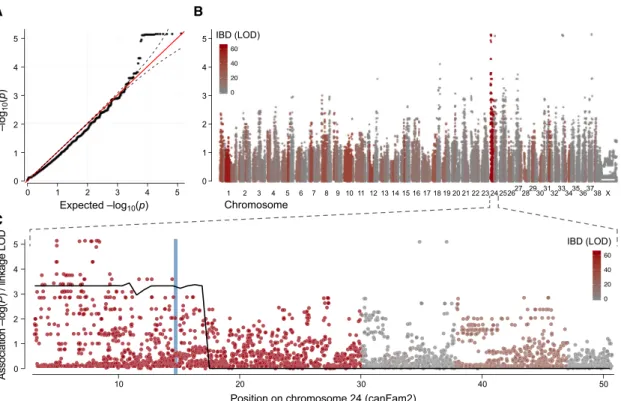
To understand the genetic basis behind the escaper phenotype in GRMD dogs, we performed a genome-wide mapping analysis comparing two related escaper GRMD dogs—the only two GRMD escapers reported to date—to 31 severely affected GRMD dogs from the same breeding population. All GRMD dogs were confirmed to carry the originally described point mu-tation (a change from adenine to guanine transition) in the intron 6 of thedystrophingene. This mutation ablates a splicing site and exon 7 is skipped from the mature mRNA. The absence of exon 7 causes a premature stop codon at exon 8 (Cooper et al., 1988; Sharp et al., 1992). Based on survival age and functional capac-ity, they were classified as escaper or affected (binary). All the dogs showing the standard range of phenotypic variability seen in GRMD dogs were classified as affected in this study. Our aim was to identify a single gene responsible for the milder phenotype seen in the two escaper dogs. We performed a two-step mapping analysis. First, we carried out an association study, utilizing the power of the many severely affected dogs ex-pected to lack the modifier locus. This was followed by segrega-tion analysis, taking advantage of the fact that the two escapers came from a well-defined pedigree in which a transmission-based test could be used. All dogs were genotyped using the Illumina CanineHD 170K SNP array. We tested for association genome wide using the mixed model approach implemented in EMMAX (Kang et al., 2010) to correct for population structure (Figure 1A) and identified strongly associated SNPs (p < 1x10⁵) on chromosomes 24, 33, and 37 (Figure 1B). We then
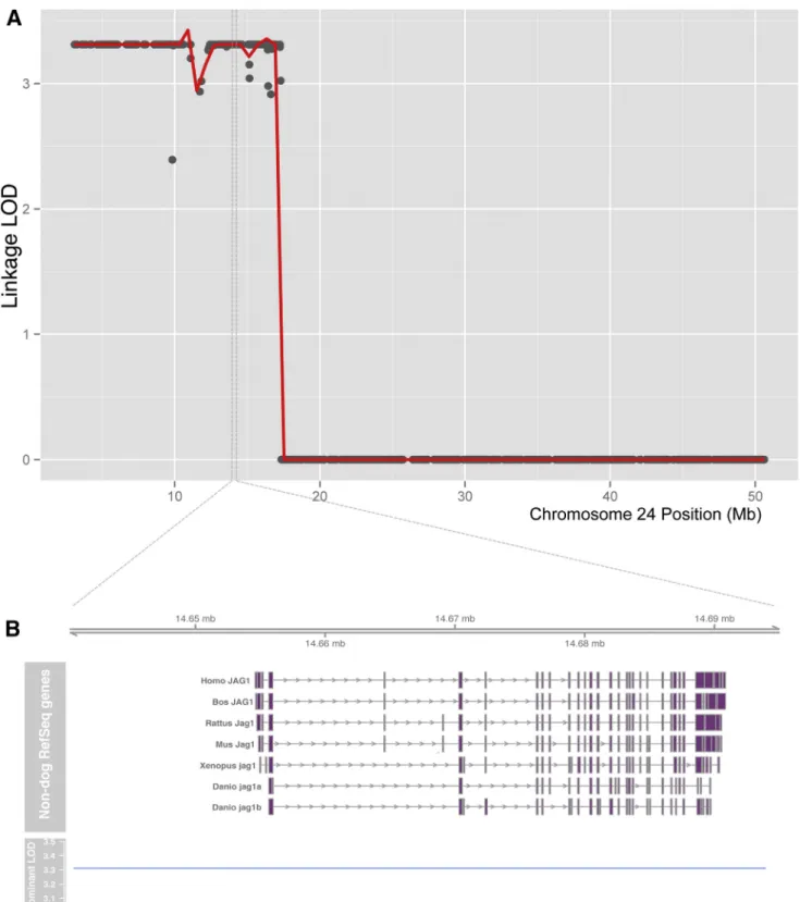
measured identity by descent (IBD) across the genome between the two escapers using Beagle (Browning and Browning, 2007). Only the associated SNPs on chromosome 24 also overlapped a segment of IBD in the two escapers, consistent with a single origin of the causative mutation (Figure 1B). The 27 Mb segment showing both IBD and association with the escaper pheno-type (CanFam2, cfa24:3,073,196-30,066,497) contains approxi-mately 350 protein-coding genes. Linkage analysis using Merlin (Abecasis et al., 2002) strongly confirmed this region, with a maximal parametric LOD score of 3.31 (dominant inheritance model with complete penetrance,Figure S1). No other genomic regions showed any signs of linkage (Figure S2). Thus, conver-gent IBD, association, and linkage analyses all pointed to the same 27 Mb region on chromosome 24 (Figure 1C).
Muscle Gene Expression Profile of Escaper and Affected GRMD Dogs
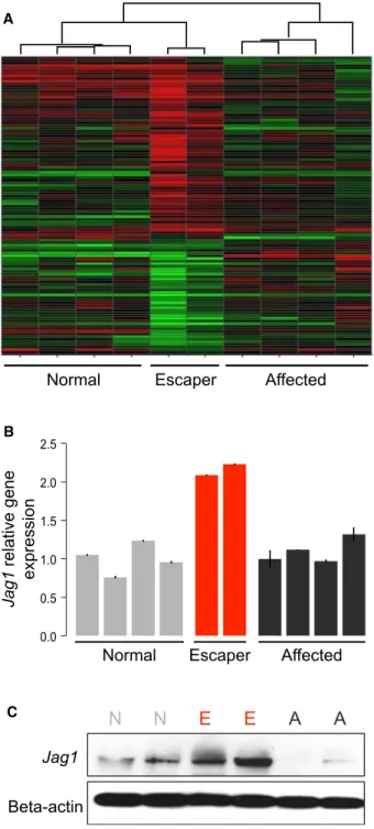
we compared muscle gene expression of the two escapers, four affected, and four wild-type dogs at two years of age. We found very similar muscle gene expression patterns in the two escaper GRMD dogs, which were more similar to muscle from wild-type dogs than from the affected dogs. In total, 114 genes were found to be differentially expressed between escapers and affected GRMD dogs, as shown by unsupervised hierarchical clustering of all ten samples (Figure 2A). Of these, 65 genes were also differ-entially expressed between escapers and wild-type dogs (Table S1), implicating them in a possible compensatory mechanism active in only the escaper dogs. Only one of these 65 genes, Jag-ged1, is located under the association peak on chromosome 24.
Jagged1mRNA levels were two times higher in the escapers when compared to both wild-type and severely affected dogs (Figure 2B). Further protein level analysis confirmed the mRNA findings (Figure 2C).
Whole-Genome Sequence of Escaper Dogs
To identify potentially causative variants behind the differential gene expression pattern observed in the escaper dogs, we performed whole-genome sequencing on three dogs (the two escapers and one severely affected related dog). We hypothe-sized that the compensatory variation would be novel, as the escaper phenotype had not previously been seen in GRMD
dogs worldwide. We looked for variants located under the association peak on chromosome 24 and focused on theJagged1
locus (including 3 KB upstream and downstream of the gene) in search for a variant present only in the escapers and not in the affected GRMD dogs. A total of1,300 variants were detected within the escaper-associated region on chromosome 24. All variants were lifted over to the human genome, and those present in muscle enhancer regions near the promoters of the two isoforms ofJagged1expressed in skeletal muscle (Figure 3A) (Hoeppner et al., 2014) were further analyzed. Since the escaper variant was hypothesized to be novel, all variants detected in previous extensive canine sequencing efforts (Axelsson et al., 2013) were excluded. After this filtering, only a single point variant was found to follow the escaper haplotype: a heterozygote G>T change in the promoter region ofJagged1(cfa24:11655709,Figure 3A). Sanger sequencing of theJagged1candidate escaper variant was per-formed in the escaper extended pedigree, including the first escaper (M1M4), his offspring, and a sibling’s offspring (M1M5) (Figure S3). We also sequenced key breeders of the kennel and found that the variant is specific to the escapers’ pedigree and was introduced in a single outcross (B1F3 mate). All affected dogs lacked theJagged1variant, while both escapers were het-erozygous. Thus, the novel Jagged1mutation segregates with the escaper phenotype in this family. Four additional individuals
A B
C
Figure 1. Combining Association, Linkage, and Identity-By-Descent Analysis Identifies a 30 Mb Candidate Region on Chromosome 24
carried the candidate variant: three were stillborn puppies and the fourth was a GRMD puppy that died at 6 months of age from an accidental ingestion of a foreign object. This puppy (K2M11) was fully ambulatory with a similar phenotype to the two escaper dogs, but he was classified as affected in the mapping analysis since we cannot predict his adult phenotype with confidence.
Functional Analysis of Jagged1 Variant
To understand the effects of the escaper variant, we performed different functional analyses. This candidate variant was found to be conserved across 29 eutherian mammals, suggesting a reg-ulatory potential for this region (Figures 3A and 3B). Transcription factor binding site analysis, using TRAP (Manke et al., 2010) and TRANSFAC (Matys et al., 2006), revealed that this G>T change creates a novel myogenin binding site (Figure 3C) with a high information content for the mutant allele (T) in the myogenin consensus binding motif (Figure 3D). Myogenin is a muscle-spe-cific transcription factor involved in muscle differentiation and repair (Wright et al., 1989). To determine whether the variant af-fects DNA binding by myogenin, we carried out electrophoretic mobility shift assays (EMSAs) using muscle cell nuclear extracts and biotin-labeled oligonucleotide probes containing either the wild-type (WT) or escaper (E) genotype. The oligonucleotide probe containing the escaper T allele robustly bound the myoge-nin protein, whereas an oligonucleotide probe contaimyoge-ning the WT G allele did not bind at all (Figure 3E). A competition assay showed that an unlabeled escaper probe efficiently competed with the binding of the labeled escaper probe. In contrast, the unlabeled WT probe had no effect on the binding activity of the labeled escaper probe, indicating a specific interaction between the escaper allele and myogenin (Figure 3E). To evaluate whether the novel myogenin binding site found in the escaper dogs was driving the increased expression ofJagged1,we performed a luciferase reporter assay using Jagged1 upstream promoter sequences containing either the WT sequence or the escaper variant fused to a luciferase reporter. Luciferase vectors contain-ing either WT or escaper sequence were transfected into muscle cells (myoblasts) and human embryonic kidney cells (HEK293T) along with constructs that overexpress either myogenin or another E-box myogenic factor (MyoD) as control. On HEK293K cells, overexpression of myogenin was able to activate the expression of the escaperJagged1reporter 3-fold, but showed no activation of the WT reporter (Figure 3F). As predicted, the overexpression of MyoD did not activate either the WT or escaper
Jagged1luciferase reporter (Figure 3F). Similarly, myoblasts (that endogenously express myogenin) transfected with the escaper vector showed a similar luciferase activation that was three times higher than the WT vector, notwithstanding the presence of overexpression vectors (Figure 3F). These results demonstrate that the creation of the novel myogenin binding site in the escaper
Jagged1promoter is essential for driving the increase ofJagged1
expression in the escaper dog skeletal muscles.
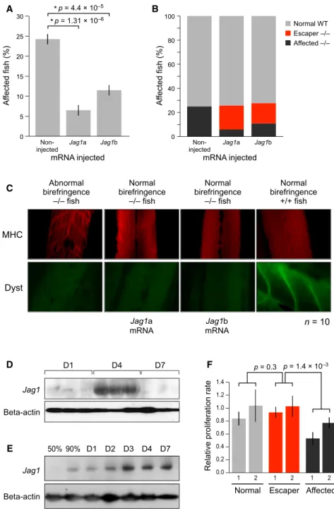
In Vivo Overexpression of Jagged1 Rescues sapje Muscle Phenotype
To evaluate if the overexpression ofJagged1can ameliorate the dystrophic muscle phenotype in other species, we used the severely affected dystrophic sapje zebrafish DMD model.
A
B
C
Figure 2. AlteredJagged1Expression in Escaper GRMD Dogs
(A) mRNA microarray comparing muscle gene expression of escaper GRMD dogs with related severely affected and WT dogs.
(B) mRNA expression of escaper dogs confirming the expression array find-ings. RelativeJagged1gene expression in muscle samples of escaper GRMD dogs as compared to related severely affected and WT dogs; bars indicate SD from the mean.
Muscle phenotype was assayed using birefringence, where fish are placed under a polarized light and dystrophin-negative fish show a decrease in the amount of light, indicative of muscle
tearing or muscle fiber disorganization. In four separate experi-ments, we injected approximately 200 fertilized one-cell stage eggs fromsapjeheterozygous fish matings with mRNA of either
A
B
C
D
F
E
Figure 3. Variant Located in theJagged1Promoter of Escaper GRMD Dogs
(A) Dog and HumanJagged1locus. Box: variant at dog chr24:11,644,709. (B) Conservation of the variant position.
(C) Predicted transcription factor binding site at the region with the base pair change.
(D) Consensus sequence of myogenin binding site, demonstrating the high information content of the T allele. (E) Electromobility shift assay (EMSA) showing myogenin binding to mutated probe (E) and not to the WT probe.
one of the zebrafishjagged1genetic copies of the mammalian
Jagged1 gene:jagged1a or jagged1b. In all experiments, an average of 24% of the non-injectedsapjefish exhibited a typical affected dystrophic, patchy birefringence phenotype. This pro-portion is within the 21%–27% expected range of affected fish of a heterozygoussapjemating. In contrast, fish injected with eitherjagged1aorjagged1bshowed a significantly lower percentage of fish with poor birefringence (p = 1.313106
for
jagged1a, p = 4.43105
for jagged1b,Figure 4A). Genotypic analysis revealed that about 75% of dystrophin-null fish injected withjagged1aand 60% of of those injected withjagged1bhad normal birefringence, which demonstrated a common rescue
A B
C
D
E
F
Figure 4. Functional Analysis of jagged1
Expression
(A) Percent affectedsapjefish as determined by birefringence assay at 4 dpf. Note fewer affected fish in the jagged1 injected sapjecohort. Four separate injection experiments were performed. (B) Genotype ofsapjeinjected fish withjagged1a and jagged1b as compared to non-injected sapjefish. In red are dystrophin-null fish with a WT phenotype, recovered byjagged1overexpression. (C) Immunofluorescence ofjagged1aandjagged1b overexpression in thesapjefish. WT, phenotypi-cally affected homozygous fish for the dystrophin mutation andjagged1aandjagged1binjected with normal birefringence (recovered) were stained for myosin heavy chain (MCH) and dystrophin anti-bodies. Note the organization of the muscle fibers in the recovered fish muscle comparable to the WT fish(n = 10)even without dystrophin. Photographs were taken at 20x magnification.
(D)Jagged1protein levels in the muscle of car-diotoxin injured mice one, four, and seven days after injury.
(E)Jagged1protein levels in muscle cells during in vitro muscle differentiation.
(F) Muscle cell proliferation rate, as measured by MTT, of two WT, two escaper, and two affected GRMD dogs. Error bars indicate SEM (n = 2, three replicates).
from the muscle lethality phenotype ( Fig-ure 4B). These results indicate that increasing jagged1 expression rescues most dystrophin-null fish from developing the abnormalities typically seen in dystro-phin-null muscle. To further evaluate the
jagged1a and jagged1b overexpression
sapjefish, we performed immunostaining on individual fish bodies using a myosin heavy chain (MHC) antibody to evaluate muscle structure. In WT fish, MHC was clearly expressed and showed that mus-cle fibers were normal. Interestingly, MHC staining ofjagged1mRNA-injected dystrophin-null rescued fish showed normal myofiber structure similar to that of WT fish, whereas affected, non-injected
dystrophin-null fish demonstrated clear muscle abnormalities (Figure 4C).
Jagged1 Expression during Muscle Regeneration and Cell Proliferation in Mice and Dogs
proliferation assay using myogenic cells from biopsies of age-matched dogs. Escaper dogs’ muscle showed typical dystro-phic features (Zucconi et al., 2010) as evidenced by cycles of degeneration and regeneration, which is not seen in normal muscle. Because of these cycles and consistent activation, myogenic cells from affected GRMD dogs are expected to divide less frequently. We show that muscle cells from escaper dogs divide significantly faster than those from affected dogs ( Fig-ure 4F). These results are consistent with previous findings that show that overexpression of the Notch intracellular domain (NICD) expands the proliferative capacity of activated muscle satellite cells in vitro and in vivo (Wen et al., 2012).
DISCUSSION
Animal models for DMD are important tools for developing new therapeutic approaches. Among the different animal models for muscular dystrophy, the GRMD dog is the closest to the human condition. Both GRMD dogs and DMD patients have a severe phenotype as well as many phenotypic and biochem-ical similarities, including early progressive muscle degenera-tion and atrophy, fibrosis, contractures, and elevated serum creatine kinase levels. We identified two dogs that escaped from the typical severe phenotype associated with dystrophin deficiency. Using a combined approach of mapping and identity by descent, we identified a candidate region of associ-ation with the escaper phenotype. Only one gene within this region showed altered expression in escaper and affected dogs:Jagged1. We found a candidate variant at an upstream, conserved position creating a new muscle-specific transcrip-tion factor binding site that drives Jagged1 overexpression.
Jagged1is also in the region associated to the mild pheno-type observed in a muscular dystrophy mouse model on the MRL (Murphy Roths Large) ‘‘superhealing’’ background. These mice show enhanced muscle regeneration and reduced dystrophic pathology. This healing phenotype was mapped to a region containing 49 genes that includes theJagged1 lo-cus (Heydemann et al., 2012).
The role ofJagged1in skeletal muscle development and dis-ease has yet to be fully elucidated.Jagged1is a Notch ligand (Lindsell et al., 1995). The Notch signaling pathway represents a central regulator of gene expression and is critical for cellular proliferation, differentiation, and apoptotic signaling during all stages of embryonic muscle development. The Notch pathway also plays an important role in muscle regeneration (Conboy and Rando, 2002; Wen et al., 2012), and overexpression ofNotch
has been shown to improve muscle regeneration in aged mice (Conboy et al., 2003). Moreover, Notch signaling has been shown to be dysregulated in muscle satellite cells and dystro-phin-deficient muscles frommdxmice (Jiang et al., 2014). Addi-tionally, there is an even more pronounced dysregulation of Notch signaling in the muscle satellite cell in the severemdx/utrn
double knockout mice (dKO) that have early lethality at two to four months due to a breakdown of the diaphragm muscles (Church et al., 2014; Mu et al., 2015). Here, we observed greater proliferative capacity of the escaper dogs’ myoblasts, suggest-ing thatJagged1overexpression might be involved in muscle cell proliferation and repair. These results are consistent with
previous findings, which demonstrate thatJagged1 overexpres-sion stimulates cell proliferation, suggesting thatJagged1-based therapy might be able to induce regeneration in a tissue-specific manner (Collesi et al., 2008). Our data show that Jagged1
expression is upregulated at day 4 after cardiotoxin-induced injury in mouse, a time point when myoblasts proliferate and fuse to promote muscle regeneration (Couteaux et al., 1988). Furthermore, Jagged1/Notch signaling has been shown to promote the expansion and differentiation capacity of bone marrow-derived stromal/stem cells (BMSCs) to promote skeletal regeneration (Dong et al., 2014). In endothelial cells, genetic
Jagged-1 overexpression resulted in endothelial branching of vasculature processes; while conversely,Jagged-1endothelial deletion blocked angiogenic growth inJagged-1eKO mice ( Pe-drosa et al., 2015). Indeed,Jagged-1overexpression leads to the activation of vasculature progenitor cells from quiescence, in a manner similar to that of muscle satellite cell activation (Ottone et al., 2014). Thus, it is likely that the endogenous overexpression ofJagged-1that occurs in the muscles of the escaper dogs is driving myogenic cell proliferation and potential muscle growth that occurs in mesodermal lineages. A proof-of-principle exper-iment in which the Notch downstream transcription factor Rbp-jk was deleted in muscle satellite cells demonstrated that inhibition of Notch activation was detrimental to both muscle growth and muscle satellite cell expansion (Bjornson et al., 2012). All these findings suggest thatJagged1is likely to be a mediator of the regenerative process that is disrupted in dystrophin-deficient muscles and has potential as a novel therapy target to mitigate DMD pathological progression.
Although the great majority of DMD patients show a severe course, exceptional cases of dystrophin-deficient patients with a milder phenotype have been identified. We have previously reported two patients carrying null mutations, with no skeletal muscle dystrophin present via immunofluorescent staining or western blot analysis, and a milder course including the mainte-nance of ambulation well into their second decade of life (Zatz et al., 2014). More recently, a dystrophin-negative patient who remained ambulant until age 30 was also reported ( Castro-Gago, 2015). Several other genetic modifiers are known to affect the severity of the clinical symptoms of Duchenne muscular dys-trophy (LTBP4,SPP1,TGFBR2). However, none of these genetic variants have been shown to fully restore or delay substantially the symptoms of dystrophin-deficiency in DMD boys (Bello et al., 2012; Flanigan et al., 2013; Pegoraro et al., 2011; Piva et al., 2012). Furthermore, it would be of great interest to examine the genomes of DMD boys with varying clinical symptoms and determine if variants inJagged1 or other Notch signaling factors exist and are causative for any variation of the dystrophic disease progression. The Notch signaling pathway, specifically
transcription factor that could increase expression ofJagged1in all of the skeletal muscles of DMD patient.
There is currently no cure for DMD, and existing therapies aiming to rescuedystrophinexpression are only partially effec-tive. Here, we show that the overexpression ofJagged1is likely to modulate the dystrophic phenotype in dystrophin-deficient GRMD dogs. We also show that overexpression ofjagged1 res-cues the dystrophic phenotype in a severe DMD model: thesapje
zebrafish. Our study highlights the possibilities of across-spe-cies analysis to identify and validate disease-modifying genes and associated pathways. These results suggest thatJagged1
may be a new target for DMD therapeutic efforts in a dystro-phin-independent manner, which will complement existing ap-proaches. In addition, further investigation on the gene target
Jagged1will contribute to a better understanding of the disease pathogenesis and molecular physiology.
EXPERIMENTAL PROCEDURES
GRMD dogs were classified for this study in two groups based on full ambula-tory capacity and survival age. The escapers group included the GRMD dogs that were fully ambulatory (can walk and run) at 9 years old. One escaper dog (M1M4) died at 11 years old from a cardiac arrest (Zatz et al., 2015) and the second one (H3M10) is now 9.5 years old and shows full ambulation. The affected group included the GRMD dogs that died before 5 years old with ambulatory difficulties, respiratory failure, and cardiopathy; this group includes stillbirths, neonatal death, and one dog that was full ambulatory when he died by ingesting a foreign object at 6-months-old (K2M11); all were confirmed to carry the GRMD mutation. DNA from GRMD dogs with and without the escaper phenotype was genotyped using the Illumina canine 170,000 SNP array and was compared using association, linkage, and IBD mapping. The threshold for genome-wide significance for each association analysis was defined based on the 95% confidence intervals (CIs) calculated from the beta distribution of observed p values, as previously described (Wellcome Trust Case Control, 2007). The likelihood of the two escapers being identity by descent (IBD) at each SNP was estimated based on haplotype frequencies in the full pedigree using Beagle 4 (release v4.r1274) with default parameter settings (Browning and Browning, 2007). Linkage analysis was performed using MERLIN (Abecasis et al., 2002) 1.1.2 to first remove inconsistent geno-types and then calculate LOD scores (logarithm of the odds ratios) using a dominant parametric model with complete penetrance. Expression analysis from the same dogs was performed using two-color microarray-based gene expression analysis. Genes differentially expressed between WT, escaper, and affected animals were identified with the significance analysis of microar-ray (SAM) statistical approach. False discovery rate (FDR) was 5%. Whole-genome sequencing was performed to 30x depth of three dogs (two escapers and one affected dog). Samples were sequenced on an Illumina HiSeq 2000, and sequencing reads were aligned to the CanFam 3.1 reference sequence using BWA. Following GATK base quality score recalibration, indel realign-ment, and duplicate removal, SNP and INDEL discovery was performed. To assess myogenin binding to candidate mutation, EMSA was performed using biotin labeled or unlabeled competitors probes and the LightShift Chemolumi-nescent EMSA kit (Thermo Scientific) following manufacturer’s instructions. Luciferase reporter assay was performed cloning WT and GRMD dogJagged1 promoter region containing the G>T change into the pIRES-2a-hrGFP expres-sion plasmid (Stratagene). HEK293T or C2C12 cells were transfected with affected or escaper 30UTR jagged1-luc reporter constructs,Myogenin or
MyoD overexpression plasmid, and renilla as internal control. Cells were lysed and assayed with luciferase substrate using the Dual Reporter Assay (Prom-ega). Luciferase measurements were normalized to the renilla luciferase con-trol on each well. Zebrafish were used forjagged1overexpression assay, where fertilized one-cell stage eggs from asapjeheterozygous fish mating were injected with mRNA from either one of the zebrafishjagged1gene copies: jagged1aorjagged1b. Zebrafish injected with either mRNA or non-injected
controls were assessed for phenotypic changes at 4 days post-fertilization (4dpf). Methods for cell growth assay and cardiotoxin injury are described in
Supplemental Experimental Procedures
Supplemental Experimental Procedures are available as supplemental materials.
ACCESSION NUMBERS
The accession number for the gene expression data reported in this paper is GEO: GSE69040.
SUPPLEMENTAL INFORMATION
Supplemental Information includes Supplemental Experimental Procedures, three figures, and one table and can be found with this article online at
http://dx.doi.org/10.1016/j.cell.2015.10.049.
AUTHOR CONTRIBUTIONS
N.M.V, I.E., M.S.A., Y.B.M., S.V.-A., L.M.K., K.L.T., and M.Z. designed the study. N.M.V., Y.B.M., M.S.A., and J.L.M. performed experiments. N.M.V., I.E., Y.B.M., E.K.K., A.E., and K.L.T. performed data analysis and interpreta-tion. N.M.V., I.E., K.L.T., L.M.K, and M.Z. wrote the paper with input from the other authors.
ACKNOWLEDGMENTS
Funding for this work was generously provided by the Duchenne Foundation (M.Z.), FAPESP-CEPID under award number 2013/08028-1 (M.Z., N.M.V.), CNPq under award number 705019/2009 (M.Z.), INCT under award number 2008/578997 (M.Z.), AACD (M.Z.), FID under award number 000663/2014 (M.Z.) the Bernard F. and Alva B. Gimbel Foundation (L.M.K.). Research re-ported in this publication was supre-ported by the National Institute of Arthritis and Musculoskeletal and Skin Diseases of the NIH under award number R01AR064300 (to L.M.K.). Additional funding for this project came from the, Swedish Research Council (I.E.) and ERC (K.L.-T.). N.M.V. is supported by a Muscular Dystrophy Association (MDA) Development Grant MDA352465. M.S.A. is supported by a Muscular Dystrophy Association (MDA) Development Grant MDA255059. Some of the sequencing was performed in the Molecular Genetics Core laboratory of the Boston Children’s Hospital Intellectual and Developmental Disabilities Research Center (IDDRC) supported by NIH grant (5P30 -HD18655-34). We would like to thank Munira Guilhon, Jessica Alfoldi, Peter Serafini, Marcos Valadares, Eder Zucconi, Mariane Secco, Emanuela Gussoni, Fedik Rahimov, Jose´ Visintin, and Jeremy Johnson for support and helpful suggestions, and Leslie Gaffney for help with figures. We thank the Broad Institute Genomics Platform for sequencing, and Chris Lawrence and Jason Best, who managed the fish facility at Boston Children’s Hospital. We are extremely grateful for the extraordinary care and dedication of the veteri-narians Vivian Landini, Thais Andrade, and Erica Cangussu from the Institute of Biosciences GRMD dogs kennel at the University of Sa˜o Paulo. L.M.K. has research funding from Pfizer to develop small molecules which would in-crease Jagged1 expression in mice.
Received: August 11, 2015 Revised: September 28, 2015 Accepted: October 19, 2015 Published: November 12, 2015
REFERENCES
Abecasis, G.R., Cherny, S.S., Cookson, W.O., and Cardon, L.R. (2002). Merlin– rapid analysis of dense genetic maps using sparse gene flow trees. Nat. Genet.
30, 97–101.
(2013). The genomic signature of dog domestication reveals adaptation to a starch-rich diet. Nature495, 360–364.
Bassett, D., and Currie, P.D. (2004). Identification of a zebrafish model of muscular dystrophy. Clin. Exp. Pharmacol. Physiol.31, 537–540.
Bassett, D.I., Bryson-Richardson, R.J., Daggett, D.F., Gautier, P., Keenan, D.G., and Currie, P.D. (2003). Dystrophin is required for the formation of stable muscle attachments in the zebrafish embryo. Development130, 5851–5860.
Bello, L., Piva, L., Barp, A., Taglia, A., Picillo, E., Vasco, G., Pane, M., Previtali, S.C., Torrente, Y., Gazzerro, E., et al. (2012). Importance of SPP1 genotype as a covariate in clinical trials in Duchenne muscular dystrophy. Neurology79, 159–162.
Bjornson, C.R., Cheung, T.H., Liu, L., Tripathi, P.V., Steeper, K.M., and Rando, T.A. (2012). Notch signaling is necessary to maintain quiescence in adult mus-cle stem cells. Stem Cells30, 232–242.
Browning, S.R., and Browning, B.L. (2007). Rapid and accurate haplotype phasing and missing-data inference for whole-genome association studies by use of localized haplotype clustering. Am. J. Hum. Genet.81, 1084–1097.
Bulfield, G., Siller, W.G., Wight, P.A., and Moore, K.J. (1984). X chromosome-linked muscular dystrophy (mdx) in the mouse. Proc. Natl. Acad. Sci. USA81, 1189–1192.
Bushby, K., Finkel, R., Birnkrant, D.J., Case, L.E., Clemens, P.R., Cripe, L., Kaul, A., Kinnett, K., McDonald, C., Pandya, S., et al.; DMD Care Consider-ations Working Group (2010). Diagnosis and management of Duchenne muscular dystrophy, part 2: implementation of multidisciplinary care. Lancet Neurol.9, 177–189.
Castro-Gago, M. (2015). Milder course in Duchenne patients with nonsense mutations and no muscle dystrophin. Neuromuscul. Disord.25, 443.
Chapman, V.M., Miller, D.R., Armstrong, D., and Caskey, C.T. (1989). Recov-ery of induced mutations for X chromosome-linked muscular dystrophy in mice. Proc. Natl. Acad. Sci. USA86, 1292–1296.
Church, J.E., Trieu, J., Chee, A., Naim, T., Gehrig, S.M., Lamon, S., Angelini, C., Russell, A.P., and Lynch, G.S. (2014). Alterations in Notch signalling in skel-etal muscles from mdx and dko dystrophic mice and patients with Duchenne muscular dystrophy. Exp. Physiol.99, 675–687.
Collesi, C., Zentilin, L., Sinagra, G., and Giacca, M. (2008). Notch1 signaling stimulates proliferation of immature cardiomyocytes. J. Cell Biol.183, 117–128.
Conboy, I.M., and Rando, T.A. (2002). The regulation of Notch signaling controls satellite cell activation and cell fate determination in postnatal myo-genesis. Dev. Cell3, 397–409.
Conboy, I.M., Conboy, M.J., Smythe, G.M., and Rando, T.A. (2003). Notch-mediated restoration of regenerative potential to aged muscle. Science302, 1575–1577.
Cooper, B.J., Winand, N.J., Stedman, H., Valentine, B.A., Hoffman, E.P., Kunkel, L.M., Scott, M.O., Fischbeck, K.H., Kornegay, J.N., Avery, R.J., et al. (1988). The homologue of the Duchenne locus is defective in X-linked muscular dystrophy of dogs. Nature334, 154–156.
Couteaux, R., Mira, J.C., and d’Albis, A. (1988). Regeneration of muscles after cardiotoxin injury. I. Cytological aspects. Biol. Cell62, 171–182.
Dong, Y., Long, T., Wang, C., Mirando, A.J., Chen, J., O’Keefe, R.J., and Hil-ton, M.J. (2014). NOTCH-Mediated Maintenance and Expansion of Human Bone Marrow Stromal/Stem Cells: A Technology Designed for Orthopedic Regenerative Medicine. Stem Cells Transl. Med.3, 1456–1466.
Fairclough, R.J., Wood, M.J., and Davies, K.E. (2013). Therapy for Duchenne muscular dystrophy: renewed optimism from genetic approaches. Nat. Rev. Genet.14, 373–378.
Flanigan, K.M., Ceco, E., Lamar, K.M., Kaminoh, Y., Dunn, D.M., Mendell, J.R., King, W.M., Pestronk, A., Florence, J.M., Mathews, K.D., et al.; United Dystro-phinopathy Project (2013). LTBP4 genotype predicts age of ambulatory loss in Duchenne muscular dystrophy. Ann. Neurol.73, 481–488.
Goemans, N.M., Tulinius, M., van den Akker, J.T., Burm, B.E., Ekhart, P.F., Heuvelmans, N., Holling, T., Janson, A.A., Platenburg, G.J., Sipkens, J.A., et al. (2011). Systemic administration of PRO051 in Duchenne’s muscular dys-trophy. N. Engl. J. Med.364, 1513–1522.
Guiraud, S., Aartsma-Rus, A., Vieira, N.M., Davies, K.E., van Ommen, G.J., and Kunkel, L.M. (2015). The Pathogenesis and Therapy of Muscular Dystrophies. Annu. Rev. Genomics Hum. Genet.16, 281–308.
Heydemann, A., Swaggart, K.A., Kim, G.H., Holley-Cuthrell, J., Hadhazy, M., and McNally, E.M. (2012). The superhealing MRL background improves muscular dystrophy. Skelet. Muscle2, 26.
Hoeppner, M.P., Lundquist, A., Pirun, M., Meadows, J.R., Zamani, N., John-son, J., Sundstro¨m, G., Cook, A., FitzGerald, M.G., Swofford, R., et al. (2014). An improved canine genome and a comprehensive catalogue of coding genes and non-coding transcripts. PLoS ONE9, e91172.
Hoffman, E.P., Brown, R.H., Jr., and Kunkel, L.M. (1987). Dystrophin: the pro-tein product of the Duchenne muscular dystrophy locus. Cell51, 919–928.
Im, W.B., Phelps, S.F., Copen, E.H., Adams, E.G., Slightom, J.L., and Cham-berlain, J.S. (1996). Differential expression of dystrophin isoforms in strains of mdx mice with different mutations. Hum. Mol. Genet.5, 1149–1153.
Jiang, C., Wen, Y., Kuroda, K., Hannon, K., Rudnicki, M.A., and Kuang, S. (2014). Notch signaling deficiency underlies age-dependent depletion of satel-lite cells in muscular dystrophy. Dis. Model. Mech.7, 997–1004.
Kang, H.M., Sul, J.H., Service, S.K., Zaitlen, N.A., Kong, S.Y., Freimer, N.B., Sabatti, C., and Eskin, E. (2010). Variance component model to account for sample structure in genome-wide association studies. Nat. Genet.42, 348–354.
Kayali, R., Ku, J.M., Khitrov, G., Jung, M.E., Prikhodko, O., and Bertoni, C. (2012). Read-through compound 13 restores dystrophin expression and improves muscle function in the mdx mouse model for Duchenne muscular dystrophy. Hum. Mol. Genet.21, 4007–4020.
Koenig, M., Beggs, A.H., Moyer, M., Scherpf, S., Heindrich, K., Bettecken, T., Meng, G., Mu¨ller, C.R., Lindlo¨f, M., Kaariainen, H., et al. (1989). The molecular basis for Duchenne versus Becker muscular dystrophy: correlation of severity with type of deletion. Am. J. Hum. Genet.45, 498–506.
Kornegay, J.N., Tuler, S.M., Miller, D.M., and Levesque, D.C. (1988). Muscular dystrophy in a litter of golden retriever dogs. Muscle Nerve11, 1056–1064.
Lindsell, C.E., Shawber, C.J., Boulter, J., and Weinmaster, G. (1995). Jagged: a mammalian ligand that activates Notch1. Cell80, 909–917.
Manke, T., Heinig, M., and Vingron, M. (2010). Quantifying the effect of sequence variation on regulatory interactions. Hum. Mutat.31, 477–483.
Matys, V., Kel-Margoulis, O.V., Fricke, E., Liebich, I., Land, S., Barre-Dirrie, A., Reuter, I., Chekmenev, D., Krull, M., Hornischer, K., et al. (2006). TRANSFAC and its module TRANSCompel: transcriptional gene regulation in eukaryotes. Nucleic Acids Res.34, D108–D110.
Mendell, J.R., Campbell, K., Rodino-Klapac, L., Sahenk, Z., Shilling, C., Lewis, S., Bowles, D., Gray, S., Li, C., Galloway, G., et al. (2010). Dystrophin immunity in Duchenne’s muscular dystrophy. N. Engl. J. Med.363, 1429–1437.
Mendell, J.R., Shilling, C., Leslie, N.D., Flanigan, K.M., al-Dahhak, R., Gastier-Foster, J., Kneile, K., Dunn, D.M., Duval, B., Aoyagi, A., et al. (2012). Evidence-based path to newborn screening for Duchenne muscular dystrophy. Ann. Neurol.71, 304–313.
Mendell, J.R., Rodino-Klapac, L.R., Sahenk, Z., Roush, K., Bird, L., Lowes, L.P., Alfano, L., Gomez, A.M., Lewis, S., Kota, J., et al.; Eteplirsen Study Group (2013). Eteplirsen for the treatment of Duchenne muscular dystrophy. Ann. Neurol.74, 637–647.
Monaco, A.P., Bertelson, C.J., Liechti-Gallati, S., Moser, H., and Kunkel, L.M. (1988). An explanation for the phenotypic differences between patients bearing partial deletions of the DMD locus. Genomics2, 90–95.
Mu, X., Tang, Y., Lu, A., Takayama, K., Usas, A., Wang, B., Weiss, K., and Huard, J. (2015). The role of Notch signaling in muscle progenitor cell depletion and the rapid onset of histopathology in muscular dystrophy. Hum. Mol. Genet.24, 2923–2937.
Passos-Bueno, M.R., Vainzof, M., Marie, S.K., and Zatz, M. (1994). Half the dystrophin gene is apparently enough for a mild clinical course: confirmation of its potential use for gene therapy. Hum. Mol. Genet.3, 919–922.
Pedrosa, A.R., Trindade, A., Fernandes, A.C., Carvalho, C., Gigante, J., Tavares, A.T., Die´guez-Hurtado, R., Yagita, H., Adams, R.H., and Duarte, A. (2015). Endothelial Jagged1 antagonizes Dll4 regulation of endothelial branch-ing and promotes vascular maturation downstream of Dll4/Notch1. Arterios-cler. Thromb. Vasc. Biol.35, 1134–1146.
Pegoraro, E., Hoffman, E.P., Piva, L., Gavassini, B.F., Cagnin, S., Ermani, M., Bello, L., Soraru, G., Pacchioni, B., Bonifati, M.D., et al.; Cooperative Interna-tional Neuromuscular Research Group (2011). SPP1 genotype is a determinant of disease severity in Duchenne muscular dystrophy. Neurology76, 219–226.
Piva, L., Gavassini, B.F., Bello, L., Fanin, M., Soraru, G., Barp, A., Ermani, M., Angelini, C., Hoffman, E.P., and Pegoraro, E. (2012). TGFBR2 but not SPP1 genotype modulates osteopontin expression in Duchenne muscular dystrophy muscle. J. Pathol.228, 251–259.
Sharp, N.J., Kornegay, J.N., Van Camp, S.D., Herbstreith, M.H., Secore, S.L., Kettle, S., Hung, W.Y., Constantinou, C.D., Dykstra, M.J., Roses, A.D., et al. (1992). An error in dystrophin mRNA processing in golden retriever muscular dystrophy, an animal homologue of Duchenne muscular dystrophy. Genomics
13, 115–121.
Sicinski, P., Geng, Y., Ryder-Cook, A.S., Barnard, E.A., Darlison, M.G., and Barnard, P.J. (1989). The molecular basis of muscular dystrophy in the mdx mouse: a point mutation. Science244, 1578–1580.
Vainzof, M., Pavanello, R.C., Pavanello Filho, I., Passos-Bueno, M.R., Rapa-port, D., Hsi, C.T., and Zatz, M. (1990). Dystrophin immunostaining in muscles from patients with different types of muscular dystrophy: a Brazilian study. J. Neurol. Sci.98, 221–233.
van Deutekom, J.C., Janson, A.A., Ginjaar, I.B., Frankhuizen, W.S., Aartsma-Rus, A., Bremmer-Bout, M., den Dunnen, J.T., Koop, K., van der Kooi, A.J., Goemans, N.M., et al. (2007). Local dystrophin restoration with antisense oligonucleotide PRO051. N. Engl. J. Med.357, 2677–2686.
Wellcome Trust Case Control, C.; Wellcome Trust Case Control Consortium (2007). Genome-wide association study of 14,000 cases of seven common diseases and 3,000 shared controls. Nature447, 661–678.
Wen, Y., Bi, P., Liu, W., Asakura, A., Keller, C., and Kuang, S. (2012). Consti-tutive Notch activation upregulates Pax7 and promotes the self-renewal of skeletal muscle satellite cells. Mol. Cell. Biol.32, 2300–2311.
Wright, W.E., Sassoon, D.A., and Lin, V.K. (1989). Myogenin, a factor regu-lating myogenesis, has a domain homologous to MyoD. Cell56, 607–617.
Xiao, Y., Gong, D., and Wang, W. (2013). Soluble JAGGED1 inhibits pulmonary hypertension by attenuating notch signaling. Arterioscler. Thromb. Vasc. Biol.
33, 2733–2739.
Zatz, M., Pavanello, R.C., Lazar, M., Yamamoto, G.L., Lourenc¸o, N.C., Cerqueira, A., Nogueira, L., and Vainzof, M. (2014). Milder course in Duchenne patients with nonsense mutations and no muscle dystrophin. Neuromuscul. Disord.24, 986–989.
Zatz, M., Vieira, N.M., Zucconi, E., Pelatti, M., Gomes, J., Vainzof, M., Martins-Bach, A.B., Garcia Otaduy, M.C., Bento dos Santos, G., Amaro, E., Jr., et al. (2015). A normal life without muscle dystrophin. Neuromuscul. Disord.25, 371–374.
Supplemental Figures
Figure S1. Chromosome 24 Linkage Results, Related toFigure 1
Figure S2. Genome-wide Linkage Analysis Results, Related toFigure 1
Figure S3. GRMD Dogs’ Pedigree and Genotype, Related toFigure 3