Efficacy of Recombinant Canine Distemper
Virus Expressing
Leishmania
Antigen against
Leishmania
Challenge in Dogs
Ryuichi Miura1☯, Takanori Kooriyama1☯¤a, Misako Yoneda1, Akiko Takenaka1¤b,
Miho Doki1, Yasuyuki Goto2, Chizu Sanjoba2, Yasuyuki Endo1¤c, Tomoko Fujiyuki1,
Akihiro Sugai1, Kyoko Tsukiyama-Kohara3, Yoshitsugu Matsumoto2, Hiroki Sato1, Chieko Kai1*
1Laboratory Animal Research Center, Institute of Medical Science, The University of Tokyo, Tokyo, Japan, 2Department of Molecular Immunology, School of Agriculture and Life Sciences, The University of Tokyo, Tokyo, Japan,3Joint Faculty of Veterinary Medicine, Kagoshima University, Kagoshima, Japan
☯These authors contributed equally to this work.
¤a Current Address: Department of Veterinary Science, Veterinary Medicine, Rakuno Gakuen University, Hokkaido, Japan
¤b Current Address: Laboratory of Infection Control and Disease Prevention, Veterinary Medical Sciences, Graduate School of Agricultural and Life Sciences, The University of Tokyo, Tokyo, Japan
¤c Current Address: Laboratory of Small Animal Internal Medicine, Joint Faculty of Veterinary Medicine, Kagoshima University, Kagoshima, Japan
Abstract
Canine distemper virus (CDV) vaccination confers long-term protection against CDV rein-fection. To investigate the utility of CDV as a polyvalent vaccine vector forLeishmania, we generated recombinant CDVs, based on an avirulent Yanaka strain, that expressed Leish-maniaantigens: LACK, TSA, or LmSTI1 (rCDV–LACK, rCDV–TSA, and rCDV–LmSTI1, respectively). Dogs immunized with rCDV-LACK were protected against challenge with lethal doses of virulent CDV, in the same way as the parental Yanaka strain. To evaluate the protective effects of the recombinant CDVs against cutaneous leishmaniasis in dogs, dogs were immunized with one recombinant CDV or a cocktail of three recombinant CDVs, before intradermal challenge (in the ears) with infective-stage promastigotes ofLeishmania major. Unvaccinated dogs showed increased nodules with ulcer formation after 3 weeks, whereas dogs immunized with rCDV–LACK showed markedly smaller nodules without ulceration. Although the rCDV–TSA- and rCDV–LmSTI1-immunized dogs showed little protection againstL.major, the cocktail of three recombinant CDVs more effectively sup-pressed the progression of nodule formation than immunization with rCDV–LACK alone. These results indicate that recombinant CDV is suitable for use as a polyvalent live attenu-ated vaccine for protection against both CDV andL.majorinfections in dogs.
OPEN ACCESS
Citation:Miura R, Kooriyama T, Yoneda M, Takenaka A, Doki M, Goto Y, et al. (2015) Efficacy of Recombinant Canine Distemper Virus Expressing LeishmaniaAntigen againstLeishmaniaChallenge in
Dogs. PLoS Negl Trop Dis 9(7): e0003914. doi:10.1371/journal.pntd.0003914
Editor:Ricardo Toshio Fujiwara, Universidade Federal de Minas Gerais, BRAZIL
Received:March 3, 2015
Accepted:June 16, 2015
Published:July 10, 2015
Copyright:© 2015 Miura et al. This is an open access article distributed under the terms of the Creative Commons Attribution License, which permits unrestricted use, distribution, and reproduction in any medium, provided the original author and source are credited.
Data Availability Statement:All relevant data are within the paper and its Supporting Information files.
Funding:This work was supported by Grants-in-Aid for Scientific Research (KAKENHI Kiban-A) from Japan Society for the Promotion of Science (JSPS), Japan (CK). The funders had no role in study design, data collection and analysis, decision to publish, or preparation of the manuscript.
Author Summary
More than 1 million new cases of leishmaniasis occur throughout the world every year. Leishmaniasis typically presents as one of two clinical forms, either cutaneous or visceral. Dogs harboringLeishmaniaact as reservoirs, and are closely associated with human infec-tions in South America and southern Europe. Therefore, immunization of dogs with effec-tive vaccines againstLeishmaniawill also effectively preventLeishmaniainfection in humans. In this study, we have evaluated the utility of recombinant canine distemper viruses (CDVs) that expressLeishmaniaantigen as effective polyvalent candidate vaccines against CDV and cutaneousLeishmaniainfections. The results indicated that recombinant CDV completely protected against challenge with a virulent strain of CDV. Furthermore, mixed immunization with three recombinant CDVs that express different antigens that mediate distinct immune responses, significantly reduced the nodule size afterLeishmania majorchallenge. These results strongly suggest that a cocktail of multiple antigens confers more effective immunity throughout the life cycle ofLeishmania. We propose that a com-bination of recombinant CDV-based vaccines expressing different antigens has utility as a polyvalent vaccine for the prevention of leishmaniasis epidemics by inhibiting the trans-mission of the parasites through dogs.
Introduction
Leishmaniasis is a major infectious disease caused by the parasitic protozoanLeishmaniain both humans and dogs. It occurs across 88 countries and affects 12 million people in tropical and subtropical regions. The World Health Organization reported that in 1993, leishmaniasis was one of the six major tropical diseases in developing countries. Leishmaniasis is a complex disease with various symptoms, and includes cutaneous, mucocutaneous, and visceral forms, displaying a broad spectrum of zoonotic diseases in humans and animals [1]. More than 1 mil-lion new cases of leishmaniasis occur throughout the world every year, predominantly as the cutaneous form (along with one million cases of cutaneous leishmaniasis and 300,000 cases of visceral leishmaniasis) [2]. The parasites are naturally transmitted by blood-sucking sand flies among reservoir animals, including rodents and dogs, and are accidentally transmitted to humans by these animals.
Leishmaniasis in humans is caused by several species ofLeishamania, which lead to strik-ingly different pathological responses. The cutaneous form of the disease, which is caused by species such asL.majorandL.tropicaaccounts for more than 50% of new cases of leishmania-sis. It results in formation of skin ulcers at the site of the sand fly bite, usually on exposed parts of the body. The disease is most often self-limiting, but the time period to lesion resolution var-ies between specvar-ies and between individuals. Visceral leishmaniasis, also known as kala-azar, is the most severe and often fatal form of the disease. Visceral species such asL.donovani,L.
infantumandL.chagasi, target visceral organs and result in a pentad of syndromes comprising fever, weight loss, splenomegaly, hepatomegaly and anemia. Because of the lack of effective therapy, it is difficult to cure patients with late-stage infections.
with effective vaccines againstLeishmaniawill also effectively preventLeishmaniainfection in humans [5].
Most studies of canine leishmaniasis have focused on the visceral form, with observations of both naturally and experimentally infected animals [6–9]. However, experimental models of canine cutaneous leishmaniasis are scarce, although the cutaneous form of the disease occurs in the majority of cases [10,11].
There is presently no vaccine against leishmaniasis, although extensive evidence from stud-ies in animal models indicates that protection can be conferred by immunization with antigens (reviewed in [6–9]). A variety of different molecules have been tested, and some have shown protective activity in animal models, and vaccines against canine visceral leishmaniasis such as Leishmune (FML antigen), Leish-Tec (A2 antigen), Canileish (LieSap antigen), LbSap (L. bra-ziliensisantigen) have previously been published.
Canine distemper (CD) is a lethal infectious disease of dogs and other members of the fam-ily Canidae, presenting as fever, pneumonia, bronchitis, leukopenia, severe diarrhea, and some-times encephalitis [12]. Canine distemper virus (CDV), the causative agent, is a member of the familyParamyxoviridaeand the genusMorbillivirus, which includes measles virus and rinder-pest virus. Live attenuated CDV vaccines were developed and introduced in the 1950s, rapidly reducing the incidence of CD in dogs. However, CD outbreaks, even involving vaccinated dogs, have been reported worldwide since the 1990s [13–18]. implying that these vaccines are insufficiently efficacious to protect dogs against the currently circulating wild-type CDV strains.
We previously isolated a recently prevalent CDV strain, the Yanaka strain [19], that is aviru-lent in dogs and induces a high titer of neutralizing antibodies [20]. We demonstrated that dogs inoculated with CDV-Yanaka are completely protected against challenge with both old and recent virulent CDV strains [20], strongly suggesting that the Yanaka strain is a potential novel vaccine strain. We successfully established a reverse genetics system for CDV-Yanaka [21] that allows us to generate recombinant viruses expressing foreign genes. This technique can be used to develop new polyvalent vaccines based on CDV. CDV vaccination usually induces life-long immunity against CDV infection in dogs after a single injection. Therefore, a recombinant CDV (rCDV) carrying a foreign gene encoding a neutralizing epitope against a specific pathogen should induce long-term immunity against both CDV and the pathogen.
In the present study, we attempted to generate recombinant CDV-Yanaka expressing Leish-maniaantigens. We selected three protein antigens: LACK (Leishmaniahomologue for recep-tors of activated C kinase receptor), TSA (L.majorhomologue of eukaryotic thiol-specific antioxidant), and LmTSI1 (L.majorhomologue of eukaryotic stress-inducible protein 1). LACK, which is expressed throughout theLeishmanialife cycle, has been extensively studied. Vaccination with either LACK DNA or LACK protein and interleukin 12 (IL-12) DNA induced long-term protection [6–9]. TSA was discovered by screening expression libraries to characterize the immune responses elicited by proteins isolated from filtrates ofL.major pro-mastigote cultures [22]. Immunizing BALB/c mice with recombinant TSA protein formulated with either IL-12 or TSA DNA induced strong cellular immune responses and conferred pro-tective immunity againstL.majorinfection [6–9,22,23]. LmSTI1 was identified when anL.
majoramastigote cDNA library was screened with sera from BALB/c mice infected withL.
coadministration of TSA and LmSTI1 or a TSA–LmSTI1 fusion protein has been reported to enhance this protective immunity [6–9].
Based on our ability to generate rCDV and our knowledge of candidate vaccines against leishmaniasis, we generated rCDVs expressingLeishmaniaantigens and evaluated their effi-cacy as polyvalent vaccines against CDV andLeishmaniainfections.
Methods
Cells, viruses and parasite
Human embryonic kidney (HEK) 293 cells (RIKEN BioResource Center: RCB1637) were maintained in Dulbecco’s modified Eagle’s medium containing 5% fetal calf serum (FCS). B95a (marmoset lymphoblastoid) cells [27] were gifted from Dr. F. Kobune (National Institute of Health, Japan), and were maintained in RPMI1640 containing 5% FCS. The CDV-Yanaka strain [28] and rescued viruses were grown on B95a cells. The recombinant vaccinia virus, MVA–T7, which expresses T7 RNA polymerase, was a gift from Dr. T. Barrett and Dr. M Baron (Institute for Animal Health, UK). The virulent CDV strain, Snyder Hill, was passaged in dog brains, as described previously, and the brain homogenates were stored at−80°C [20].
Infective promastigotes ofL.majorstrain PM2 were prepared as described previously [29]. In brief, promastigotes ofL.majorPM2 were maintained at 25°C in 199 medium (Nissui Pharma-ceutical, Tokyo, Japan) containing 10% FCS and 25 mM HEPES, pH7.4. Late log-phase para-sites were harvested and used in the experiments.
Plasmid construction
A full-length LACK cDNA was isolated fromL.donovani[29]. The cDNAs for TSA and LmSTI1 were isolated fromL.major[22,24]. After confirmation by sequencing, the cDNAs were reamplified by PCR using a forward primer containing theFseI restriction site and the CDV transcription signal unit (aaactcattataaaaaacttagggctcaggtagtccaaca) at its 5’end and a reverse primer containing theFseI site at its 5’end (Fig 1A). The amplified cDNA fragments were inserted into theFseI site in pCDV(Yanaka), which encodes the full-length cDNA of the Yanaka strain RNA genome, previously established by our group [21] (Fig 1A).
Viral rescue
CDV rescue was performed as described previously [21]. In brief, HEK293 cells infected with MVA–T7 were transfected with the full-genome plasmid described above, together with expression plasmids encoding viral nucleoprotein (N), phosphoprotein (P), and large protein (L) (pKS–N, pKS–P, and pGEM–L, respectively), using FuGENE6 Transfection Reagent (Invi-trogen, Carlsbad, CA, USA). Two days later, the transfected HEK293 cells were overlain with B95a cells. Syncytia formed by the rescued viruses were observed approximately 3 days later. The viruses were harvested, and their titers determined with the limiting dilution method and expressed as the 50% tissue culture infective dose (TCID50).
Preparation of recombinant
Leishmania
antigens and their polyclonal
antibodies
LACK, TSA and LmSTI1 cDNAs were ligated into theEscherichia coliprotein expression vec-tor pET32a (Novagen, Darmstadt, Germany), for expression as proteins fused to an N-terminal histidine tag. Competent BL21 cells were transformed with the plasmids, and 1-L cell cultures were induced to express the recombinant proteins at mid-log phase of growth (OD600= 0.2) by
collected and washed with PBS. The bacteria were lysed in lysis buffer (1% Triton X-100, 50 mM Tris-HCl [pH7.5], 50 mM NaCl, 1 mM EDTA, 1 mM dithiothreitol) and centrifuged at 15,000 ×gfor 30 min. Recombinant TSA was mainly produced in the soluble fraction and recombinant LACK and LmSTI1 in the insoluble fraction. The TSA protein in the supernatant was affinity purified with a Ni–nitrilotriacetic acid (NTA) resin column (GE Healthcare, Amer-sham, UK), according to the manufacturer’s protocol, with the AKTA Prime FPLC chromatog-raphy system (GE Healthcare). LACK and LmSTI1 in the insoluble pellets were lysed with 6 M guanidine-HCl, applied to NTA resin column, and eluted with AKTA Prime FPLC chromatog-raphy system according to the manufacturer’s instructions. Each antigen (100μg) was mixed
with RIBI adjuvant (Corixa Corporation, Seattle, WA, USA) and used to immunize rabbits twice at a 1-month interval. Sera were collected 55 days after the first immunization.
Immunoblotting analysis
B95a cells infected with each of the CDVs were washed once with PBS and lysed with 1% Tri-ton X-100 in 10 mM Tris–HCl (pH 7.5), 5 mM EDTA, 1 mM dithiothreitol, 0.25 mM PMSF. Each sample was separated by 10% SDS-PAGE and blotted onto Immobilon-P membrane (Millipore, Billerica, MA, USA). After the membranes were blocked with PBS containing 4%
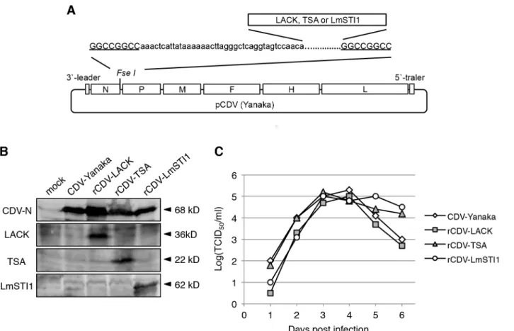
Fig 1. Generation andin vitrocharacterization of rCDVs.(A) Schematic model of pCDV (Yanaka) with theFseI site introduced between the N and P genes. EachLeishmaniaantigen gene carrying the CDV transcription signal unit (lower case) andFseI site (underlined) was inserted at theFseI site. (B) The rescued recombinant viruses were identified by immunoblotting analysis. Cell lysates were examined with anti-CDV-N, anti-LACK, anti-TSA and anti-LmSTI1 antibodies. The bands corresponding to CDV-N (68 kDa), LACK (38 kDa), TSA (22 kDa) and LmSTI1 (62 kDa) are indicated by arrowheads. (C) Kinetics of recombinant viral growth. B95a cells were inoculated with a recombinant virus at a multiplicity of infection of 0.01, and then harvested on the indicated day. The titers of the viruses were determined using a TCID50assay.
BLOCK ACE Reagent (DS Pharma Biomedical, Osaka, Japan) and 0.05% Tween 20, they were incubated with rabbit LACK antibody, rabbit TSA antibody, rabbit LmSTI1 anti-body (described above), or rabbit anti-N protein antianti-body for 1 h at room temperature. After the membranes were washed, they were incubated with goat anti-rabbit IgG antibody conju-gated with horseradish peroxidase (Dako Cytomation, Glostrup, Denmark) and then treated with ECL western blotting detection reagent (GE Healthcare). The reaction was visualized on an LAS 1000 Image Analyzer (Fujifilm, Tokyo, Japan).
Growth kinetic analysis
Monolayers of B95a cells in a 24-well plate were infected with virus at a multiplicity of infection (MOI) of 0.01. The infected cells and medium were harvested daily, frozen and thawed three times, and centrifuged at 15,000 ×gfor 10 min. Virus titers of the supernatants were deter-mined as TCID50values using standard methods.
Ethics statement
All animal experiments followed the laws of Japan: The Law for the Humane Treatment and Management of Animals (Act No. 105 of October 1, 1973) and the Law Concerning the Con-servation and Sustainable Use of Biological Diversity through Regulations on the Use of Living Modified Organisms (Act No. 97 of June 18, 2003). All animal experiments were approved by the Animal Experiment Committee at the University of Tokyo (approval numbers: 13–56, 18–
23, A11-41), and were performed in accordance with the Regulations for Animal Care and Use of the University of Tokyo, which were developed under the two laws stated above and nine guidelines including Fundamental Guidelines for Proper Conduct of Animal Experiment and Related Activities in Academic Research Institutions under the jurisdiction of the Ministry of Education, Culture, Sports, Science and Technology, and Standards Relating to the Care and Management of Laboratory Animals and Relief of Pain under the jurisdiction of the Ministry of the Environment. All surgery was performed under anesthesia with Dormicum and Domitor All efforts were made to minimize animal suffering. At the end of the experiments, the dogs were euthanized by exsanguination under anesthesia induced with ketamine–xylazine.
Experimental animals
Female beagle puppies, 5 weeks of age and confirmed free of CDV infection by an anti-CDV antibody enzyme-linked immunosorbent assay (ELISA), were purchased from Nihon Nosan (Yokohama, Japan). The dogs were group-housed in cages with ample space for exercise. The groups of dogs were kept in strict isolation to prevent viral cross-contamination during the course of all experiments.
Immunization and challenge with virulent CDV
The animal experiments were conducted using two dogs per group. The dogs were subcutane-ously inoculated with 500μl of rCDV–LACK (titer of 104.5TCID50per ml) on days 0 and 14.
Unimmunized mock-treated control dogs were inoculated with 500μl of phosphate-buffered
saline (PBS). The dogs were challenged intracerebrally with 500μl of 10% brain homogenate
Immunization and challenge with
L
.
major
The animal experiments were conducted using two dogs per group. The dogs were subcutane-ously inoculated with 500μl of parental CDV-Yanaka, rCDV–LACK, rCDV–TSA, rCDV–
LmSTI1 (all titers 104.5TCID50per ml), or a cocktail of three rCDVs (500μl each) on days 0
and 14. Unimmunized mock-treated control dogs were inoculated with 500μl of PBS. The
dogs’body temperatures, bodyweights, and clinical signs were checked daily for 21 days. Their leukocyte counts were checked 0, 7, 14 and 21 days after the first vaccination. The dogs were inoculated intradermally (in the ears) with infective promastigotes ofL.majorPM2 (5 × 10⁷ parasites per spot) 42 days after the first vaccination. Every week after challenge, the sizes of the nodules on the ears were measured (mm2) with calipers.
ELISA
The production of antibodies against CDV, LACK, TSA and LmSTI1 in dog sera were deter-mined with an ELISA. When anti-CDV antibodies were checked, the extracts of either the CDV-Yanaka-infected B95a cells or mock infected cells were used. Recombinant LACK, TSA and LmSTI1 described above were utilized for the detection of respective antibodies. ELISA was performed using 96-well plates with a standard method. In brief, the plates were consecu-tively incubated with various dilutions of dog sera and sheep anti-dog IgG conjugated with HRP (Cappel Lab., Cochranville, PA, USA) and then with the ELISA substrate (Bio Rad, Her-cules, CA, USA), and optical density values at 492 nm (OD492) were measured.
Detection of
L
.
major
At 74 days after challenge, the dogs were euthanized, and the ears, spleen, bone marrow, liver and parotid lymph node were collected and subjected to a detection test. In brief, these tissues were suspended in C-M199 medium, and an aliquot of the suspension was combined with blood agar plates and incubated at 26°C for 7 days. The presence of parasites was observed using an inverted microscope.
Results
Rescue and growth characteristics of rCDVs expressing
Leishmania
genes
We first generated rCDV-Yanaka expressingLeishmaniaantigens. We selected three protein antigens: LACK, TSA and LmTSI1. TheLACK,TSAorLmTSI1gene was inserted into the cDNA clone of the CDV-Yanaka genome between the N and P genes (Fig 1A). The plasmids were then used in our CDV rescue system [21]. Three days after HEK293 cells were overlain with B95a cells, a typical cytopathic effect was observed.
Protein expression by the rescued viruses was confirmed with immunoblotting (Fig 1B), indicating that the recombinant viruses were successfully rescued. The rescued viruses were designated rCDV-LACK, rCDV-TSA and rCDV-LmSTI1, respectively. These rCDVs showed similar viral growth to the parental CDV-Yanaka strain (Fig 1C).
Vaccine efficacy against lethal CDV challenge in dogs
the parental CDV-Yanaka strain, dogs inoculated with rCDV showed no clinical signs of CD, including leucopenia or pyrexia, demonstrating that the rCDVs are safe for dogs, even when expressingLeishmaniaantigens. The antibody responses were analyzed by ELISA. An increase of anti-CDV antibodies was found in all dogs’sera on Day 21, while interestingly, no LACK antibodies were observed for more than 50 days after the first vaccination by ELISA.
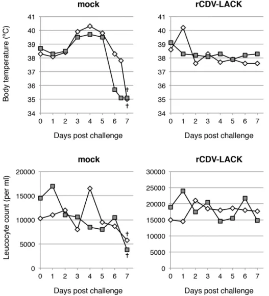
To confirm the protective effects of the rCDVs against virulent CDV, dogs inoculated with PBS or rCDV-LACK were challenged with the virulent CDV strain, Snyder Hill. The mock-inoculated control dogs showed severe pyrexia and leucopenia, and the body temperatures of the dogs rapidly fell below 35°C (Fig 2). They were euthanized 7 days after challenge in a mori-bund state. In contrast, the rCDV-LACK-vaccinated dogs showed no specific clinical signs of distemper (Fig 2). This result indicated that the expression of a foreign gene does not affect the protective immunity against CDV conferred by CDV-Yanaka.
Fig 2. Changes in body temperatures and leukocyte counts after virulent CDV challenge.The body temperatures and leucocyte counts of mock- and rCDV-LACK-immunized dogs after challenge with the virulent Snyder Hill strain of CDV were measured daily. At 7 days after challenge, the mock-treated dogs were in a moribund state and were euthanized (†). Each symbol represents one animal.
L
.
major
challenge in dogs
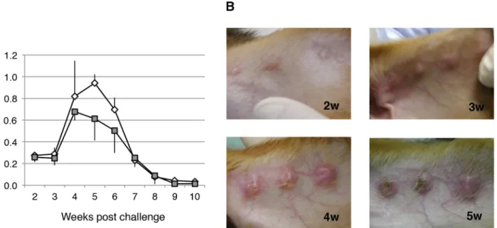
Next we attempted to establish experimental models for canine cutaneous leishmaniasis. To evaluate the utility in this model ofL.major, a major species responsible for cutaneous leish-maniasis, dogs were inoculated intradermally (three spots in the ears) with infective promasti-gotes (5 × 10⁷per spot) of the parasite. As shown inFig 3, the parasites proliferated at the sites of inoculation, and formed nodules in the skin lesions. The nodules first appeared in the second week and ulcers were observed in the third week (Fig 3B). The nodules became enlarged, reach-ing their maximum size in the fourth or fifth week, with typical crater-like lesions, and then regressed (Fig 3A).
Protective effect of rCDVs expressing
Leishmania
antigens against
L
.
major
challenge
Using this animal model, we evaluated the efficacy of the rCDVs as vaccines againstL.major. Two dogs each were immunized twice with PBS (mock), parental CDV-Yanaka, or each rCDV. An increase in anti-CDV antibodies was found in all dogs, while anti-TSA and anti-LmSTI1 antibodies levels were not as readily detectable as anti-LACK antibody. Four weeks after the second immunization,L.majorwas challenged, as described above. As shown inFig 4A, nod-ule formation in the mock-inoculated dogs displayed reproducible progression (Fig 3). Nodule formation was slightly slower in the dogs immunized with parental CDV-Yanaka, but the sizes of the nodules were similar to those in the mock-immunized dogs. Interestingly, in the rCDV-LACK-vaccinated dogs, the nodules were smaller than those in the mock-immunized dogs, particularly up to 5 weeks after challenge (Fig 4A). Furthermore, none of the nodules in the rCDV-LACK-vaccinated dogs were ulcerated (Fig 4B). This result indicated that vaccina-tion with rCDV-LACK conferred marked protective immunity, effectively suppressing the pro-liferation ofL.majorat an early stage of infection. Dogs immunized with rCDV-TSA or rCDV-LmSTI1 showed similar nodule growth to that observed in the
CDV-Yanaka-Fig 3. Time course of skin lesion development in dogs infected withL.major.(A) Beagle dogs were infected intradermally (in the ears) with 5 × 10⁷
infective promastigotes ofL.majorper spot, and the lesion sizes were measured weekly. Parasite growth was evaluated as nodule size. Three independent spots per dog were determined and followed-up. Data are shown as means±SEM, and the error bars reflect the three inoculated spots. (B) Images of lesions
at 2 to 5 weeks after infection.
immunized dogs (Fig 4A), suggesting that the expression of TSA or LmSTI1 alone produced only a weak immunogenic effect.
Therefore, we also vaccinated dogs with a cocktail of rCDV-LACK, rCDV-TSA, and rCDV-LmTSI1. The nodules in the cocktail-immunized dogs were significantly smaller than those in the single-vaccine-immunized dogs (Fig 4A). In particular, the nodules in the cocktail-immunized dogs had decreased rapidly in size after the sixth week of challenge. This suggested that the period of low-level parasite proliferation observed in the rCDV-LACK-vaccinated dogs was significantly suppressed in the cocktail-vaccinated dogs. All dogs were euthanized at the 10thweek after the challenge. The tissues of ears, spleen, bone marrow, liver and the parotid lymph node were collected and the parasites were detected. As shown inTable 1, no parasite was detected in these tissues of the unvaccinated dogs. By contrast, in the CDV-LACK vacci-nated dogs,L.majorwas detected in several tissues. These results showed that CDV-LACK vaccination suppressed proliferation of parasites, and caused a delay in parasite clearance. In cocktail-immunized dogs, no parasite was detected. Therefore, the cocktail immunization sup-pressed the proliferation ofL.majorat all stages of infection more effectively than immuniza-tion with the single rCDV-based vaccines.
This is the first report of a dog model of cutaneous leishmaniasis that is appropriate for test-ing vaccines. We also demonstrated that rCDVs expresstest-ingLeishmaniaantigens confer protec-tive immunity against both virulent CDV andL.majorchallenge in dogs.
Discussion
We previously examined the CDV-Yanaka strain as a potential novel live vaccine against recently prevalent CDV strains. In addition, we also previously established a reverse genetics system for the Yanaka strain [21], and the data presented here show that CDV-Yanaka is a safe and effective viral vector (Fig 2). The technique described here can simultaneously induce immunity against CDV and other pathogens.
Fig 4. Protective efficacy of immunization with rCDV-LACK, rCDV-TSA and rCDV-LmSTI1 againstL. majorchallenge.(A) Dogs were inoculated intradermally (in the ears) with 5 × 10⁷infective promastigotes of
L.majorper spot, 42 days after their first vaccination with the indicated inoculum. Parasite growth was evaluated every week as nodule size. Three independent spots were inoculated per dog and followed-up. Data are shown as the mean±SEM, and the error bars reflect the three inoculated spots. (B) Photographs of the nodules on the ears of mock- and rCDV-LACK-immunized dogs in the fourth week afterL.major challenge.
doi:10.1371/journal.pntd.0003914.g004
Table 1. Detection ofL.majorat the 10th week post challenge.
Immunized virus Tissue type
Ear Parotid lymph node Spleen Bone marrow Liver
Mock - - - -
-- - - -
-rCDV-LACK - + - -
-+ + - -
-Cocktail - - - -
-- - - -
--: not detected +: detected
Although visceral leishmaniasis has been studied extensively in dogs and various models have been described [30–35], little is known about canine cutaneous leishmaniasis, and only experimental infections withL.(Viannia) braziliensis[10] andL.mexicana[11] have been reported. In the present study, we presented an animal model of experimental infection withL.
majorin beagle dogs. The infection of dogs withL.majorcaused typical ulcerated skin lesions to develop, with similar sizes in all dogs and a rapid onset 3 to 5 weeks after infection (Figs3
and4). This infection model is highly reproducible. The progression of the lesions ofL.major
was similar to those ofL.mexicana[11]. Therefore, challenging dogs withL.majorgenerates a suitable animal model of cutaneous leishmaniasis. In particular, the progression of nodules slo-wed at about 10 weeks in control dogs, which is desirable compared with canine visceral leish-maniasis which usually takes over 1 year.
Based on this information, we evaluated the utility of our rCDVs as effective polyvalent can-didate vaccines against CDV andL.majorinfections. The results of this study indicated that vaccination with rCDV-LACK markedly reduced the nodule size afterL.majorchallenge, par-ticularly in the early phase of infection (Fig 4). Previous studies ofL.majorinfection in a mouse model demonstrated that the protective efficacy of LACK is mainly observed in cutane-ous leishmaniasis [6–9], so we consider our results to be reproducible. Studies with a mouse model also demonstrated that plasmid DNA encoding TSA or LmSTI1 partially or markedly protected the mice againstL.majorchallenge, respectively [6–9]. In contrast, rCDV-TSA and rCDV-LmSTI1 showed little immunogenic efficacy in the present study (Fig 4A), suggesting that TSA and LmSTI1 alone are weakly immunogenic in dogs. In contrast, combined immuni-zation with rCDV-LACK, rCDV-TSA, and rCDV-LmSTI1 produced a more effective result againstL.majorchallenge than immunization with each construct alone (Fig 4A). Previous studies have demonstrated that combinations of TSA and LmSTI1 proteins conferred strong protective immunity againstL.majorchallenge in mice and monkeys [23]. Immunization with a fusion protein, designated“Leish 111”, composed of TSA, LmSTI1 and LeIF (Leishmania
elongation initiation factor), or its derivative Leish 110, together with an adjuvant, conferred significant protection againstLeishmaniachallenge, producing smaller lesions in mice [6–9]. These results strongly suggest that a cocktail of multiple antigens confers more effective immu-nity throughout the life cycle ofLeishmaniathan single antigens. In particular, the cocktail vac-cine reduced the time period between challenge and cure compared with that achieved with the rCDV-LACK vaccine (Fig 4A). The reduction in nodule size may be mainly attributable to rCDV-LACK, and the shortened duration of the disease to rCDV-TSA and/or rCDV-LmSTI1.
tested as a vaccine adjuvant, and enhanced the efficiencies of various vaccines in mammals [43–46]. Recently, we generated a rCDV that secretes bioactive canine IL-18 and induces
IFN-γproduction by canine peripheral blood mononuclear cells [47]. This recombinant virus can potentially be used as an immunoadjuvantin vivo.
We propose that a combination of rCDV-based vaccines expressing different antigens with different effects on the immune response, has utility as a polyvalent vaccine for the prevention of leishmaniasis epidemics by inhibiting the transmission of the parasites through dogs.
Acknowledgments
We thank Dr. T. Barrett and Dr. M Baron (Institute for Animal Health, UK) for provision of MVA–T7.
Author Contributions
Conceived and designed the experiments: CK. Performed the experiments: RM TK MY AT MD YG CS YE AS KTK. Analyzed the data: RM TK TF HS. Contributed reagents/materials/ analysis tools: YM. Wrote the paper: RM TK TF HS.
References
1. Herwaldt BL (1999) Leishmaniasis. Lancet 354: 1191–1199.
2. World Health Organization. Leishmaniasis.http://wwwwhoint/leishmaniasis/burden/magnitude/ burden_magnitude/en/indexhtml.
3. Zerpa O, Ulrich M, Negron E, Rodriguez N, Centeno M, Rodriguez V, et al. (2000) Canine visceral leish-maniasis on Margarita Island (Nueva Esparta, Venezuela). Transactions of the Royal Society of Tropi-cal Medicine & Hygiene 94: 484–487.
4. Dietze R, Barros GB, Teixeira L, Harris J, Michelson K, Falqueto A, et al. (1997) Effect of eliminating seropositive canines on the transmission of visceral leishmaniasis in Brazil. Clinical Infectious Diseases 25: 1240–1242. PMID:9402389
5. Gradoni L (2001) An update on antileishmanial vaccine candidates and prospects for a canine Leish-mania vaccine. Veterinary Parasitology 100: 87–103. PMID:11522409
6. Kobets T, Grekov I, Lipoldova M (2012) Leishmaniasis: prevention, parasite detection and treatment. Current Medicinal Chemistry 19: 1443–1474. PMID:22360481
7. Das A, Ali N (2012) Vaccine Development Against Leishmania donovani. Frontiers in Immunology 3: doi:10.3389/fimmu.2012.00099
8. Palatnik-de-Sousa CB (2008) Vaccines for leishmaniasis in the fore coming 25 years. Vaccine 26: 1709–1724. doi:10.1016/j.vaccine.2008.01.023PMID:18295939
9. Kedzierski L (2010) Leishmaniasis Vaccine: Where are We Today? Journal of Global Infectious Dis-eases 2: 177–185. doi:10.4103/0974-777X.62881PMID:20606974
10. Pirmez C, Marzochi MC, Coutinho SG (1988) Experimental canine mucocutaneous leishmaniasis (Leishmania braziliensis braziliensis). Memorias do Instituto Oswaldo Cruz 83: 145–151. PMID:
2687621
11. Cruz-Chan J, Aguilar-Cetina Adel C, Villanueva-Lizama L, Martínez-Vega P, Ramírez-Sierra M, Rosado-Vallado ME, et al. (2014) A canine model of experimental infection with Leishmania (L.) mexi-cana. Parasites & Vectors 7: doi:10.1186/1756-3305-1187-1361
12. Appel MJ (1969) Pathogenesis of canine distemper. American Journal of Veterinary Research 30: 1167–1182. PMID:4894003
13. Blixenkrone-Moller M, Svansson V, Have P, Orvell C, Appel M, Pedersen IR, et al. (1993) Studies on manifestations of canine distemper virus infection in an urban dog population. Veterinary Microbiology 37: 163–173. PMID:8296445
14. Calderon MG, Remorini P, Periolo O, Iglesias M, Mattion N, La Torre J. (2007) Detection by RT-PCR and genetic characterization of canine distemper virus from vaccinated and non-vaccinated dogs in Argentina. Veterinary Microbiology 125: 341–349. PMID:17628358
16. Haas L, Martens W, Greiser-Wilke I, Mamaev L, Butina T, Maack D, et al. (1997) Analysis of the hae-magglutinin gene of current wild-type canine distemper virus isolates from Germany. Virus Research 48: 165–171. PMID:9175255
17. Jozwik A, Frymus T (2002) Natural distemper in vaccinated and unvaccinated dogs in Warsaw. Journal of Veterinary Medicine Series B 49: 413–414. PMID:12489707
18. Simon-Martinez J, Ulloa-Arvizu R, Soriano VE, Fajardo R (2008) Identification of a genetic variant of canine distemper virus from clinical cases in two vaccinated dogs in Mexico. Veterinary Journal 175: 423–426.
19. Iwatsuki K, Miyashita N, Yoshida E, Gemma T, Shin YS, Mori T, et al. (1997) Molecular and phyloge-netic analyses of the haemagglutinin (H) proteins of field isolates of canine distemper virus from natu-rally infected dogs. Journal of General Virology 78: 373–380. PMID:9018060
20. Takenaka A, Yoneda M, Seki T, Uema M, Kooriyama T, Nishi T, et al. (2014) Characterization of two recent Japanese field isolates of canine distemper virus and examination of the avirulent strain utility as an attenuated vaccine. Veterinary Microbiology 174: 372–381. doi:10.1016/j.vetmic.2014.10.024
PMID:25465179
21. Fujita K, Miura R, Yoneda M, Shimizu F, Sato H, Muto Y, et al. (2007) Host range and receptor utiliza-tion of canine distemper virus analyzed by recombinant viruses: Involvement of heparin-like molecule in CDV infection. Virology 359: 324–335. PMID:17055025
22. Webb JR, Campos-Neto A, Ovendale PJ, Martin TI, Stromberg EJ, Badaro R, et al. (1998) Human and murine immune responses to a novel Leishmania major recombinant protein encoded by members of a multicopy gene family. Infection & Immunity 66: 3279–3289.
23. Campos-Neto A, Porrozzi R, Greeson K, Coler RN, Webb JR, Seiky YA, et al. (2001) Protection against cutaneous leishmaniasis induced by recombinant antigens in murine and nonhuman primate models of the human disease. Infection & Immunity 69: 4103–4108.
24. Webb JR, Kaufmann D, Campos-Neto A, Reed SG (1996) Molecular cloning of a novel protein antigen of Leishmania major that elicits a potent immune response in experimental murine leishmaniasis. Jour-nal of Immunology 157: 5034–5041.
25. Mendez S, Belkaid Y, Seder RA, Sacks D (2002) Optimization of DNA vaccination against cutaneous leishmaniasis. Vaccine 20: 3702–3708. PMID:12399198
26. Ahmed SB, Touihri L, Chtourou Y, Dellagi K, Bahloul C (2009) DNA based vaccination with a cocktail of plasmids encoding immunodominant Leishmania (Leishmania) major antigens confers full protection in BALB/c mice. Vaccine 27: 99–106. doi:10.1016/j.vaccine.2008.10.013PMID:18951941
27. Kobune F, Sakata H, Sugiura A (1990) Marmoset lymphoblastoid cells as a sensitive host for isolation of measles virus. Journal of Virology 64: 700–705. PMID:2153236
28. Gemma T, Watari T, Akiyama K, Miyashita N, Shin YS, Iwatsuki K, et al. (1996) Epidemiological obser-vations on recent outbreaks of canine distemper in Tokyo area. Journal of Veterinary Medical Science 58: 547–550. PMID:8811624
29. Okuno T, Goto Y, Matsumoto Y, Otsuka H, Matsumoto Y (2003) Applications of recombinant Leish-mania amazonensis expressing egfp or the beta-galactosidase gene for drug screening and histopath-ological analysis. Experimental Animals 52: 109–118.
30. Manna L, Reale S, Viola E, Vitale F, Foglia-Manzillo V, Pavone LM, et al. (2006) Leishmania DNA load and cytokine expression levels in asymptomatic naturally infected dogs. Vet Parasitol 142: 271–280. PMID:16920264
31. Carrillo E, Ahmed S, Goldsmith-Pestana K, Nieto J, Osorio Y, Travi B, et al. (2007) Immunogenicity of the P-8 amastigote antigen in the experimental model of canine visceral leishmaniasis. Vaccine 25: 1534–1543. PMID:17178178
32. Reis AB, Giunchetti RC, Carrillo E, Martins-Filho OA, Moreno J (2010) Immunity to Leishmania and the rational search for vaccines against canine leishmaniasis. Trends in Parasitology 26: 341–349. doi:10. 1016/j.pt.2010.04.005PMID:20488751
33. Poot J, Rogers ME, Bates PA, Vermeulen A (2005) Detailed analysis of an experimental challenge model for Leishmania infantum (JPC strain) in dogs. Veterinary Parasitology 130: 41–53. PMID:
15893068
34. Nogueira FS, Moreira MA, Borja-Cabrera GP, Santos FN, Menz I, Parra LE,. et al. (2005) Leishmune vaccine blocks the transmission of canine visceral leishmaniasis: absence of Leishmania parasites in blood, skin and lymph nodes of vaccinated exposed dogs. Vaccine 23: 4805–4810. PMID:16011864
36. Kemp K (2000) Cytokine-producing T cell subsets in human leishmaniasis. Archivum Immunologiae et Therapiae Experimentalis 48: 173–176. PMID:10912621
37. Heinzel FP, Sadick MD, Holaday BJ, Coffman RL, Locksley RM (1989) Reciprocal expression of inter-feron gamma or interleukin 4 during the resolution or progression of murine leishmaniasis. Evidence for expansion of distinct helper T cell subsets. Journal of Experimental Medicine 169: 59–72. PMID:
2521244
38. Locksley RM, Scott P (1991) Helper T-cell subsets in mouse leishmaniasis: induction, expansion and effector function. Immunology Today 12: A58–61. PMID:1829891
39. Scott P, Natovitz P, Coffman RL, Pearce E, Sher A (1988) Immunoregulation of cutaneous leishmania-sis. T cell lines that transfer protective immunity or exacerbation belong to different T helper subsets and respond to distinct parasite antigens. Journal of Experimental Medicine 168: 1675–1684. PMID:
2903212
40. Dinarello CA, Novick D, Puren AJ, Fantuzzi G, Shapiro L, Muhl H, et al. (1998) Overview of interleukin-18: more than an interferon-gamma inducing factor. Journal of Leukocyte Biology 63: 658–664. PMID:
9620656
41. Trinchieri G, Gerosa F (1996) Immunoregulation by interleukin-12. Journal of Leukocyte Biology 59: 505–511. PMID:8613697
42. Gracie JA, Robertson SE, McInnes IB (2003) Interleukin-18. Journal of Leukocyte Biology 73: 213– 224. PMID:12554798
43. O'Donovan LH, McMonagle EL, Taylor S, Bain D, Pacitti AM, Golder MC, et al. (2005) A vector express-ing feline mature IL-18 fused to IL-1beta antagonist protein signal sequence is an effective adjuvant to a DNA vaccine for feline leukaemia virus. Vaccine 23: 3814–3823. PMID:15893619
44. Suo S, Ren Y, Li G, Zarlenga D, Bu RE, Su D, et al. (2012) Immune responses induced by DNA vac-cines bearing Spike gene of PEDV combined with porcine IL-18. Virus Research 167: 259–266. doi:
10.1016/j.virusres.2012.05.007PMID:22643071
45. Siva Reddy K, Muralidhar Rao D, Badrinaryana N, Suryanaryana VV, Reddy GR (2010) Enhancement of DNA vaccine (P12A3C-pcDNA) efficacy against foot-and-mouth disease by coadministration of inter-leukin-18-expressing (IL18 pcDNA) plasmid in guinea-pigs. FEMS Immunology & Medical Microbiology 60: 261–269.
46. Shen G, Jin N, Ma M, Jin K, Zheng M, Zhuang T, et al. (2007) Immune responses of pigs inoculated with a recombinant fowlpox virus coexpressing GP5/GP3 of porcine reproductive and respiratory syn-drome virus and swine IL-18. Vaccine 25: 4193–4202. PMID:17418456