online | memorias.ioc.fiocruz.br
Comparative evaluation of phenol and thimerosal as preservatives for
a candidate vaccine against American cutaneous leishmaniasis
Wilson Mayrink1, Carlos Alberto Pereira Tavares2, Rosangela Barbosa de Deus3, Melina Barros Pinheiro4, Tânia Mara Pinto Dabés Guimarães4, Hélida Monteiro de Andrade1,
Carlos Alberto da Costa4, Vicente de Paulo Coelho Peixoto de Toledo4 /+
1Departamento de Parasitologia 2Departamento de Bioquímica e Imunologia, Instituto de Ciências Biológicas 4Departamento de Análises
Clinicas e Toxicológicas, Faculdade de Farmácia, Universidade Federal de Minas Gerais, Av. Antônio Carlos 6627, 31270-901 - Belo Horizonte, MG, Brasil 3Departamento de Farmácia, Escola de Farmácia, Universidade Federal de Ouro Preto, Ouro Preto, MG, Brasil
For decades thimerosal has been used as a preservative in the candidate vaccine for cutaneous leishmaniasis, which was developed by Mayrink et al. The use of thimerosal in humans has been banned due to its mercury content. This study addresses the standardization of phenol as a new candidate vaccine preservative. We have found that the proteolytic activity was abolished when the test was conducted using the candidate vaccine added to merthiolate (MtVac) as well as to phenol (PhVac). The Montenegro’s skin test conversion rates induced by MtVac and by PhVac was 68.06% and 85.9%, respectively, and these values were statistically significant (p < 0.05). The proliferative response of peripheral mononuclear blood cells shows that the stimulation index of mice immunized with both can-didate vaccines was higher than the one in control animals (p < 0.05). The ability of the cancan-didate vaccines to induce protection in C57BL/10 mice against a challenge with infective Leishmania amazonensis promastigotes was tested and the mice immunized with PhVac developed smaller lesions than the mice immunized with MtVac. Electrophoresis of phenol-preserved antigen revealed a number of proteins, which were better preserved in PhVac. These results do in fact encourage the use of phenol for preserving the immunogenic and biochemical properties of the candidate vaccine for cutaneous leishmaniasis.
Key words: leishmaniasis - candidate vaccine - phenol - merthiolate
Financial support: FAPEMIG
+ Corresponding author: [email protected] Received 3 August 2009
Accepted 1 December 2009
Leishmaniasis currently threatens more than 350 mil-lion men, women and children in 88 countries around the world. It is estimated that 2 million new cases occur each year, with at least 12 million people presently infected worldwide (WHO 2002). The disease is endemic in Af-rica, Southwest Asia, the Middle East, Southern Europe and Central and South America (Sinha et al. 2005).
Leishmaniasis in general, but particularly cutaneous leishmaniasis is one of a few parasitic diseases that will most likely become controllable through the use of vac-cines. Given the relatively simple life cycle and the fact that recovery from a primary infection renders the host resistant to subsequent infections, indicates that a suc-cessful vaccination is feasible. During the past several decades, extensive efforts have been made to search for an effective Leishmania candidate vaccine (Khamesi-pour et al. 2006). Despite several efforts towards can-didate vaccine development, no effective recombinant vaccine currently exists in humans. The induction of protective immunity using either inactivated or attenu-ated live vaccines (first generation vaccines) would be a significant step towards controlling the disease
The Brazilian researcher, Mayrink and his coworkers conducted trials of different preparations of killed para-sites in the 1970s. They developed a pentavalent candidate vaccine, which after two intramuscular injections was able to convert the Montenegro’s leishmanin skin test [also known as Montenegro skin test (Mst)], resulting in the successful vaccination of 78.4% of volunteers within three months (Mayrink et al. 1979). However, the difficulties of standardization and establishing the safety of this candi-date vaccine are potential obstacles to its registration.
In 2006, permission was granted (25000.045522/99-10) by the Brazilian National Health Surveillance Agen-cy (Brazil) to use the leishmaniasis candidate vaccine developed by Mayrink et al. (1979) as a therapeutic agent in the treatment of leishmaniasis. It is still used in Brazil and some other countries in Central America in its origi-nal formulation with thimerosal.
This study investigated the standardization of phenol as a new preservative for the anti-leishmaniasis candi-date vaccine to replace thimerosal. It is critical to know the effect of proteolysis on immunogenicity and to deter-mine the stability of different preparations of a single-strain killed candidate vaccine conserved by thimerosal and phenol and whether phenol can stabilize candidate vaccine proteins more effectively than thimerosal. If so, it may be advisable to license the candidate vaccine as a therapeutic agent using the new proposed formulation with phenol as a preservative.
SUBJECTS, MATERIALS AND METHODS
Study subjects - A group of 247 Mst negative healthy subjects were immunized against American tegumentar leishmaniasis (ATL) using either mer-thiolated (mtVac) or phenolated (PhVac) candidate vaccine preparations. One hundred and nineteen indi-viduals received the merthiolated preparation and 128 received the phenolated preparation. All of the study subjects resided in the endemic area of ATL in Cara- tinga, Minas Gerais, Brasil. This research was regis-tered in the Conselho Nacional de Saúde, Comissão Nacional de Ética em Pesquisa, Ministério da Saúde, Brasil (13,085 - 02/09/2007).
Preparation ofcandidate vaccines - The candidate vaccine preparations used in this study were produced using a well defined WHO reference Leishmania ama-zonensis strain (IFLA/BR/67/PH8) in the Departamento de Parasitologia, Instituto de Ciências Biológicas (ICB), Universidade Federal de Minas Gerais (UFMG), Belo Horizonte, Minas Gerais, Brazil, according to a meth-odology described in detail elsewhere (Mayrink et al. 1979, Antunes et al. 1986, De Luca et al. 1999, Armijos et al. 2004). Briefly, promastigotes of L. amazonensis
IFLA/BR/67/PH8 were grown in liver infusion medium and harvested at stationary phase (7 days of culture) us-ing centrifugation. After washus-ing with sterile saline, the pellet was divided in two: one part was disrupted by sonication and the other was then left particulated. The final concentration of total nitrogen was 240 µg/mL. To preserve this candidate vaccine preparation, merthio-late was used in saline at 1:10,000 (v/v) or a solution of phenol in saline at 0.35% (v/v). After being produced, the candidate vaccines were stored at 4°C. All candidate vaccines used had been stored for four months.
Volunteer vaccination - Two different groups of vol-unteers were vaccinated with mtVac and PhVac in two 1.5-mL doses injected intramuscularly in 21 day inter-vals. All the volunteers were Mst-negative and all the personnel involved in the trial were blinded with regard to which candidate vaccine preparation was used.
Mst - All vaccinated volunteers were skin-tested twice: two days before the administration of the first candidate vaccine dose (for screening of volunteers to be vaccinated) and 40 days after the administration of the second dose. The antigen was composed of the same strain used in the candidate vaccine preparation [L. ama-zonensis strain (IFLA/BR/67/PH8)] according to Costa
et al. (1996). The test result was considered positive when induration with a diameter greater than or equal to 5 mm was detected 48 h after the injection.
Mice - Female C57BL/10 mice (8-12 week old) were obtained from the breeding facilities at the ICB-UFMG.
Immunization and challenge of mice - Forty isogenic C57BL/10 mice per group were vaccinated according to Costa et al. (1992). Twenty animals received two subcu-taneous inoculations in seven-day intervals. Each dose contained 100 mg of mtVac plus 250 mg of Coryne-bacterium parvum as adjuvant. The other 20 animals received the phVac according to the same vaccination scheme. Twenty-eight days after the second dose, the animals received an additional 10 mg of candidate vac-cine without adjuvant. Seven days after the booster, 10 mice from both groups were challenged with 1 x 10⁵L.
amazonensis promastigotes inoculated subcutaneously into the base of the tails. These animals were examined at two-week intervals for a total of 180 days after the challenge to observe the appearance of lesions and their subsequent development and for measurement of lesion diameters. Animals were considered protected when no lesions were observed. The control group consisted of 10 other mice immunized with C. parvum alone. The 10 remaining vaccinated and unchallenged mice in each group were kept non-infected to be used in the evalua-tion of a lymphoproliferative response. All the animals reserved for this study were sacrificed and their spleens were removed aseptically to obtain the lymphocytes used in the lymphoproliferation assay.
Lymphocyte proliferation assay - The lymphocyte proliferation assay were performed as described by Cos-ta et al. (1992). Briefly, spleen cells were harvested in a RPMI 1640 (SIGMA-USA) medium containing 10% fetal calf serum (SIGMA, USA), 2 Mm L-Glutamine, 50
mM 2-β-Mercaptoethanol, 10,000 units of penicillin, 10
mg streptomycin and 250 mg fungizone (SIGMA-USA) per mL. Red blood cells were removed by lysis in 0.144 mM NH4Cl. The cells (4 x 10⁵/well) were then incubated
in triplicate in 96-well plates for five days either with 20 mg/mL of each L. amazonensis antigen or 0.5 µg/ mL of Concanavalin A (SIGMA, USA) in an atmosphere of 5% CO2 at 37°C. The antigens used in the prolifera-tive assays were obtained by the same candidate vaccine preparation procedures (De Luca et al. 1999, Armijos et al. 2004). The antigen concentration was adjusted to 1 mg/mL. The proliferation was assayed by [3H] thymi-dine incorporation (0.2 mCi/well, specific activity 5.0 mCi/mL - Dupont, NEN Research Products, USA). The results were expressed as a stimulation index. A positive response showed a stimulation index equal to or higher than 2.5 (Mendonça et al. 1986).
the volume was brought to 1 mL with 0.075 M Tris-HCL, pH 7.6, containing 25 mM CaCl2. After incubation at 30°C for 15-120 min with the synthetic substrate, Bapna (N-benzoyl-L-arginine-p-nitroanilide), at a final concen-tration of 1 x 10⁻⁵ M, the reaction was stopped with the
addition of 60% acetic acid. The extent of hydrolysis was measured by a spectrophotometer (Shimadzu) at 405 nm; each sample was assayed in triplicate.
Electrophoresis of candidate vaccine antigens -
Twenty micrograms of antigen were submitted to elec-trophoresis in a 10% polyacrilamide gel (SDS-PAGE) (Laemmli 1970). Then the gel was fixed overnight in 50% methanol and stained using the silver nitrate method (Merril et al. 1981).
Statistical analysis - One way ANOVA was used for the evaluation of proteolytic activity in mtVac and phVac experiments and for the lymphocyte proliferation assay. To analyze responses to Mst, the Mann-Whitney test was employed. For the immunization and challenge of mice, we used bootstrapping with a probability function, ac-cording to the central limit theorem to assume normality followed by means comparison by one way ANOVA.
RESULTS
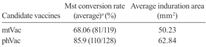
Mst - The volunteers who received the mtVac had a mean age of 28.89 ± 20.45, while the phenol vacci-nated volunteers had a mean age of 30.32 ± 21.41. All study subjects had negative results for the first skin test. The induration area of positivity for the second skin test in the groups who had received either mt-Vac and phmt-Vac is shown in mm2 in the Table. The Mst
rates induced by mtVac and by phVac were 68.06% and 85.9%, respectively, and these values were statis-tically significant (p < 0.05). No difference was found between the average vertical and transversal diam-eters of the induration area (p > 0.05).
Evaluation of immunoprotection in mice - The abil-ity of the candidate vaccines to induce persistent pro-tection in C57BL/10 mice against a challenge with in-fective L. amazonensis promastigotes was tested (Fig. 1). Mice were immunized with each candidate vaccine according to the schedule described in the materials and methods. The animals were examined for the appear-ance of lesions at the site of inoculation and/or other manifestations along the course of 180 days. Animals were considered protected when no lesions were ob-served. As shown in Fig. 1, all the control mice devel-oped a progressive infection at the base of the tail con-sisting of ulceration and necrosis between the 7th-17th weeks after the challenge. Along the same time range, the immunized mice with phenolated vaccine devel-oped smaller lesions than the mice immunized with mtVac. Around week 17 after the challenge, about 50% of the immunized mice from both groups developed le-sions, which were about 10 times smaller than those in the control group. No visceralization was observed in either the control or vaccinated animals.
Lymphocyte proliferation assay - Spleen cells from each immunized and control group were incubated with the homologous candidate vaccine antigens. The
pro-liferative response was measured by [3H] - thymidine incorporation. Fig. 2 shows that the stimulation index of mice immunized with candidate vaccine antigens was at least 4.9 times higher than the one in control animals (non-vaccinated) and this proliferative response was sig-nificantly higher (p < 0.05). The control consisted of non-vaccinated mouse spleen cells stimulated with candidate vaccine antigens. No difference was observed between the groups (mtVac and phVac) in this regard. All the ConA-stimulated spleen cells from the non-vaccinated mice showed a stimulation index above 7.0, suggesting that the culture cell system was sufficiently viable.
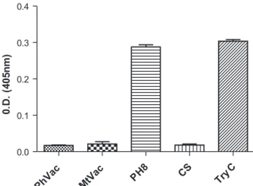
Evaluation of proteolytic activity - Fig. 3 shows that the test for determining the enzymatic activity was ef-ficient because both the controls of substrate and the en-zyme trypsin (TryC) had the expected results. The pro-tease activity in the Leishmania extract (strain PH8) was similar to that seen in the control enzyme (TryC), which was expected because the preparation, in addition to be-ing recently prepared, had no protease inhibitor added. We also found that the proteolytic activity was abolished when the test was conducted using MtVac or PhVac.
Electrophoresis of candidate vaccine antigens - A reducing SDS-PAGE was performed on a slab gel ap-paratus with the discontinuous system. The separated protein bands were stained using silver stain. In Fig. 4,
TABLE
Montenegro’s skin test (Mst) conversion rates after vaccination
Candidate vaccines
Mst conversion rate (average)a (%)
Average induration area (mm2)
mtVac 68.06 (81/119) 50.23
phVac 85.9 (110/128) 62.84
a: significant difference between the merthiolated (mtVac) and phenolated (phVac) candidate vaccines ( p < 0.05).
Fig. 1: the course of Leishmania amazonensis infection in control
C57BL/10 mice (♦) and mice immunized with phenolated candidate
vaccine plus Corynebacterium parvum (▼) or with merthiolated
candidate vaccine plus C.parvum (●). The adjuvant C. parvum was included in all immunizations. The control group consisted of mice immunized with C. parvum alone (all the control animals developed lesions). The data are the mean of 10 vaccinated mice per group.
0 50 100 150 200
0 10 20 30 40 50 60 70 80 90 100
TIME(DAYS)
M
IC
E
W
IT
H
L
E
S
IO
N
(
%
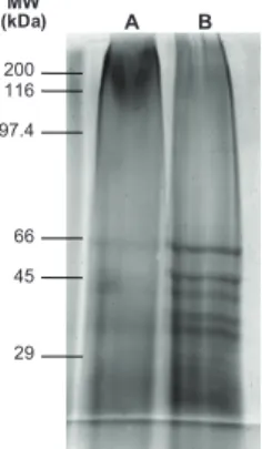
qualitative and quantitative differences were observed in the total protein profiles of the two samples. The electrophoresis of phenol-preserved antigen revealed a number of proteins, including those already described in the literature as immunogenically more immuno-genic: gp63, gp46 and a 30-kDa protein, among oth-ers (Nascimento et al. 1990). Protein degradation was more evident in the merthiolated antigen since all of the above-mentioned antigens were not as evident as in the phVac. Moreover, the merthiolated antigen had fewer bands and they were more diffuse.
DISCUSSION
Therapeutic trials of first generation leishmaniasis candidate vaccines have shown very encouraging results. From Convit’s trials for the treatment of patients in
Vene-zuela and Machado Pinto’s results in Brazil, to the results of Musa et al. (2008) on therapy of post Kalazar dermal leishmaniasis in Sudan, immunochemotherapy seems a promising mode of treatment (Convit et al. 1987, 2003, Machado Pinto et al. 2002, Musa et al. 2008). This justi-fies further investigation of first generation leishmaniasis candidate vaccines for therapeutic purposes. The accumu-lated evidence suggests that protective immunity followed by healing is attributed to the development of a strong
Leishmania-specific CD4+ Th1 cell response, which pro-duces and releases Th1 cytokines, mainly Interferon-γ,
which has the ability of activating macrophages to inhibit and/or kill parasites (Amaral et al. 2002).
In our study, we assessed the ability of a candidate vaccine, preserved by phenol, to induce immunity against Leishmania. Thus, we measured delayed hyper-sensitivity to the Mst antigen and evaluated the spleen cell proliferative response. In trials where post vaccina-tion Mst was measured, responses were notably larger in the candidate vaccine group, but this evidence of immunogenicity induced by the candidate vaccine was not carried over to having a protective effect. Neverthe-less, conversion from a negative Mst reaction to Mst > 5 mm after vaccination has been observed to be associ-ated with a significantly lower incidence of infection in Brazil, Iran and Sudan, suggesting that Mst conversion may be associated with an immune response that pro-vides some level of protection (Noazin et al. 2008). The percentage of Mst conversion is essentially the average described by Mayrink et al. (1978), which was 78.4%. In our study, the subjects were examined by the study clinicians on the day of vaccination and on post-vacci-nation days for the presence of lymphadenopathy, fever, ulcerations, redness, bleeding and swelling at the vacci-nation site. No major side effects were registered in the volunteers who received phVac or mtVac. No individual aspects of the subjects or possible correlations with age, gender or nutritional status were significantly different based on variance analysis.
A skin test conversion was observed in the majority of vaccinated volunteers. The conversion rates induced by phVac and mtVac were 85.9% and 69%, respectively, and this difference was statistically significant (p < 0.03). The Mst demonstrates the immunogenicity of the monos-train PH8 candidate vaccine, independent of the type of preservative used. We can infer that the higher Mst conversion rate in the group that received phVac (85.9%) could be explained by the improved preservation of anti-gens, as observed from the vaccines protein silver stain gel (Fig. 4). Therefore, we can rule out the possibility of hypersensitivity to phenol reactogenicity, since Mayrink et al. (2006) demonstrated its absence in Montenegro’s antigens preserved by phenol as well as in merthiolate. Furthermore, in both groups the averages of the Mst in-duration diameters were not statistically significant.
In regard to the spleen cell proliferative response, our data show that both candidate vaccines elicit an antigen-specific recall response in spleen cells from mice im-munized with these candidate vaccines. Compared to Fig. 2: the lymphoproliferative response (shown as a stimulation
in-dex) of spleen cells from mice immunized with the phenolated (PhVac) and merthiolated (MtVac) candidate vaccines after in vitro stimula-tion with homologous antigen. Spleen cells from non-vaccinated mice were used as the control and were stimulated with phVac antigens or mtVac antigens. The stimulation indexes greater than or equal to 2.5 were considered positive (horizontal line).
PhVac Control MtVac Control 0
1 2 3 4 5 6 7 8 9 10
S. I.
Fig. 3: evaluation of proteolytic activity using BApNA as substrate. One hundred micrograms of protein was used for the control for trypsin activity (TryC). The vertical lines represent the standard de-viation for each analysis. The data are the mean of three experiments. The top bar refers to SD. CS: control for substrate activity; MtVac: merthiolated candidate vaccine; PhVac: phenolated candidate vac-cine; PH8: fresh extract from Leishmania.
PhVac MtVac P
H8 CS
Try C 0.0
0.1 0.2 0.3 0.4
0
.D
.
(4
0
5
n
the control groups, there was a significant increase in the antigen-stimulated spleen cell proliferative response in vaccinated mice, demonstrating the immunogenic-ity of both candidate vaccines. Thus, merthiolate and phenol preserve the immunogenicity of the Leishma-nia antigens in this single strain candidate vaccine. To supplement this investigation, we also investigated the immunogenicity and efficacy of an L. amazonensis can-didate vaccine. Groups of C57BL/10 mice were immu-nized with both candidate vaccines plus C. parvum as an adjuvant. Both mtVac and phVac induced protective immunity against infective promastigotes. The animals presenting lesions of any size were considered pro-tected, but the lesions observed in the vaccinated, non-protected mice were always much smaller than those in the unvaccinated animals. No self healing was observed in the control or non-protected mice. Interestingly, near week 17 the lesions in the group that received the phVac had a delayed onset and a smaller size than those in the group that received mtVac. The data obtained from this experiment, combined with the significant increase in the antigen-stimulated spleen cell proliferative response after vaccination demonstrate the immunogenic efficacy of the single strain candidate vaccine.
The current results confirm those conducted by other groups working with candidate vaccines formulated with different Leishmania strains that showing the immuno-genicity of the candidate vaccine is maintained with both merthiolate and phenolate preservatives (Mayrink et al. 1979, Mendonça et al. 1995, Armijos et al. 1998, 2004).
The antigen stability of these candidate vaccines was analyzed by polyacrilamide gel electrophoresis. We ob-served the same number of bands in both the mtVac and phVac, but the latter were more intense. This phenom-enon was also observed in Mst when it was conserved by phenol (Mayrink et al. 2006).
Thimerosal not only helps to prevent the growth of bacteria and fungi in vaccines but can also prevent pro-teolysis. According to the protease activity test (Fig. 3), both MtVac and PhVac present low protease activity when
compared to TryC and to a Leishmania extract without protease inhibitor. These data demonstrate that candidate vaccines prepared with merthiolate or phenol do not suf-fer from degradation by proteolytic enzymes, indicating these substances are efficient for preserving vaccines.
Tests conducted with the soluble fraction of candi-date vaccines showed that the phVac had its proteolytic activity reduced by 90% and the mtVac by 34%. This result indicates that phenol is a more efficient preserva-tive agent than merthiolate (data not shown).
A meningococcal vaccine stabilized with 0.25% of phenol was reported to be stable for two years (WHO 1981). Considering the diminished protease activity through the use of phenol in leishmaniasis candidate vaccine, we expect a similar storage time for a pheno-lated leishmaniasis candidate vaccine.
Taken together, this study encourages the use of ph-Vac as a means for preserving the immunogenic and bio-chemical properties of leishmaniasis candidate vaccine.
ACKNOWLEDGEMENTS
To the Secretaria Municipal de Saúde of Caratinga, state of Minas Gerais, Brazil, and Mr. Jair Cecílio de Paula, for his invaluable support during the field work.
REFERENCES
Amaral VF, Teva A, Oliveira-Neto MP, Silva AJ, Pereira MS, Cupo-lillo E, Porrozzi R, Coutinho SG, Pirmez C, Beverly SM, Grimal-di GJr 2002. Study of the safety, immunogenicity and efficacy of attenuated and killed Leishmania (Leishmania) major vaccines in a rhesus monkey (Macaca mulatta) model of the human disease.
Mem Inst Oswaldo Cruz97: 1041-1048.
Antunes CMF, Mayrink W, Magalhães PA, Costa CA da, Melo MN, Dias M, Michalick MSM, Williams P, Oliveira-Lima A, Vieira JBF, Schettini APM 1986. Controlled field trials of a vaccine against new cutaneous leishmaniasis. Int J Epidemiol 15: 147-154.
Armijos RX, Weigel MM, Aviles H, Maldonado R, Racines J 1998. Field trial of a vaccine against New World cutaneous leishma-niasis in an at-risk child population: safety, immunogenicity and efficacy during the first 12 months of follow-up. J Infect Dis 177: 1352-1357.
Armijos RX, Weigel MM, Calvopina M, Hidalgo A, Cevallos W, Cor-rea J 2004. Safety, immunogenicity and efficacy of an autoclaved
Leishmania amazonensis vaccine plus BCG adjuvant against New World cutaneous leishmaniasis. Vaccine 22: 1320-1326.
Convit JO, Castellanos PIO, Rondon AO, Pinardi MEO, Ulrich MO, Castes M 1987. Immunotherapy versus chemotherapy in loca-lised cutaneous leishmaniasis. Lancet 1: 401-405.
Convit JO, Ulrich MO, Zerpa OO, Borges RO, Aranzazu NO, Valera MO, Villarroel HO, Zapata Z, Tomedes I 2003. Immunotherapy of American cutaneous leishmaniasis in Venezuela during the pe-riod 1990-1999. Trans R Soc Trop Med Hyg 97: 469-472.
Costa CA da, Afonso LCC, Toledo VPCP, Guimarães TMPD, Nasci-mento E, Tavares CAP, Mayrink W 1992. Immune responses and protection induced in mice by an industrialized vaccine against American cutaneous leishmaniasis. Parassitologia 34: 45-51.
Costa CA da, Toledo VPCP, Genaro O, Williams P, Mayrink W 1996. Montenegro skin test evaluation of the composition and stability of the antigen preparation. Mem Inst Oswaldo Cruz 91: 193-194.
Fig. 4: electrophoresis in 10% polyacrilamide gel silver stained using merthiolated and phenolated candidate vaccines. Lane A: protein pro-file for merthiolated-preserved antigens: B: protein propro-file for phenol-preserved antigens. MW molecular weight markers (kDa).
A B
200 116
97.4
66
45
29
De Luca PM, Mayrink W, Alves CR, Coutinho SG, Oliveira MP, Bertho AL, Toledo VPCP, Costa CA, Genaro O, Mendonça SC 1999. Evaluation of the stability and immunogenicity of auto-claved and non autoauto-claved preparations of a vaccine against American tegumentary leishmaniasis. Vaccine 17: 1179-1185.
Erlanger BF, Kokowsky N, Cohen W 1961. The preparation and prop-erties of two new chromogenic substrates of trypsin. Arch Bio-chem Biophys 95: 271-278.
Geier DA, Sykes LK, Geier MR 2007. A review of thimerosal (merthi-olate) and its ethylmercury breakdown product: specific histori-cal considerations regarding safety and effectiveness. J Toxicol Environ Health B Crit Rev 10: 575-596.
Khamesipour A, Rafati S, Davoudi N, Maboudi F, Modabber F 2006. Leishmaniasis vaccine for development: a global overview. In-dian J Med Res 123: 423-438.
Laemmli UK 1970. Cleavage of structural protein. The assembly of the head of bacteriophage T4. Nature 27:680-685.
Machado-Pinto J, Pinto J, da Costa CA, Genaro O, Marques MJ, Modabber F, Mayrink W 2002. Immunochemotherapy for cuta-neous Leishmaniasis: a controlled trial using killed Leishmania (Leishmania) amazonensis vaccine plus antimonial. Int J Der-matol 41: 73-78.
Mayrink W, Costa CA da, Magalhães PA 1979. A field trial of a vac-cine against American dermal leishmaniasis. Trans R Soc Trop Med Hyg 73: 385-387.
Mayrink W, Machado-Coelho GLL, Guimarães TMPD, Andrade HM, Perez EC, Costa CA da, Toledo VPCP 2006. Immuno-bio-chemical evaluations of phenol and merthiolate as antigens pre-servatives in Montenegro skin test. Acta Trop 98: 87-93.
Mayrink W, Magalhães PA, Dias M, Costa CA da, Melo MN, Olivei-ra-Lima A 1978. Responses to Montenegro antigen after immu-nization with killed Leishmania promastigotes. TransR Soc Trop Med Hyg 72: 676.
Mendonça SC, Coutinho SG, Amendoeira RR, Marzochi MC, Pirmez C 1986. Human American cutaneous leishmaniasis (Leishmania b. braziliensis) in Brazil: lymphoproliferative responses and in-fluence of therapy. Clin Exp Immunol 64: 269-276.
Mendonça SCF, De Luca PM, Mayrink W, Restom TG, Conceição-Silva F, da-Cruz AM, Bertho AL, Costa CA da, Genaro O, To-ledo VPCP, Coutinho SG 1995. Characterization of the human T lymphocyte-mediate immune response induced by a vaccine against American tegumentary leishmaniasis. Am J Trop Med Hyg 53: 195-201.
Merril CR, Goldman D, Sedman SA, Ebert MH 1981. Ultrasensitive strain for proteins in polyacrilamide gels shows regional varia-tion in cerebrospinal fluid proteins. Science 211: 1437-1438.
Musa AM, Khalil EA, Mahgoub FA, Elgawi SH, Modabber FO, Elka-daru AE 2008. Immunochemotherapy of persistent post-kala-azar dermal leishmaniasis: a novel approach to treatment. Trans R Soc Trop Med Hyg 102: 58-63.
Nascimento E, Mayrink W, Costa CA da, Michalick MSM, Melo MN, Barros GC, Dias M, Antunes CMF, Lima MS, Taboada DC, Liu TY 1990. Vaccination of humans against cutaneous leishmaniasis: cel-lular and humoral immune responses. Infect Immun 58: 2198-2203.
Noazin S, Moddaber F, Khamesipour A, Smith PS, Moulton LH, Nas-seri K, Sharifi I, Khalil EAG, Bernal IDV, Antunes CMF, Kieny MP, Tanner M 2008. First generation leishmaniasis vaccines: a review of field efficacy trials. Vaccine 26: 6759-6767.
Sinha PK, Pandey K, Bhattacharya SK 2005. Diagnosis & management of Leishmania/HIV co-infection. Indian J Med Res 121: 407-414.
WHO 1981. Report of Expert Committee on Biological Standartiza-tion. Requirements for meningococcal vaccine. Technical Report Series 658. Available from: www.who.int/biologicals/publica-tions/trs/areas/en/.