GASTROENTEROLOGIA PEDIÁ
TRICA / PEDIA
TRIC GASTROENTEROLOGY
INTRODUCTION
Diarrhea is one of the most frequent symptoms in Human Immunodeiciency Virus (HIV) infected chil-dren. Enteropathy described in those patients may be due to HIV itself or due to common enteropathogenic or opportunistic microorganisms. HIV enteropathy has a negative impact on nutritional status, quality of life, and patient survival. However, diarrhea, intestinal villous atrophy and absorption tests are not always well correlated. Therefore, the mechanism responsible for HIV enteropathy remains unclear.
After a little more than a quarter century of the irst descriptions of AIDS cases(12), there has been increasing resurgence of scientiic interest in the role of mucosa in HIV infection. Particular attention has been paid to the gut-associated lymphoid tissue (GALT). Indeed, most HIV transmission occurs
EVALUATION OF THE ULTRASTRUCTURE
OF THE SMALL INTESTINE OF HIV
INFECTED CHILDREN BY TRANSMISSION
AND SCANNING ELECTRONIC MICROSCOPY
Christiane Araujo Chaves
LEITE
1, Ulysses
FAGUNDES-NETO
2and
Edna Freymüller
HAAPALAINEN
3ABSTRACT - Objectives - To describe HIV children’s small intestinal ultrastructural indings. Methods - Descriptive, observational study of small intestine biopsies performed between August 1994 and May 1995 at São Paulo, SP, Brazil. This material pertained to 11 HIV infected children and was stored in a laboratory in parafin blocks. Scanning and transmission electronic microscopy were used to view those intestine samples and ultrastructural indings were described by analyzing digitalized photos of this material. Ethical Committee approval was obtained. Results - In most samples scanning microscopy showed various degrees of shortening and decreasing number of microvilli and also completes effacements in some areas. Derangement of the enterocytes was seen frequently and sometimes cells well deined borders limits seemed to be loosened. In some areas a mucous-ibrin like membrane with variable thickness and extension appeared to partially or totally coat the epithelial surface. Fat drops were present in the intestinal lumen in various samples and a bacterium morphologically resembling bacilli was seen in two occasions. Scanning microscopy conirmed transmission microscopy microvilli indings and also showed little “tufts” of those structures. In addition, it showed an increased number of vacuoles and multivesicular bodies inside various enterocytes, an increased presence of intraepithelial lymphocytes, mitochondrial vacuolization and basement membrane enlargement in the majority of samples analyzed. However, some samples exhibited normal aspect. Conclusions - Our study showed the common occurrence of various important intestinal ultrastructural alterations with variable degrees among HIV infected children, some of them in our knowledge not described before.
HEADINGS - Microscopy, electron. HIV. Intestine, small. Child.
Declared conflict of interest of all authors: none
Acknowledgement of grants and other financial support: Coordenação de Aperfeiçoamento de Pessoal de Ensino Superior (CAPES), Ministry of Education, Brazil, for supporting Doctorat of Christiane Araujo Chaves Leite.
Departamento de Pediatria da Universidade Federal de São Paulo – Escola Paulista de Medicina
1Pediatrics, Universidade Federal do Ceará, Fortaleza, CE; 2Pediatrics, Universidade Federal de São Paulo – Escola Paulista de Medicina (UNIFESP-EPM); 3Centro de
Microscopia Eletrônica, UNIFESP-EPM, São Paulo, SP, Brasil.
Correspondence: Dr. Christiane Araujo Chaves Leite - Rua Coronel Linhares, 511 – apt. 1001 – Meireles – 60170-240 – Fortaleza, CE, Brazil. E-mail: [email protected]
through vaginal or rectal mucosa. This also happens during the vertical transmission of HIV when the virus is inoculated in the upper gastrointestinal tract during the ingestion of infected amniotic fluid in the uterus, ingestion of infected blood and vaginal secretions during delivery or through infected breast milk(13, 27, 28, 29). In summary, HIV infection is related to villous atrophy of the small bowel mucosa, hyperpla-sia of crypts, disruption of epithelial barriers with apoptosis of enterocytes, depletion of large numbers of CD4+ T cells and infection of a large proportion of CD4+ T cells by the release of virions, translocation of microbes, and increased electron permeability(4).
However, to date, there have been few studies conducted on the ultrastructure of the small intestine in children infected with HIV. Thus, the purpose of this study is to describe the indings of transmission electron microscopy and biopsy specimens from the small intestines of HIV infected children.
METHODS
Materials
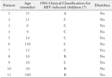
We analyzed small intestines biopsies of 11 HIV-infect-ed children between 6-149 months of age (mean age: 35.1 months, six females) classiied in 1994 Centers for Diseases Control clinical categories (7) as A (1), B (3) and C (7), as can be seen in Table 1. The samples were obtained by endoscopic biopsy or by using Watson capsule attached to a polyethylene probe and were collected between August 1994 and May 1995 in São Paulo, Brazil.
TABLE 1. Age, HIV clinical category and report of diarrhea among children with HIV/AIDS
Patient Age
(months)
1994 Clinical Classiication for
HIV infected children (7) Diarrhea
1 35 A No
2 11 C No
3 6 C Yes
4 9 C No
5 14 C Yes
6 149 C No
7 12 C Yes
8 10 B No
9 10 C Yes
10 30 B No
11 100 B Yes
METHODS
Preparation of intestinal samples for viewing by transmission microscopy
Intestinal fragments were washed in a sodium cacodylate buffer-0.1 M HCl and ixed with Karnovsky solution. Then, they were washed with sodium cacodylate buffer-HCl and ixed with osmium tetroxide 1%. After washing with water, they were placed in a solution of 0.5% sucrose uranyl acetate. Then, they were dehydrated in 70%, 90%, and 100% ethanol and iniltrated with propylene oxide and Araldite® (Joinville, SC, Brazil) resin. After iniltration, fragments were placed in a mixture of pure Araldite® resin and iniltrated under a vacuum in a desiccator. After this they were placed in silicone molds and incubated at 60°C. To prepare each block, ultra-thin 90 nm sections were obtained in an MT2 ultramicrotome (SORVALL) with diamond knife (LADD). The sections, collected on copper screens (SEM), were stained with 0.88% lead citrate solution and examined in a transmission electron microscope operating at 80 kV.
Preparation of the intestinal samples for viewing by scanning microscopy
After ixing the fragments of intestine as described above, two further washes with sodium cacodylate buffer-0.1 M HCl were performed. Then, the fragments were transferred to permeable baskets, which were placed in 50% ethanol, and 100% p. a. absolute ethanol (MERCK) for dehydration. Subsequently, fragments were placed in the drying chamber of the apparatus (Balzers CPD 030) using the critical point of carbon dioxide method. With the aid of a stereoscopic microscope, the fragments were oriented with the villi direct-ed upward, placdirect-ed in a sample to support port scanning, and attached with colloidal silver (SEM) to improve conductivity. After assembly, the fragments were subjected to a metabolizer (Balzers - Sputter Coater SCD 050) for the deposition of a thin layer of gold (the “Sputtering” method) to improve con-ductivity. Observations were made with a scanning electron microscope (JEOL JSM - 5300), operating at 10 kV.
Ethical aspects
The study was approved by the Ethics Committee of Universidade Federal de São Paulo – Escola Paulista de Medicina, and a written consent form was obtained from the parents or legal guardians of all participating children.
RESULTS
Electron transmission microscopy
Some microvilli were preserved in number with apparently normal height (Figure 1). Others showed patterns suggesting
that they had been destroyed and signiicantly decreased (Figure 2andFigure 3) with a tendency to form tufts as if they had been compressed at the base, resembling a “bouquet of lowers” (Figure 3). Additionally, we noticed focal areas with microvilli disruption as compared to other areas, which showed reasonably preserved microvilli, but with changes in the number and height of microvilli and the presence of tufts. Differences in the height and number of microvilli were also seen near each other (Figure 3).
Intracytoplasmic vacuoles (Figure 3 and Figure 4) and intense vacuolization of enterocytes were present in some samples (Figure 2).
Multivesicular bodies were frequently identiied in large numbers and were present in most specimens (Figure 1).
Mitochondria were preserved in some samples and vac-uolated in others (Figure 2).
The structure of the epithelium showed membrane thickening in some samples (Figure 3), and the extrusion of enterocytes into the intestinal lumen was also observed (Figures 1, Figure 2 and Figure 3). The intercellular space was well deined, and desmosomes of normal appearance could be observed (Figure 1).
Intraepithelial lymphocytes of varying frequency and quantity were observed (Figure 4).
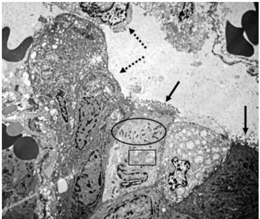
FIGURE 2. Small intestine transmission electron microscopy of an HIV-infected child. Enterocytes with intense vacuolization. Microvilli destruction and tufts formation (black arrows). Vacuolization of mitochon-dria (ellipse) and of Golgi apparatus (rectangle). Extrusion of enterocytes into the intestinal lumen (dashed arrow). 2,500X
FIGURE 3. Small intestine transmission electron microscopy of an HIV-infected child. Microvilli with differences in height and number showing tufts formation (dashed arrows) Cellular extrusion also including the presence of goblet cells (GC). Multiple multivesicular bodies (black arrows). Intracytoplasmatic vacuolization (large arrow). 2,500X
Electron scanning microscopy
The architecture of the villi showed signiicant breakdown (Figure 5) in some samples, and lattening of these structures was observed in others (Figure 6). A ibrin-mucoid crust of variable thickness (Figure 7) often partially or completely obscured the enterocytes. In addition, fat droplets were ob-served at a variable quantity in the lumen (Figure 7).
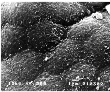
Enterocytes were observed both with well-deined con-tours and architecture, as well as with disorganized archi-tecture (Figure 5 and Figure 8); in some samples, partial or FIGURE 5. Small intestine scanning electronic microscopy of an HIV-in-fected child. Microvilli are decreased in number and height and showing total effacement in some areas. Focal areas with enterocytes showing villous architecture disruption. 2,000X
FIGURE 6. Small intestine scanning electronic microscopy of an HIV-in-fected child. Villi lattening and loss of cellular structures delineation. Some areas showing more severe changes in epithelial surface (arrow). 2,000X
FIGURE 7. Small intestine scanning electronic microscopy of an HIV-in-fected child. Mucosa covered with ibrin-crust (arrow) and presence of fat drops. 7,500X
FIGURE 8. Small intestine scanning electronic microscopy of an HIV-in-fected child. Denudated epithelium showing loss of cellular architecture, villi lattening and no mucus. 3.500X
complete loss of boundaries between the cells was observed (Figure 9).
Microorganisms with structures that were morpholog-ically similar to bacilli were occasionally identiied in the intestinal lumen (Figure 10).
DISCUSSION
It is important to emphasize that the intestine samples examined in this study were obtained in the period preced-ing the introduction of highly active antiretroviral therapy (HAART). Therefore, this is a unique study that almost relects the natural history of the disease. Moreover, the current research is one of the exceedingly rare studies that
use transmission electron microscopy to analyze the small intestine of HIV infected children.
We observed signiicant effects on microvilli, including complete denudation (Figure 2), shortening (Figures 3 and Figure 4) and the formation of small “tufts” (Figure 3) that resembled a “bouquet of lowers”. These indings are similar to those described by other authors who have also exam-ined microvilli in similar patients(3, 10, 11, 25). The repetition of these patterns in different samples appears to be compatible that they are related to HIV itself, rather than a result of a co-occurring infection. This hypothesis was irst suggested by Kotler et al.(16). However, one should consider the limita-tions of this methodology. Intestinal samples were obtained by biopsy and represent tiny fragments of tissue; thus, they may represent only focal alterations.
Another prominent inding was the increase of intracyto-plasmic vacuoles in the enterocytes in many of the fragments (Figures 3 and Figure 4). In some samples, this change was so signiicant that it promoted a structural derangement of the involved enterocytes (Figure 2). It was previously report-ed(17, 21, 24, 25) and may be indicative of cellular degeneration or inlammatory activity, particularly when irregularly shaped and located in the periphery of the cytoplasm. Like Kotler et al.(17) we did not detect any intracellular or extracellular viral particles in the enterocytes with increased vacuoles(17). This may suggests that if HIV was involved in this process, it may have occurred through molecular signaling and not by a direct cytopathic effect.
Our observation of the thickening of the basal membrane (Figure 3) constituted a unique and very important change that, to our knowledge, has not been previously described. This com-ponent of the epithelia, in addition to promoting the adhesion of epithelial cells to underlying connective tissue, plays a role in the migration, proliferation, and differentiation of cells(15). Thus, that inding could be the result of ongoing inlammation in the gut or a facilitator of detachment of cells to the intestinal lumen, as observed in some samples (Figures 1, 2, 3).
We often observed intraepithelial lymphocytes (Figure 4), which were also previously described in a study that used light microscopy to examine the intestinal mucosa(18). Along with the lymphocytes of the lamina propria, intraepithelial lymphocytes are part of the effector arm of the GALT im-mune response. They are the irst imim-mune cells to recognize pathogens that have invaded the epithelial surface and are comprised of a large population of oligoclonal resting cells able to display phenotypic and functional cytolytic T cells when activated(1). The increased presence of intraepithelial lymphocytes is well established in celiac disease, a condition usually followed by villous atrophy(20) due to excessive apop-tosis of enterocytes(23). Considering that several histological changes observed in the intestinal mucosa during HIV infec-tion are similar to those seen in celiac disease, it is reasonable FIGURE 9. Small intestine scanning electronic microscopy of an
HIV-in-fected child. Areas denuded of microvilli (arrows). Others areas with presence of microvilli showing differences in height and tendency to form tufts (ellipse). 7,500X
to assume that the intraepithelial lymphocytes may have a cytolytic effect on the enterocytes of patients with HIV/AIDS. Weber Jr. and Dobbins III(30) have suggested it comparing the ultrastructural characteristics of these lymphocytes in both diseases and demonstrating their relative scarcity in healthy patients(30). The present paper is novel in its demonstration of the presence of intraepithelial lymphocytes in the intestinal mucosa of young HIV patients through electron microscopy. Currently, it is known that intraepithelial lymphocytes are represented predominantly by CD8+ T cells and cells-T γ/δ(14). More recently, CD8+ T cells have garnered interest because some of them have demonstrated the capacity to control the replication of HIV in the bloodstream and gastrointestinal tracts and preserve CD4+ T cells given the capacity of various effector cytokines such as interferon-γ, macrophage inlam-matory protein 1β, TNF-α, and interleukin-2 (IL-2). These “multifunctional” CD8+ T cells are speciic for HIV and are rarely found among individuals who present progression of HIV infection(2). Considering that our patients were progres-sors, they probably would be better categorized as having a “mono-functional” immune response proile with ineffective CD8+ T cells unable to control viral replication at the level of intestinal mucosa(6). This information is relevant because it suggests that an effective HIV vaccine should be able to induce a response in CD8+ T cells speciic for the virus that it the “multifunctional” proile in the gastrointestinal tract. Such a vaccine would be capable of inhibiting the entry of HIV into the mucosa and controlling viral replication thereby ultimately, preserving immune function(4).
Other structures observed frequently in our samples were the multivesicular bodies (Figure 1), intracytoplasmic organelles that are rich in the cholesterol, formed during endocytosis. The increased presence of these structures could be indicative that the intestinal changes occurring in these patients were large such to promote absorption of potentially antigenic macromolecules. This could provoke local chronic inlammatory processes and the consequent recruitment of CD4+ targets of HIV infection. These processes could contribute to the systemic spread of the virus from the gas-trointestinal tract, which acts as a major viral reservoir. It is interesting to note the similarity of our indings with those previously described in children with chronic diarrhea due to milk protein intolerance(8). We hypothesize that it may represent the occurrence of similar pathogenic mechanisms or, alternatively, that different form of injury to the gut could culminate in similar pathological responses. Moreover, it seems reasonable to think that children with HIV/AIDS are at an increased risk of developing intolerance to various components of their diet. This could happen as a result of increased intestinal permeability caused by HIV infection, by co-infection with other pathogens or even by early exposure to cow’s milk protein due to breastfeeding contraindication.
It is also estimated that the multivesicular bodies may play an important role in virus entry from their presence in the intestinal lumen, as occurs in HIV transmission. Like other retroviruses, HIV gag proteins contain small binding sites that mediate the interactions between multivesicular bodies and other components related to endocytosis, suggesting that these viral proteins are directly involved in this process(19).
The mitochondria were normal in appearance in most cases, as shown in Figure 4. This differs from the indings of Oktedalen et al.(24), who noted a reduction in the number of these organelles. According to these authors, this reduction could cause possible enterocyte dysfunction and could ex-plain the occurrence of pronounced changes in the intestinal absorption of D-xylose in HIV patients as in celiac disease. Nevertheless, in our study, we also observed the vacuolization of mitochondria and Golgi complex (Figure 2), which could be indicative of enterocyte dysfunction.
Analyses using scanning electron microscopy also recog-nized of several abnormalities on the epithelial surface of the small intestine. Thus, in most images, we could clearly see the impressive extent and variable intensity of microvilli shortening and could often view extensive areas of complete destruction of these structures (Figure 5). Furthermore, tufts of microvilli was also observed in some samples (Figure 9), as were areas showing microvilli derangement. Although these changes were prominent, other areas of mucosa remained essentially unchanged. Thus, our indings revealed a pattern of variable epithelial changes, which differs from the more diffuse pattern of changes and uniformity characteristic of celiac disease(26).
In several samples, it was possible to observe a crust of mucus and ibrin, which varied in thickness and in extent, covering the epithelial surface of the intestine (Figure 7). This crust was often suficiently large to completely prevent the viewing of enterocytes. Apparently, this finding has not been described previously in relation to ultrastructural changes of the intestine in patients with HIV/AIDS. How-ever, there is a close resemblance between this inding and the description given by Fagundes-Neto et al.(9) of children with persistent diarrhea. Given the frequency and the extent of this phenomenon, it is feasible to postulate that this crust results in diminished intestinal absorption through the me-chanical blockage of nutrients. Under these circumstances, the presence of unabsorbed solutes in the intestinal lumen could trigger osmotic diarrhea.
Alternatively, fat solubilization could be impaired due to primary bile salts disconjugation and 7-a-dehydroxilation secondary to bacterial overgrowth in the intestine. Such a process has been described in environmental enteropathy(22), or as a result of pancreatic dysfunction due to malnutrition(7).
Sometimes we observed the presence of bacteria with ba-cilli-like morphology in the intestinal lumen (Figure 10). This may be similar to the occurrence of bacterial overgrowth in the small intestine that occurs in environmental enteropathy, which shares functional and ultrastructural similarities with the pathology observed in our study(22).
CONCLUSION
Signiicant ultrastructural changes varying in intensity and frequency were found through scanning and transmission electron microscopy in the intestine of HIV infected children. Some of these changes, to our knowledge, had not yet been described. Given their magnitude and the frequency with which recurred in the analyzed samples, they could intensely impact intestinal function and permeability and could there-fore profoundly affect children’s nutritional status, quality of life and life expectancy.
Leite CAC, Fagundes-Neto U, Haapalainen EF. Avaliação da ultra-estrutura do intestino delgado de crianças infectadas pelo HIV por microscopia eletrônica de transmissão e varredura. Arq Gastroenterol. 2013,50(1):70-7.
RESUMO - Objetivos - Descrever achados ultra-estruturais do intestino delgado de crianças infectadas pelo HIV. Métodos - Estudo descritivo, observa-cional de biopsias do intestino delgado, realizada entre agosto de 1994 e maio de 1995 em São Paulo - Brasil. Este material pertencia a 11 crianças infectadas pelo HIV e foi armazenado em um laboratório em blocos de paraina. As amostras de intestino delgado foram analisadas por microscopia eletrônica de transmissão e de varredura e achados os achados ultra-estruturais foram descritos por meio da análise de fotos digitalizadas desse material. Foi obtida aprovação pelo Comitê de Ética. Resultados - Na maioria das amostras a microscopia de varredura mostrou vários graus de encurtamento e diminuição do número das microvilosidades e até o completo apagamento dessas estruturas em algumas áreas. O desarranjo dos enterócitos foi visto com freqüência e, por vezes, os limites celulares estavam imprecisos. Em algumas áreas uma membrana ibrino-mucosa com espessura e extensão variáveis aparentava revestir parcial ou totalmente a superfície epitelial. Gotas de gordura no lúmen intestinal estavam presentes em várias amostras e bactérias morfologicamente semelhantes a bacilos foram observadas em duas amostras. A microscopia eletrônica de varredura conirmou as observações constatadas nas microvilosidades através da microscopia de transmissão e também mostrou pequenos “tufos” dessas estruturas. Além disso, mostrou aumento do número de vacúolos e de formações multivesiculares dentro de vários enterócitos, aumento da presença de linfócitos intraepiteliais, vacuolização mitocondrial e alargamento da membrana basal na maioria das amostras analisadas. No entanto, algumas amostras apresentavam-se com aspecto normal. Conclusões - Nosso estudo mostrou a ocorrência comum de várias alterações ultra-estruturais intes-tinais importantes e de intensidade variável entre crianças infectadas pelo HIV, algumas delas, ao nosso conhecimento, não descritas anteriormente.
REFERENCES
1. Beagley KW, Husband AJ. Intraepithelial lymphocytes: origins, distribution, and function. Crit Rev Immunol. 1998;18:237-54.
2. Betts MR, Nason MC, West SM, De Rosa SC, Migueles SA, Abraham J, Leder-man MM, Benito JM, Goepfert PA, Connors M, Roederer M, Koup RA. HIV nonprogressors preferentially maintain highly functional HIV-speciic CD8+ T cells. Blood. 2006;107:4781-9.
3. Blanshard C, Ellis DS, Tovey G, Gazzard BG. Electron microscopy of rectal biopsies in HIV-positive individuals. J Pathol. 1993;169:79-87.
4. Brenchley JM, Douek DC. HIV infection and the gastrointestinal immune system. Mucosal Immunol. 2008;1:23-30.
5. Centers for Disease Control and Prevention. 1994 revised Classiication System for Human Immunodeiciency Virus Infection in Children Less than 13 Years of Age. MMWR Recomm Rep. 1994;43(RR-12):1-10.
6. Critchield JW, Lemongello D, Walker DH, Garcia JC, Asmuth DM, Pollard RB, Shacklett BL. Multifunctional human immunodeiciency virus (HIV) gag-speciic CD8+ T-cell responses in rectal mucosa and peripheral blood mononuclear cells during chronic HIV type 1 infection. J Virol. 2007;81:5460-71.
7. Descos L, Duclieu J, Minaire Y. Exocrine pancreatic insuficiency and primitive malnutrition. Digestion. 1977;15:90-5.
8. Fagundes-Neto U, Wehba J, Viaro T, Machado NL, Patricio FR. Protracted diarrhea in infancy: clinical aspects and ultrastructural analysis of the small intestine. J Pediatr Gastroenterol Nutr. 1985;4:714-22.
9. Fagundes-Neto U, De Martini-Costa S, Pedroso MZ, Scaletsky IC. Studies of the small bowel surface by scanning electron microscopy in infants with persistent diarrhea. Braz J Med Biol Res. 2000;33:1437-42.
10. Fontana M, Boldorini R, Zuin G, Tosoni A, Costanzi G, Principi N. Ultra-structural changes in the duodenal mucosa of HIV-infected children. J Pediatr Gastroenterol Nutr. 1993;17:255-9.
11. Giovanni B, Calabrese C, Manfredi R, Pisi AM, Di Febo G, Hakim R, Cenacchi G, Basco G. HIV enteropathy: undescribed ultrastructural changes of duodenal mucosa and their regression after triple antiviral therapy. A case report. Dig Dis Sci 2005; 50: 617-22.
12. Gottlieb MS, Schroff R, Schanker HM, Weisman JD, Fan PT, Wolf RA, Saxon A. Pneumocystis carinii pneumonia and mucosal candidiasis in previously healthy
homosexual men: evidence of a new acquired cellular immunodeiciency. N Engl J Med. 1981;305:1425-31.
13. Haase AT. Perils at mucosal front lines for HIV and SIV and their hosts. Nat Rev Immunol. 2005;5:783-92.
14. Hein WR. Organization of mucosal lymphoid tissue. Curr Top Microbiol Im-munol. 1999;236:1-15.
15. Kierszenbaum AL. Histology and Cell Biology: an Introduction to Pathology. 1st. ed. St Louis, Mo: Mosby Inc.;2002.
16. Kotler DP, Gaetz HP, Lange M, Klein EB, Holt PR. Enteropathy associated with the acquired immunodeiciency syndrome. Ann Intern Med. 1984;101:421-8. 17. Kotler DP, Weaver SC, Terzakis JA. Ultrastructural features of epithelial
cell degeneration in rectal crypts of patients with AIDS. Am J Surg Pathol. 1986;10:531-8.
18. Leite CA, Succi RC, Patricio FR, Fagundes-Neto U. Functional, microbiological and morphological intestinal indings among human immunodeiciency virus infected children. Arq Gastroenterol. 2006;43:310-5.
19. Lindwasser OW, Resh MD. Human immunodeiciency virus type 1 Gag contains a dileucine-like motif that regulates association with multivesicular bodies. J Virol. 2004;78:6013-23.
20. Marsh MN, Crowe PT. Morphology of the mucosal lesion in gluten sensitivity. Baillieres Clin Gastroenterol. 1995;9:273-93.
21. Mathan MM, Grifin GE, Miller A, Batman P, Forster S, Pinching A, Harris W. Ultrastructure of the jejunal mucosa in human immunodeiciency virus infection. J Pathol. 1990;161:119-27.
22. Morais MB, Fagundes-Neto U. Enteropatia Ambiental. Estudos Avançados. 2003;17:137-48.
23. Moss SF, Attia L, Scholes JV, Walters JR, Holt PR. Increased small intestinal apoptosis in coeliac disease. Gut. 1996;39:811-7.
24. Oktedalen O, Skar V, Dahl E, Serck-Hanssen A. Changes in small intestinal structure and function in HIV-infected patients with chronic diarrhoea. Scand J Infect Dis. 1998;30:459-63.
25. Orenstein JM, Kotler DP. Diarrheogenic bacterial enteritis in acquired immune deiciency syndrome: a light and electron microscopy study of 52 cases. Hum Pathol. 1995;26:481-92.
26. Owens SR, Greenson JK. The pathology of malabsorption: current concepts. Histopathology. 2007;50:64-82.
27. Paiardini M, Frank I, Pandrea I, Apetrei C, Silvestri G. Mucosal immune dys-function in AIDS pathogenesis. AIDS Rev. 2008;10:36-46.
28. Smith PD, Meng G, Salazar-Gonzalez JF, Shaw GM. Macrophage HIV-1 infection and the gastrointestinal tract reservoir. J Leukoc Biol 2003; 74: 642-9. 29. Van de Perre P. Mother-to-child transmission of HIV-1: the ‘all mucosal’
hypoth-esis as a predominant mechanism of transmission. AIDS. 1999;13:1133-8. 30. Weber JR, Jr., Dobbins WO 3rd. The intestinal and rectal epithelial lymphocyte
in AIDS. An electron-microscopic study. Am J Surg Pathol. 1986;10:627-39.