Ectopic Bone Formation by Mesenchymal
Stem Cells Derived from Human Term
Placenta and the Decidua
Gina D. Kusuma1,2☯, Danijela Menicanin3,4☯, Stan Gronthos3, Ursula Manuelpillai2,5, Mohamed H. Abumaree6, Mark D. Pertile7,8, Shaun P. Brennecke1,2, Bill Kalionis1,2*
1Department of Obstetrics and Gynaecology, Royal Women’s Hospital, The University of Melbourne, Parkville, Victoria, Australia,2Pregnancy Research Centre, Department of Perinatal Medicine, Royal Women’s Hospital, Parkville, Victoria, Australia,3Mesenchymal Stem Cell Laboratory, Faculty of Health Sciences, School of Medical Sciences, University of Adelaide, Adelaide, Australia,4Colgate Australian Clinical Dental Research Centre, School of Dentistry, University of Adelaide, Adelaide, Australia,5Centre for Genetic Diseases, Monash Institute of Medical Research-Prince Henry’s Institute of Medical Research and Monash University, Clayton, Victoria, Australia,6King Abdullah International Medical Research Center/ King Saud Bin Abdulaziz University for Health Sciences, College of Science and Health Professions, King Abdulaziz Medical City-National Guard Health Affairs, Riyadh, Kingdom of Saudi Arabia,7Victorian Clinical Genetics Services (VCGS), Murdoch Children’s Research Institute, Royal Children's Hospital, Parkville, Victoria, Australia,8Department of Paediatrics, Royal Children’s Hospital, The University of Melbourne, Parkville, Victoria, Australia
☯These authors contributed equally to this work. *bill.kalionis@thewomens.org.au
Abstract
Mesenchymal stem cells (MSCs) are one of the most attractive cell types for cell-based bone tissue repair applications. Fetal-derived MSCs and maternal-derived MSCs have been isolated from chorionic villi of human term placenta and thedecidua basalisattached
to the placenta following delivery, respectively. Chorionic-derived MSCs (CMSCs) and decidua-derived MSCs (DMSCs) generated in this study met the MSCs criteria set by Inter-national Society of Cellular Therapy. These criteria include: (i) adherence to plastic; (ii)
>90% expression of CD73, CD105, CD90, CD146, CD44 and CD166 combined with<5% expression of CD45, CD19 and HLA-DR; and (iii) ability to differentiate into osteogenic, adi-pogenic, and chondrogenic lineages.In vivosubcutaneous implantation into SCID mice
showed that both bromo-deoxyuridine (BrdU)-labelled CMSCs and DMSCs when implanted together with hydroxyapatite/tricalcium phosphate particles were capable of forming ectopic bone at 8-weeks post-transplantation. Histological assessment showed expression of bone markers, osteopontin (OPN), osteocalcin (OCN), biglycan (BGN), bone sialoprotein (BSP), and also a marker of vasculature, alpha-smooth muscle actin (α-SMA). This study provides
evidence to support CMSCs and DMSCs as cellular candidates with potent bone forming capacity.
a11111
OPEN ACCESS
Citation:Kusuma GD, Menicanin D, Gronthos S, Manuelpillai U, Abumaree MH, Pertile MD, et al. (2015) Ectopic Bone Formation by Mesenchymal Stem Cells Derived from Human Term Placenta and the Decidua. PLoS ONE 10(10): e0141246. doi:10.1371/journal.pone.0141246
Editor:Michiya Matsusaki, Osaka University, JAPAN
Received:May 4, 2015
Accepted:October 5, 2015
Published:October 20, 2015
Copyright:© 2015 Kusuma et al. This is an open access article distributed under the terms of the
Creative Commons Attribution License, which permits unrestricted use, distribution, and reproduction in any medium, provided the original author and source are credited.
Data Availability Statement:All relevant data are within the paper.
Introduction
Mesenchymal stem cells (MSCs), which are also referred to as multipotent stromal cells, are found in many tissues. MSCs are capable of multipotent differentiation, allowing them to con-tribute to bone regeneration and repair since MSCs can readily differentiate into osteocytic
lin-eages [1]. Moreover, MSCs are readily isolated, their numbers can be greatly expanded in
culture, cryopreserved for later use, and importantly, they display low immunogenicity, are immunomodulatory and have a good safety profile. According to the International Society for Cellular Therapy (ISCT), MSCs must; (i) adhere to untreated plastic surfaces; (ii) express
CD105, CD73, and CD90 but not CD34, CD14, CD19, CD11b, CD79αor HLA-DR, and (iii)
differentiate into osteogenic, adipogenic and chondrogenic lineagesin vitro[2].
The human term placenta is an abundant, readily accessible and non-controversial source of MSCs. MSCs have been isolated from fetal derived placental tissues including the amnion, chorion and chorionic villi, and from maternal derived tissues that are attached to placental
tis-sue following delivery i.e. thedecidua parietalisanddecidua basalis[3–7]. The peripheral
region of the placenta on the maternal side that is in contact with the uterine wall (called the
basal plate) comprises the chorionic villi on one side, and maternaldecidua basalison the
other. Following delivery of the placenta, thedecidua basalisremains attached to the maternal
side of the placenta. Thus, careful preparation and characterization needs to be carried out to
confirm the maternal origins ofdecidua basalisMSCs (DMSCs) and the fetal origins of the
chorionic villous MSCs (CMSCs). As such, in addition to the criteria stipulated by the ISCT,
Parolini et al. proposed that CMSCs should have<1% maternal cells in the population [8,9].
The significant issue of the presence of maternal cells in human placental MSCs cultures was
reviewed recently [10]. Therefore, the first aim of this study was to isolate and characterize
CMSCs and DMSCs according to the criteria described above and to confirm the respective fetal and maternal origins of these cells. This characterization was an essential prerequisite to
the use of CMSCs and DMSCs forin vivoassays.
While studies have reported osteogenesis by CMSCs and DMSCsin vitro, bone formation
in vivohas not been investigated. Such studies are essential for evaluating the functional capac-ity of CMSCs and DMSCs and their potential for clinical applications. Therefore, we initiated the study using a mouse model of ectopic bone formation to explore the possibility that isolated
human CMSCs and DMSCs were capable of regenerating ectopic bone-like structurein vivo.
The orthotopic bone formation assay is commonly used to study osteogenesisin vivo.
Com-pared to the orthotopic assay, the ectopic bone forming assay has unique advantages since there is no requirement for bone cytokine stimulation and cell-to-cell interaction with endogenous
bone-forming cells [11]. In addition, a variety of ectopic locations can be used for cell
implanta-tion, including subcutaneous and intramuscular sites and the kidney capsule [11]. Subcutaneous
implantation is the simplest experimental model of ectopic bone formation. Mouse models are preferable and most widely used due to their low cost, loose skin folds that can accommodate large implants, and the availability of immunodeficient mice that will accept implanted human cells. Another important consideration is the lack of naturally occurring bone-forming stem cells within the intradermal compartment and therefore newly-formed bone can be confidently attributed to the exogenous stem cells. The most pertinent concern regarding subcutaneous implantation is the lack of robust bone growth which may be due to poor blood flow. However, subcutaneous bone formation can be stimulated by incorporating hydroxyapatite and tricalcium phosphate (HA/TCP) together with stem cells. HA/TCP are currently used as bone graft
substi-tutes, are biocompatible and form bonds between bone and ceramic implants [12]. The second
and principal aim of this study was to evaluatein vivobone formation capacity of CMSCs and
DMSCs following subcutaneous implantation together with HA/TCP.
Materials and Methods
Tissue collection
Placental samples were collected from healthy women with normal pregnancies following elec-tive Caesarean section or vaginal delivery at term (n = 6). The placental tissue had no obvious signs of calcification, infarcts or meconium staining. Exclusion criteria were women who smoked or had a twin or triplet pregnancy, drug dependency, intrauterine infection, prolonged rupture of the fetal membranes or placental abruption. Informed written consent was obtained
from all participants before delivery. The study was approved by the Royal Women’s Hospital
Human Research Ethics Committee.
Isolation of CMSCs
CMSCs were isolated using the explant method as described previously [7] with the following
modifications. Briefly, an incision was made through the fetal membranes near the umbilical
cord insertion site and 1 g of chorionic villous tissue was obtained from approximately 1–2 cm
below the chorionic plate. Pieces of chorionic tissue with typical villous morphology were cleaned with a 21 gauge needle under a dissecting microscope to remove non-villous tissue. Cleaned villi were finely diced and digested in 0.25% trypsin for 40 min at 37°C. The trypsin was inactivated with FBS and tissues were washed in PBS. The digested villi were cultured in
Amniomax C100 complete medium (Life Technologies) in 25 cm2tissue culture flasks
main-tained at 37°C in a humidified 5% CO2incubator. After 7 days, villous tissues were removed
from the flask and the adherent cells arising from the explants (P0 cells) were grown until at least 80% confluent before expanding to reach P5.
Isolation of DMSCs
We have previously reported the isolation of DMSCs from thedecidua basalisadhering onto
the maternal side of the placenta [13]. About eight grams of placental tissue was dissected from
the basal plate, washed four times in PBS, finely minced and digested in trypsin (0.25%; Life
Technologies, CA, USA) and DNAse 1 (50μg/mL; Worthington, NJ, USA) at 4°C overnight.
Fetal bovine serum (FBS; Thermo Scientific, MA, USA) was added to inactivate the trypsin and
the digest was centrifuged at 200gfor 5 min. The pelleted tissue was digested in type 1
collage-nase (10 mg/mL; Worthington) and DNAse 1 (50μg/mL, Worthington) for 30 min at 37°C
and strained through a 100μm stainless steel sieve. The filtrate was layered over Histopaque
(Sigma-Aldrich, MO, USA) and separated by density gradient centrifugation at 400gfor 30
min. Mononuclear cell layers containing the DMSCs were aspirated and centrifuged at 200g
for 5 min. DMSCs were maintained inα-MEM medium (Sigma-Aldrich) with 10% FBS,
peni-cillin/streptomycin (100 U/mL and 100 mg/mL, respectively; Life Technologies) and 2 mM L-glutamine (Sigma-Aldrich). P0 DMSCs were passaged after reaching 80% confluence and cells were expanded up to P5.
Fluorescence in situ hybridisation (FISH)
FISH was used to determine whether the DMSCs were maternal and the CMSCs fetal in origin
as described elsewhere [6,13]. Briefly, term placentae delivered from pregnancies carrying
male babies (n = 3) were used to prepare DMSCs and CMSCs. Cells were lifted with TrypLE
Express, washed in Hank’s buffered saline solution (HBSS; Life Technologies) and placed on
(Spectrum Green) and chromosome Y (Spectrum Orange) probes (Abbott Molecular, MO, USA). Approximately 200 cells per slide were examined.
Flow cytometry
To determine whether expanded cells expressed positive and negative markers characteristic of MSCs, cells were analyzed by flow cytometry for CD73, CD105, CD90, CD146, CD44 and CD166 and the absence of CD45, CD19 and HLA-DR. Cells were incubated with each of the
primary antibodies or equivalent concentrations of matched isotype controls (Table 1) as
described previously [13]. Cells were then washed in HBSS containing 2% FBS and centrifuged
at 1000 rpm for 5 min. Cell pellets were resuspended in 500μL of HBSS with 2% FBS and 1μg/
mL DAPI (Sigma-Aldrich). The cells were analyzed on a LSRII flow cytometer with FACS Diva software (BD Biosciences, CA, USA).
In vitro
differentiation into mesenchymal lineages
Differentiation of DMSCs and CMSCs into adipogenic, osteogenic, and chondrogenic lineages
was assessedin vitro.In vitrodifferentiation was carried out with bullet kits as described [5,6,
13]. Adipogenic and osteogenic differentiation was carried out in Mesencult basal medium
together with the respective differentiation supplements according to manufacturer’s
instruc-tions (Stem Cell Technologies, BC, Canada). Chondrogenic differentiation was carried in DMEM/F12 medium (Life Technologies) with 1% ITS and chondrogenic supplements (both
from R&D Systems, MN, USA) according to the manufacturer’s instructions. Cells were
stained with Oil Red O solution, Alizarin Red solution and Safranin O (Sigma-Aldrich) to visu-alize adipogenesis, osteogenesis and chondrogenesis respectively.
In vivo
ectopic bone formation assay
CMSCs and DMSCs (n = 3 each) were expanded to reach approximately 1x10⁷cells per sample
(P2-P3). Approximately 2x10⁶MSCs from each donor were mixed with 40 mg hydroxyapatite/
tricalcium phosphate (HA/TCP) ceramic particles (Zimmer Inc., IN, USA) and then subcuta-neously transplanted into the dorsal surface of eight-week-old SCID mice (National Institutes
of Health-bg-nu-xid; Harlan Sprague-Dawley, IN, USA) as described previously [14,15]. Each
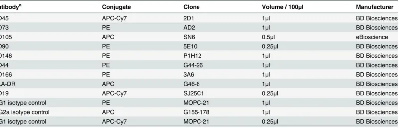
Table 1. Antibodies used for characterizing CMSCs and DMSCs by flow cytometry.
Antibodya Conjugate Clone Volume / 100
μl Manufacturer
CD45 APC-Cy7 2D1 1μl BD Biosciences
CD73 PE AD2 1μl BD Biosciences
CD105 APC SN6 0.5μl eBioscience
CD90 PE 5E10 0.25μl BD Biosciences
CD146 PE P1H12 1μl BD Biosciences
CD44 PE G44-26 1μl BD Biosciences
CD166 PE 3A6 1μl BD Biosciences
HLA-DR APC G46-6 1μl BD Biosciences
CD19 APC-Cy7 SJ25C1 0.25μl BD Biosciences
IgG1 isotype control PE MOPC-21 1μl BD Biosciences
IgG2a isotype control APC G155-178 1μl BD Biosciences
IgG1 isotype control APC-Cy7 MOPC-21 0.25μl BD Biosciences
aanti-human antibodies raised in mice.
mouse received two implants. These procedures were performed in accordance with guidelines of an approved small-animal protocol (South Australia Pathology Animal Ethics Committee #139/09). After 8 weeks, the implants were removed, fixed in 10% formalin overnight at 4°C, and then decalcified for 2 weeks in 0.5 M EDTA, prior to paraffin embedding. For histological
analysis, 5μm sections of the implants were prepared and stained with haematoxylin and eosin
(H&E). Expression of the specific osteogenesis markers osteocalcin (OCN), osteopontin (OPN), biglycan (BGN), and bone sialoprotein (BSP), were analyzed by
immunohistochemis-try using previously published methods [16]. The implanted cells were labelled with BrdU at
24 and 48 hrs prior to implantation to evaluate the localization of the transplanted cells. Immu-nohistochemical staining using an anti-BrdU antibody was carried out as previously described [17].
Results
Isolation and expansion of CMSCs and DMSCs
CMSCs were isolated using the explant method [5,7]. CMSCs migrated from the explants
approximately 7 days after plating. P0 and expanded CMSCs exhibited the characteristic the
fibroblast-like morphology of MSCs (Fig 1Ai). DMSCs isolated from thedecidua basalis
attached to chorionic villi of term placentae adhered onto tissue culture flasks within 24 h of
plating. Consistent with previous findings [18–20], P0 DMSCs were initially heterogeneous
and became more homogeneous following expansion and had the fibroblastic morphology
characteristic of MSCs (Fig 2Ai). Given that CMSCs and DMSCs were morphologically
indis-tinguishable after passaging, it was crucial that CMSCs and DMSCs used in these experiments were well-characterized with respect to their surface markers expression, origin, and differenti-ation potential.
Phenotypic characterization of CMSCs and DMSCs
CMSCs and DMSCs at P3-P5 were analyzed by flow cytometry for cell surface markers present
on expanded MSCs [2,9]. More than 90% of CMSCs (Fig 1Band DMSCs (Fig 2B) expressed
the MSC markers CD90, CD146, CD166, CD44, CD73 and CD105, while<5% were CD45,
CD19 and HLA-DR positive. These findings were consistent with the expression profile stipu-lated for MSCs. Cells beyond P5 were not analyzed since studies have reported that MSCs
undergo cell death or senescence after five passages [4,21–23].
Given the risk of cross contamination between fetal and maternal cells, firstly we analyzed CMSCs and DMSCs by FISH to verify that pure cell populations had been isolated. CMSCs and DMSCs were isolated from placentae of women delivering a male baby, and approximately 200 cells of each type were analyzed for signals in interphase nuclei using X/Y chromosome probes. CMSCs showed the XY phenotype (Spectrum Green and Orange labelled
chromo-somes, respectively) and were therefore male (Fig 1Aii). Evaluation of 200 interphase nuclei
revealed that CMSCs used in this study had 94% XY (6% XXYY), 100% XY, and 99.5% XY (0.5% XXYY). Two Spectrum Green labelled X chromosomes were visible in DMSCs and
therefore DMSCs were female (Fig 2Aii). Evaluation of 200 interphase nuclei showed that
DMSCs used in this study had 100% XX, 92.5% XX (7.5% XXXX), and 99% XX (1% XXXX). Cases of tetraploidy were always XXYY from fetal CMSCs and XXXX from maternal DMSCs preparations. Tetraploidy is a common artefact of cell culturing and does not preclude the use
of cell preparations for further analysis [24].
Differentiation of expanded CMSCs and DMSCs into osteogenic, adipogenic and
chondro-genic lineages was also examined to further verify theirin vitroMSC properties. Alizarin Red
Fig 1. CMSC phenotypic characterization.A. (i) Bright field microscopy image of CMSCs at P0. Magnification is 100X and scalebar is 100μm. (ii) CMSCs
osteogenic induction medium (Fig 1Ci). Oil Red O stained lipid droplets were observed around
cell nuclei in CMSCs stimulated in adipogenic differentiation medium (Fig 1Cii). CMSCs
aggregated into a three-dimensional spherical structure after approximately 24 h stimulation in chondrogenic differentiation medium. Safranin O staining in sections taken from different regions of the cell pellet after 3 weeks stimulation showed the presence of proteoglycans, which
are normally secreted into the extracellular matrix by cartilage cells (Fig 1Ciii). Control CMSCs
cultures maintained in the appropriate basal medium did not show evidence of differentiation
into these lineages (Fig 1Ci–1Ciiiinsets). DMSCs also differentiated into the osteogenic,
adipo-genic and chondroadipo-genic mesenchymal lineages (Fig 2Ci, 2Cii and 2Ciiirespectively) [13].
In vivo
ectopic bone formation assay
CMSCs and DMSCs (n = 3 per group) were assayed for their capacity to develop bone-like tis-sue following ectopic transplantation into SCID mice with HA/TCP particles as a vehicle. All
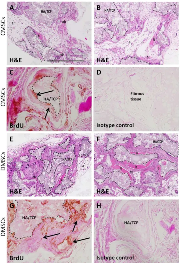
implants showed new bone formation throughout the implants.Fig 3A, 3B, 3E and 3Fshow
representative sections of CMSC and DMSC implants, respectively stained with H&E. The H&E staining was performed with bright pink H&E staining is indicative of mineralized tissue
[17]. New bone was formed (area of pink staining) in HA/TCP (indicated by dashed lines) and
directly interfaces the ceramic surface. The new bone contains osteocytes embedded within the matrix indicating that bone formation was active and progressive. Further histological exami-nation showed that new bone formation was surrounded by the presence of adipocytes (honey-comb-like structures), fibrous tissue, and small blood vessels. Anti-BrdU staining
demonstrated the presence of implanted cells associated with areas of mineral formation and
areas of dense fibrous tissue formation (Fig 3C, 3D, 3G and 3H). The transplanted CMSCs and
DMSCs exhibited the capacity to form mineralized and fibrous tissuesin vivo.
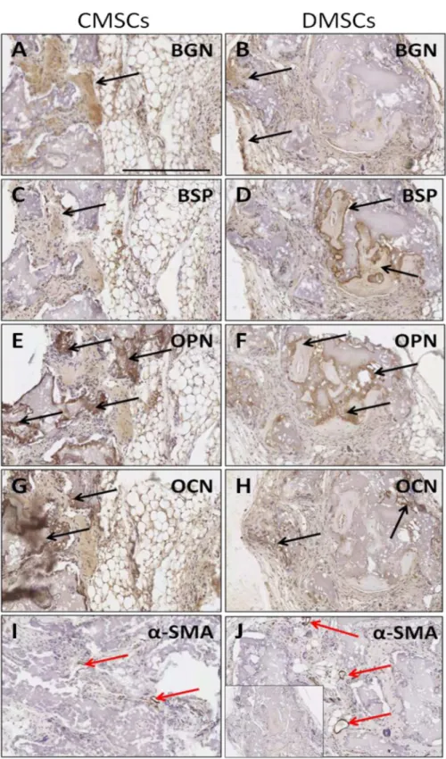
Immunohistochemical staining with several osteogenesis markers was performed to con-firm that cells with osteocyte properties were formed in the transplants. Implanted CMSCs and DMSCs lining or embedded within the mineralized surfaces expressed the bone-related
mark-ers: BSP, OCN, OPN, and BGN (Fig 4A–4H). The reactivity of these markers showed that the
implanted CMSCs and DMSCs contributed to the generation of osteogenic cells. In addition, the presence of newly formed vessels in the transplants was also indicated by immunostaining
withα-SMA (Fig 4I and 4J). Overall, the data demonstrated the presence of new bone
forma-tion with no obvious qualitative differences between CMSCs and DMSCs.
Discussion
We demonstrated the isolation and expansion of two different MSC populations obtained from human term placenta, fetal-derived CMSCs and maternal-derived DMSCs. Both CMSCs and DMSCs are shown to have typical MSC features: (a) adherence to plastic; (b) ability to
dif-ferentiatein vitrointo adipocytes, osteocytes, and chondrocytes; and (c) expression of MSC
surface markers (CD90, CD146, CD166, CD44, CD73, and CD105). FISH analysis confirmed that pure populations of fetal CMSCs and maternal DMSCs could be obtained using the isola-tion methods employed. Thus, the CMSCs and DMSCs adhered to the criteria stipulated for
placental MSCs [2,9].
differentiation into mesenchymal lineages. (i) Osteogenic differentiation, Alizarin Red staining in cells after 5weeks growth in osteogenic induction medium. Arrows show calcium depositions. (ii) Adipogenic differentiation, Oil Red O staining in cells after 14 days growth in adipogenic induction medium. Arrows show fat droplets. (iii) Chondrogenic differentiation, Safranin O staining for proteoglycans depositions in cells after 21 days growth in chondrogenic induction medium. Inset shows control uninduced CMSCs. Scalebar is 100μm.
Fig 2. DMSC phenotypic characterization.A. (i) Bright field microscopy image of DMSCs at P0. Magnification is 100X and scalebar is 100μm. (ii) DMSCs
MSCs from different sources such as adipose tissue, bone marrow, and dental pulp have
been subjected to transplantation in the ectopic bone formation assay [14,15,25,26]. This
study investigated CMSCs and DMSCs for the first time, and found that HA/TCP implanted with these cells into the subcutaneous space of immunodeficient mice could differentiate into new tissue with characteristics of bone. This indicates that donor MSCs has the capacity for
long-term survival and could contribute to the generation of different tissue typesin vivo. The
efficacy of thisin vivoassay was demonstrated in another study where HA/TCP were
implanted with human foreskin fibroblasts and HA/TCP only controls were tested and showed
only fibrous tissue growth with no indication of osteogenesis or haematopoiesis [26].
In this study, we set out to confirm that the implanted cells had survived and contributed to tissue formation with BrdU staining. In principle, BrdU stain incorporates into the DNA of dividing cells and diminishes as those cells further divide. Following 8 weeks post-implanta-tion, a proportion of MSCs may have lost their BrdU expression due to their initial prolifera-tionin vivo, or due to the physical location within tissue (i.e. osteocytes in lacunae), antigen retrieval and DNA denaturing protocol may not have sufficiently exposed the antigen for its detection by immunohistochemistry. Whilst we acknowledge the limitation of using BrdU to
detect implanted cells, this protocol has been widely used and accepted [27–29].
It is also of interest to note that development potential of BMMSCsin vivowas similar to
CMSCs and DMSCs in which the cells contributed to new bone formation together with
sur-rounding fibrous and adipocytes accumulation [14,15,30]. Our findings further support other
studies demonstrating placenta-derived MSCsin vivoosteogenesis capacity. Intrabone, but not
subcutaneous, injection of placenta adherent cells into a mouse model of myeloma-associated
bone loss promoted bone formation by stimulating differentiation of the host’s osteoblasts
[31]. Furthermore, placenta-derived MSCs grown in a silk fibroin/HA scaffold were
trans-planted in a rabbit radius defect model and improved bone repair as evidenced by formation of
new lamellar bone, trabecular bone and a number of osteoblasts [32]. In addition, there was
evidence of angiogenesis with evidence of new blood vessel formation. The presence of newly formed vasculature is in agreement with previous studies that have reported dental pulp stem
cells when implanted using the similarin vivomouse model [14,16]. Another study showed
amnion-derived MSCs promoted neovascularisation in anin vivomouse model [33].
Porous HA/TCP showed good tissue tolerance with no immunological or toxic reaction, and that bone tissue could directly deposit upon their surfaces. This property is important for bone graft substitutes because without it, fibrous tissue can intervene at the interface between
bone tissue and the graft, and cause loosening of the graft [12,26]. The choice of osteoinductive
biomaterials (HA/TCP) combined with the appropriate choice of animal model are potentially
crucial to determinein vivodifferentiation ability of CMSCs and DMSCs. To conclude, this is
the first evidence ofin vivodifferentiation potential of DMSCs and CMSCs following
trans-plantation in the mouse model of ectopic bone formation.
Conclusions
In this study, we have isolated human CMSCs and DMSCs and both cell types demonstrated the characteristic MSC phenotype. Subcutaneous transplantation of CMSCs and DMSCs embedded in a HA/TCP biomatrix, into a mouse model of ectopic bone formation led to the formation of a bone-like structure. BrdU labelling indicated that transplanted cells were differentiation, Oil Red O staining in cells after 14 days growth in adipogenic induction medium. Arrows show fat droplets. (iii) Chondrogenic differentiation, Safranin O staining for proteoglycans depositions in cells after 21 days growth in chondrogenic induction medium. Inset shows control uninduced DMSCs. Scalebar is 100μm.
Fig 3. Histology of CMSCs and DMSCs transplants.Cross sections are representative of CMSCs transplants (A-B) and DMSCs transplants (E-F) after 8 weeks stained with Haematoxylin and Eosin (H&E). In the transplant, the HA/TCP carrier surfaces (dashed lines) are lined with new bone formation (b), areas of immature bone (ob) together with the surrounding fibrous and hematopoietic tissue (a) and blood vessel (bv). Representative BrdU staining for localization of implanted CMSCs (C-D) and DMSCs (G-H). BrdU-stained implanted cells were found lining the mineralized matrix (black arrows) and surrounding fibrous tissue. Brown nuclear staining is indicative of DAB reactivity. There was no immunoreactivity present in sections stained with isotype-matched antibodies. HA/TCP: hydroxyapatite/tricalcium phosphate particles. Magnification is 100X and scalebar is 500μm.
Fig 4. Immunoreactivity of osteogenesis markers afterin vivotransplantation of primary CMSCs and DMSCs into immunocompromised mice.(A-B) BGN: biglycan expression. (C-D) BSP: bone sialoprotein expression. (E-F) OPN: osteopontin expression. (G-H) OCN: osteocalcin expression. (I-J)α-SMA:
alpha-smooth muscle actin as negative control. Inset shows representative control sections stained with isotype-matched antibodies. Colour detection was performed using DAB reaction. Magnification is 200X and scalebar is 300μm. Black arrow show bone-forming surfaces and red arrows show blood vessels.
retained in the structure and contributed to tissue formation. Bone-specific markers such as OPN, OCN, BGN, and BSP were present in both transplants without any qualitative difference.
These data suggest that human CMSCs and DMSCs have potentin vivobone forming capacity
and may be worthwhile candidates forin vivobone tissue repair.
Acknowledgments
We acknowledge the patients who consented to provide placental samples and the clinical
research midwives at the Royal Women’s Hospital, Sue Duggan and Moira Stewart for tissue
collection. We thank Melissa Duggan and Debora Singgih for their technical assistance.
Author Contributions
Conceived and designed the experiments: GDK BK. Performed the experiments: GDK DM MDP. Analyzed the data: GDK DM SG BK. Contributed reagents/materials/analysis tools: GDK DM SG MHA BK MDP. Wrote the paper: GDK UM BK SPB MHA DM SG MDP.
References
1. Pittenger MF, Mackay AM, Beck SC, Jaiswal RK, Douglas R, Mosca JD, et al. Multilineage potential of adult human mesenchymal stem cells. Science. 1999; 284(5411):143–7. PMID:10102814
2. Dominici M, Le Blanc K, Mueller I, Slaper-Cortenbach I, Marini F, Krause D, et al. Minimal criteria for defining multipotent mesenchymal stromal cells. The International Society for Cellular Therapy position statement. Cytotherapy. 2006; 8(4):315–7. PMID:16923606
3. In 't Anker PS, Scherjon SA, Kleijburg-van der Keur C, de Groot-Swings GM, Claas FH, Fibbe WE, et al. Isolation of mesenchymal stem cells of fetal or maternal origin from human placenta. Stem Cells. 2004; 22(7):1338–45. PMID:15579651
4. Portmann-Lanz CB, Schoeberlein A, Huber A, Sager R, Malek A, Holzgreve W, et al. Placental mesen-chymal stem cells as potential autologous graft for pre- and perinatal neuroregeneration. Am J Obstet Gynecol. 2006; 194(3):664–73. PMID:16522395
5. Castrechini NM, Murthi P, Gude NM, Erwich JJ, Gronthos S, Zannettino A, et al. Mesenchymal stem cells in human placental chorionic villi reside in a vascular Niche. Placenta. 2010; 31(3):203–12. doi:
10.1016/j.placenta.2009.12.006PMID:20060164
6. Castrechini NM, Murthi P, Qin S, Kusuma GD, Wilton L, Abumaree M, et al. Decidua parietalis-derived mesenchymal stromal cells reside in a vascular niche within the choriodecidua. Reproductive sciences (Thousand Oaks, Calif). 2012; 19(12):1302–14.
7. Abumaree MH, Al Jumah MA, Kalionis B, Jawdat D, Al Khaldi A, AlTalabani AA, et al. Phenotypic and functional characterization of mesenchymal stem cells from chorionic villi of human term placenta. Stem Cell Rev. 2013; 9(1):16–31. doi:10.1007/s12015-012-9385-4PMID:22628114
8. Parolini O, Alviano F, Bergwerf I, Boraschi D, De Bari C, De Waele P, et al. Toward cell therapy using placenta-derived cells: disease mechanisms, cell biology, preclinical studies, and regulatory aspects at the round table. Stem Cells Dev. 2010; 19(2):143–54. doi:10.1089/scd.2009.0404PMID:19947828
9. Parolini O, Alviano F, Bagnara GP, Bilic G, Buhring HJ, Evangelista M, et al. Concise review: isolation and characterization of cells from human term placenta: outcome of the first international Workshop on Placenta Derived Stem Cells. Stem Cells. 2008; 26(2):300–11. PMID:17975221
10. Heazlewood CF, Sherrell H, Ryan J, Atkinson K, Wells CA, Fisk NM. High incidence of contaminating maternal cell overgrowth in human placental mesenchymal stem/stromal cell cultures: a systematic review. Stem Cells Transl Med. 2014; 3(11):1305–11. doi:10.5966/sctm.2014-0051PMID:25154781
11. Scott MA, Levi B, Askarinam A, Nguyen A, Rackohn T, Ting K, et al. Brief review of models of ectopic bone formation. Stem Cells Dev. 2012; 21(5):655–67. doi:10.1089/scd.2011.0517PMID:22085228
12. Ohgushi H, Okumura M, Tamai S, Shors EC, Caplan AI. Marrow cell induced osteogenesis in porous hydroxyapatite and tricalcium phosphate: a comparative histomorphometric study of ectopic bone for-mation. J Biomed Mater Res. 1990; 24(12):1563–70. PMID:2277053
13. Kusuma GD, Manuelpillai U, Abumaree MH, Pertile MD, Brennecke SP, Kalionis B. Mesenchymal stem cells reside in a vascular niche in the decidua basalis and are absent in remodelled spiral arteri-oles. Placenta. 2015.
15. Gronthos S, Zannettino AC, Hay SJ, Shi S, Graves SE, Kortesidis A, et al. Molecular and cellular char-acterisation of highly purified stromal stem cells derived from human bone marrow. J Cell Sci. 2003; 116(Pt 9):1827–35. PMID:12665563
16. Menicanin D, Mrozik KM, Wada N, Marino V, Shi S, Bartold PM, et al. Periodontal-ligament-derived stem cells exhibit the capacity for long-term survival, self-renewal, and regeneration of multiple tissue types in vivo. Stem Cells Dev. 2014; 23(9):1001–11. doi:10.1089/scd.2013.0490PMID:24351050
17. Hynes K, Menicanin D, Mrozik K, Gronthos S, Bartold PM. Generation of functional mesenchymal stem cells from different induced pluripotent stem cell lines. Stem Cells Dev. 2014; 23(10):1084–96. doi:10. 1089/scd.2013.0111PMID:24367908
18. Huang YC, Yang ZM, Chen XH, Tan MY, Wang J, Li XQ, et al. Isolation of mesenchymal stem cells from human placental decidua basalis and resistance to hypoxia and serum deprivation. Stem Cell Rev. 2009; 5(3):247–55. doi:10.1007/s12015-009-9069-xPMID:19590988
19. Hayati A-R, Nur Fariha M-M, Tan G-C, Tan A-E, Chua K. Potential of Human Decidua Stem Cells for Angiogenesis and Neurogenesis. Archives of Medical Research. 2011; 42(4):291–300. doi:10.1016/j. arcmed.2011.06.005PMID:21820607
20. Brooke G, Rossetti T, Pelekanos R, Ilic N, Murray P, Hancock S, et al. Manufacturing of human pla-centa-derived mesenchymal stem cells for clinical trials. Br J Haematol. 2009; 144(4):571–9. doi:10. 1111/j.1365-2141.2008.07492.xPMID:19077161
21. Soncini M, Vertua E, Gibelli L, Zorzi F, Denegri M, Albertini A, et al. Isolation and characterization of mesenchymal cells from human fetal membranes. Journal of tissue engineering and regenerative medi-cine. 2007; 1(4):296–305. PMID:18038420
22. Zhang X, Soda Y, Takahashi K, Bai Y, Mitsuru A, Igura K, et al. Successful immortalization of mesen-chymal progenitor cells derived from human placenta and the differentiation abilities of immortalized cells. Biochem Biophys Res Commun. 2006; 351(4):853–9. PMID:17094946
23. Pochampally R. Colony forming unit assays for MSCs. Methods in molecular biology (Clifton, NJ). 2008; 449:83–91.
24. Barlow S, Brooke G, Chatterjee K, Price G, Pelekanos R, Rossetti T, et al. Comparison of human pla-centa- and bone marrow-derived multipotent mesenchymal stem cells. Stem Cells Dev. 2008; 17 (6):1095–107. doi:10.1089/scd.2007.0154PMID:19006451
25. Zannettino AC, Paton S, Arthur A, Khor F, Itescu S, Gimble JM, et al. Multipotential human adipose-derived stromal stem cells exhibit a perivascular phenotype in vitro and in vivo. J Cell Physiol. 2008; 214(2):413–21. PMID:17654479
26. Kuznetsov SA, Krebsbach PH, Satomura K, Kerr J, Riminucci M, Benayahu D, et al. Single-colony derived strains of human marrow stromal fibroblasts form bone after transplantation in vivo. J Bone Miner Res. 1997; 12(9):1335–47. PMID:9286749
27. Maeshima A, Yamashita S, Nojima Y. Identification of renal progenitor-like tubular cells that participate in the regeneration processes of the kidney. Journal of the American Society of Nephrology: JASN. 2003; 14(12):3138–46. PMID:14638912
28. Chan RW, Gargett CE. Identification of label-retaining cells in mouse endometrium. Stem Cells. 2006; 24(6):1529–38. PMID:16456137
29. Kameyama H, Kudoh S, Udaka N, Kagayama M, Hassan W, Hasegawa K, et al. Bromodeoxyuridine (BrdU)-label-retaining cells in mouse terminal bronchioles. Histology and histopathology. 2014; 29 (5):659–68. PMID:24301684
30. Shi S, Gronthos S. Perivascular niche of postnatal mesenchymal stem cells in human bone marrow and dental pulp. J Bone Miner Res. 2003; 18(4):696–704. PMID:12674330
31. Li X, Ling W, Pennisi A, Wang Y, Khan S, Heidaran M, et al. Human placenta-derived adherent cells prevent bone loss, stimulate bone formation, and suppress growth of multiple myeloma in bone. Stem Cells. 2011; 29(2):263–73. doi:10.1002/stem.572PMID:21732484
32. Jin J, Wang J, Huang J, Huang F, Fu J, Yang X, et al. Transplantation of human placenta-derived mes-enchymal stem cells in a silk fibroin/hydroxyapatite scaffold improves bone repair in rabbits. Journal of bioscience and bioengineering. 2014.