Comparative Immunogenicity of HIV-1 gp140
Vaccine Delivered by Parenteral, and Mucosal
Routes in Female Volunteers; MUCOVAC2, A
Randomized Two Centre Study
Catherine A. Cosgrove1☯, Charles J. Lacey2☯, Alethea V. Cope3, Angela Bartolf1,
Georgina Morris2, Celine Yan3, Susan Baden1, Tom Cole3, Darrick Carter4,
Elizabeth Brodnicki5, Xiaoying Shen6, Sarah Joseph5, Stephen C. DeRosa8, Lili Peng9, Xuesong Yu9, Guido Ferrari6,7, Mike Seaman10, David C. Montefiori6,7, Nicole Frahm8, Georgia D. Tomaras6,7, Wolfgang Stöhr5, Sheena McCormack5, Robin J. Shattock3*
1Centre for Infection, St George’s, University of London, London, United Kingdom,2Hull York Medical School & Centre for Immunology and Infection, University of York, York, United Kingdom,3Mucosal Infection & Immunity Group, Division of Infectious Diseases, Department of Medicine, Imperial College London, London, United Kingdom,4Infectious Disease Research Institute (IDRI), Seattle, WA, United States of America,5Medical Research Council, Clinical Trials Unit at UCL, University College London, London, United Kingdom,6Duke Human Vaccine Institute, Duke University Medical Center, Durham, NC, United States of America,7Department of Surgery, Duke University Medical Center, Durham, NC, United States of America,8Vaccine and Infectious Disease Division, Fred Hutchinson Cancer Research Center, Seattle, Washington, United States of America,9Statistical Center for HIV/AIDS Research and Prevention, Fred Hutchinson Cancer Research Center, Seattle, Washington, United States of America,10 CAVD Neutralizing Antibody Laboratory, Beth Israel Deaconess Medical Center, Harvard Medical School, Boston, MA, United States of America
☯These authors contributed equally to this work. *r.shattock@imperial.ac.uk
Abstract
Background
Defining optimal routes for induction of mucosal immunity represents an important research priority for the HIV-1 vaccine field. In particular, it remains unclear whether mucosal routes of immunization can improve mucosal immune responses.
Methods
In this randomized two center phase I clinical trial we evaluated the systemic and mucosal immune response to a candidate HIV-1 Clade C CN54gp140 envelope glycoprotein vaccine administered by intramuscular (IM), intranasal (IN) and intravaginal (IVAG) routes of admin-istration in HIV negative female volunteers. IM immunizations were co-administered with Glucopyranosyl Lipid Adjuvant (GLA), IN immunizations with 0.5% chitosan and IVAG immunizations were administered in an aqueous gel.
Results
Three IM immunizations of CN54 gp140 at either 20 or 100μg elicited significantly greater systemic and mucosal antibodies than either IN or IVAG immunizations. Following
a11111
OPEN ACCESS
Citation:Cosgrove CA, Lacey CJ, Cope AV, Bartolf A, Morris G, Yan C, et al. (2016) Comparative Immunogenicity of HIV-1 gp140 Vaccine Delivered by Parenteral, and Mucosal Routes in Female Volunteers; MUCOVAC2, A Randomized Two Centre Study. PLoS ONE 11(5): e0152038. doi:10.1371/ journal.pone.0152038
Editor:Ray Borrow, Public Health England, UNITED KINGDOM
Received:January 8, 2016
Accepted:March 7, 2016
Published:May 9, 2016
Copyright:© 2016 Cosgrove et al. This is an open access article distributed under the terms of the Creative Commons Attribution License, which permits unrestricted use, distribution, and reproduction in any medium, provided the original author and source are credited.
Data Availability Statement:All relevant data are within the paper and its Supporting Information files.
additional intramuscular boosting we observed an anamnestic antibody response in nasally primed subjects. Modest neutralizing responses were detected against closely matched tier 1 clade C virus in the IM groups. Interestingly, the strongest CD4 T-cell responses were detected after IN and not IM immunization.
Conclusions
These data show that parenteral immunization elicits systemic and mucosal antibodies in women. Interestingly IN immunization was an effective prime for IM boost, while IVAG administration had no detectable impact on systemic or mucosal responses despite IM priming.
Clinical Trials Registration
EudraCT 2010-019103-27 and the UK Clinical Research Network (UKCRN) Number 11679
Introduction
The need for a vaccine capable of reducing heterosexual transmission of HIV-1 via the female genital tract remains an urgent priority for curbing the epidemic in women. A key attribute of such a vaccine will be its ability to induce protective antibodies in the vagina and cervix that could prevent transmission of HIV to women of child-bearing age, without compromising fer-tility. This approach is supported by non-human primate (NHP) studies demonstrating that neutralizing antibodies can prevent vaginal acquisition when administered by intravenous infusion or applied topically to the vagina [1–4]. However, the relative importance of antibody levels in secretions versus mucosal tissue and the role of non-neutralizing antibodies in vaginal acquisition has yet to be fully defined [1,5]. The modest reduction in risk of HIV acquisition in the human RV144“Thai”efficacy trial is thought to correlate with polyclonal non-neutralizing antibodies against the V1V2 region of gp120, in particular the IgG1 and IgG3 subclass, associ-ated with antibody dependent cytotoxicity (ADCC). Interestingly, high systemic levels of enve-lope (Env) specific IgA targeting the same epitopes were directly correlated with risk, although mucosal levels of specific IgG and IgA were not determined [6,7].
Different strategies for optimal induction of vaginal antibody responses have been explored in a number of animal models. These studies led to the concept of immunological linkage between the upper respiratory tract and lower genital tract [8]. For example, preclinical studies of intranasal (IN) immunization of mice with HIV gp140 were shown to elicit specific antibod-ies in vaginal secretions [9,10]. Nasal immunization with CTB has been associated with vaginal antibodies in humans and induced stronger responses than those seen with direct IVAG immunization [11], although no comparison was made to parenteral vaccination. Currently the only examples of vaccine-induced protection against cervico-vaginal viral infection are the two licensed parenteral vaccines against human papillomaviruses [12]. These responses are assumed to be due to transudation of neutralizing IgG from the plasma into cervico-vaginal tis-sue and/or secretions [13,14]. There has only been one previous clinical study of parenteral vaccination with recombinant gp140 alone (in the absence of DNA or viral vector priming), this included limited immunological analysis and did not assess vaginal antibody levels [15]. Data on the effects of direct vaginal vaccination in humans are extremely limited. In preclinical studies direct vaginal administration of gp140 in mice fails to induce local and systemic anti-body responses, whilst in NHP this approach is partially effective and in rabbits it appears to be funded by the Bill & Melinda Gates Foundation. The
authors gratefully acknowledge Dormeur Investment Service Ltd for providing funds to purchase equipment used in these studies. The funders had no role in study design, data collection and analysis, decision to publish, or preparation of the manuscript.
highly effective [9,16,17]. However, clinical studies of direct vaginal vaccination with gp140 in the absence of a parenteral prime have thus far failed to induce mucosal antibody responses [18,19].
To the best of our knowledge this is the first comparative Phase I clinical trial in women to investigate the safety and immunogenicity of three HIV-1 clade C gp140 immunizations deliv-ered by intramuscular (IM), intranasal (IN) and intravaginal (IVAG) routes with a specific focus on antibody responses to gp140 in cervico-vaginal secretions and in serum. The choice of a clade C immunogen was based on the high global prevalence of this HIV-1 subtype and in particular for its relevance to sub-Saharan Africa.
Methods
Vaccines
The recombinant clade C HIV-1 envelope gp140 protein (CN54gp140) is a naturally cleavage resistant, envelope clone of 97CN54 [20]. Recombinant CN54gp140 was manufactured to GMP specification [21] (Polymun Scientific, Austria) providing a product that was>80% tri-meric, with a projected mass of 420 kD and a defined glycan profile [20]. For IM immuniza-tions either 20 (IM20) or 100μg (IM100) CN54gp140 was mixed with 5μg Glucopyranosyl
Lipid Adjuvant- aqueous formulation (GLA-AF) (IDRI, Seattle USA) [22] and administered in 0.4mls into the deltoid muscle. For IN immunization, 100μg CN54gp140 was mixed with 0.5%
(w/v) chitosan in 0.2ml (NovaMatrix, Norway) and dropped into each anterior nares [23]. For each IVAG immunization 500μg CN54gp140 was given in a 1m aqueous gel vehicle (Particle
Sciences Inc., USA) administered using an applicator directly into the vagina [19].
Study design and conduct
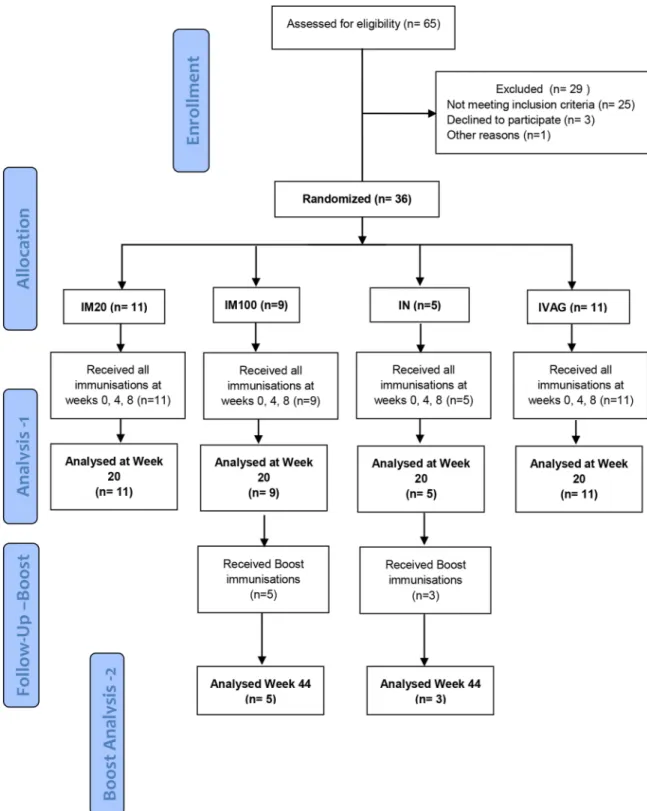
Mucovac2 was a Phase I randomized trial conducted at two centers; (St Georges University of London (SGUL) and Hull York Medical School, Experimental Medicines Unit (HYMS EMU). Volunteers were enrolled 17thNovember 2011 to 7thAugust 2012, follow-up for the study was completed on 11thJanuary 2013. All participants gave fully informed written consent and all relevant approvals were in place, the trial being conducted according to the UK Clinical Trials Regulations and GCP guidelines; reviewed and approved by NRES London Bridge Research Ethics Committee (S1 CONSORT Checklist). The trial was registered with the UK Clinical Research Network (UKCRN) assigned No. 11679 and the European Union Drug Regulating Authorities for Clinical Trials and assigned the EudraTC No. 2010-019103-27. The primary objective was to determine the safety and immunogenicity of the vaccination regimes in healthy female volunteers aged 18–45 years who were at low risk for HIV infection. The trial was open-label with laboratory assessment staff blind to regimen throughout and the partici-pants blind to the dose administered in the intramuscular regimen. Eligible female participartici-pants were randomised centrally into one of the four groups using a computer-generated algorithm based on random permuted blocks stratified over one factor (clinical centres) aiming to enroll 10 individuals into each of the IM20, IM100 and IVAG groups and 6 into the IN group (as there were no responses following IN immunization in a preceding macaque experiment; at SGUL), 36 in total (Fig 1). Statistical Software Stata (StataCorp. 2011. Release 12. College Sta-tion, TX) was used by the trial statistician to create the randomization sequence with variable block sizes (3 and 6 for HYMS; 4, 8 and 12 for SGUL). Women in the IVAG group received a single IM prime (100ug) followed by two IVAG immunizations. Volunteers from the IM100 and IN groups were invited back to receive two further boosts of 100μg gp140 adjuvanted with
Fig 1. CONSORT Flow diagram.Numbers of participants recruited into the trial“Analysis 1”is the original protocol schedule to week 20 incorporating the planned main analysis. Five from IM100 and 3 from the IN group also received additional boost vaccinations following an amendment to the protocol, and these individuals were included in the exploratory“Boost Analysis -2”.
Safety, reactogenicity and adverse events evaluation
Information on adverse events was collected at every visit. Information on solicited adverse events was collected one hour post-immunization, one week later, and recorded in diary cards by the participants in the seven days following each immunization. The primary safety out-come was a severe or worse local or systemic adverse event or an event that led to discontinua-tion of the vaccine schedule.
Clinical specimens
Serum, PBMC and mucosal samples were collected for immunogenicity at baseline, and weeks 4, 5, 8, 12, 16 and 20. Genital tract secretions were collected using the Instead Softcup™(Evofem Inc) or Weck-Cel1surgical spears (Medtronic). Weck-Cel samples were also taken from the cervical os and vaginal fornices from all volunteers at screening and weeks 5 and 12. Mucosal secretion samples were acquired and processed as detailed inS1 Text.
Assays of humoral immune responses
Serum and mucosal binding antibodies against recombinant CN54gp140 were measured using a standardized ELISA with minor modifications as described in theS1 Text. Serum specificity mapping was performed using a peptide epitope array of heterologous strains as previously described [24]. Neutralizing titers were measured in TZM-bl and ADCC activity was assessed as described [25,26].
Assays of cellular immune response
Intracellular cytokine staining (ICS) was performed as described previously [27]. Vaccine-induced T cell responses were analyzed using the mixture models for single-cell assays (MIMOSA) [28] statistical analysis to delineate vaccine-specific T-cell induced profiles. Multi-plex bead array (MBA) to measure cytokine profiles of responding cells was conducted as pre-viously described [29].
Statistical analysis
Categorical variables were described by number, percentage and, where indicated, their 95% confidence interval (Wilson CI suitable for small sample sizes). Comparisons of categorical variables were made using Fisher’s exact test. The magnitude of an antibody response and other continuous variables were compared between groups using non-parametric tests (Wil-coxon matched-pairs signed rank test, or Mann-Whitney test). Association between continu-ous variables was examined using rank correlation. The level of statistical significance was 5% for all analyses, without adjustment for multiple comparisons. Statistical analyses were per-formed using Stata (Version 12.1).
Results
Participant accrual, safety and reactogenicity
moderate solicited adverse event (Table 1). There were no significant differences in the median number of events per participant (p = 0.27) or in the maximum grade between groups (p = 0.61).
Immunogenicity
CN54gp140-specific serum antibody responses. Systemic CN54gp140-specific IgG responses were detectable one week after the second vaccination (Fig 2A) in the two IM groups. The highest number of responders with a detectable serum IgG response across all groups (20/ 36; 55%) was at Week 12, 4 weeks after the third immunization. 9/11 responded (detectable IgG) in the IM20 group (median 5.32μg/ml; range 0.01–23.47μg/ml) and 9/9 (median 4.18μg/
ml; 1.23–12.01μg/ml) in the IM100 group, with no significant difference between doses
(p = 0.71, Mann Whitney test). One individual in the IVAG group had a detectable serum IgG response at Week 12 only and there were no detectable responses in the IN group. At Week 20, 6/11 (54.5%) and 6/9 (60%) still had detectable serum IgG in the IM20 and IM100 groups respectively. Serum IgG antibody concentrations in responders from these two groups were comparable throughout the vaccination schedule and during the follow-up period (Fig 3A). There were no systemic vaccine-induced CN54gp140-specific IgA responses detected in any of the samples from any of the participants during the trial.
CN54gp140-specific cervico-vaginal antibodies. CN54gp140-specific IgG antibodies were detected in the cervico-vaginal secretions of eight individuals at Week 12 (Fig 2BandS1 Table). Of these, 4/11 (36%; 95% CI 15–65%) were in the IM20 group, and 4/9 (44%; 95% CI 19–73%) in the IM100 group. There were no responders in the IN or IVAG groups at Week 12.
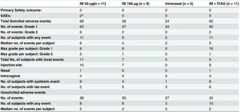
Table 1. Summary safety data–adverse events during main trial¶.
IM 20μg(n = 11) IM 100μg (n = 9) Intranasal (n = 5) IM + IVAG (n = 11)
Primary Safety outcome: 0 0 0 0
SAEs: 2* 0 0 0
Total Solicited adverse events: 69 39 24 62
No. of events;Grade 1 63 37 24 60
No. of events: Grade 2 6 2 0 2
No. of subjects with any event 11 9 5 11
Median no. of events per subject 6 4 2 5
Max grade per subject: Grade1 8 8 5 10
Max grade per subject: Grade 2 3 1 0 1
Total No. of subjects with local events 11 7 5 9
Injection-site 10 7 0 9
Nasal 1 0 5 1
Intravaginal 4 0 0 4
No. of subjects with systemic event 8 4 1 8
No. of subjects with lab event 2 5 3 6
Unsolicited adverse events
No. of events: 36 33 27 34
No. of subjects with any event 8 8 5 10
Median no. of events per subject 2 2 5 2
Table 1 footnotes:
¶before boosting phase; there were no grade 3 or 4 solicited adverse events.
*Two serious adverse events were reported, thefirst was a suspected (accidental) paracetamol overdose, the second, a Bartholin abscess. Since this individual was not in the IVAG group the cyst was considered unlikely vaccine related.
Responses were more reliably detected in Softcup samples than Weck-Cel (S1 Table). Across all time-points, CN54gp140-specific IgG was detected in only 1/11 participants in the IVAG group, and none of the participants in the IN group. At Week 20, 12 weeks after the third vacci-nation, 2 individuals in the IM20 group, 3 in the IM100 group, and 1 in the IVAG group still had detectable CN54gp140-specific IgG antibody (Fig 2B).
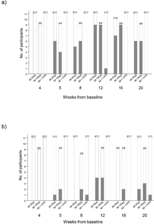
Fig 2. Participant response rates in systemic and mucosal compartments;Panel a) shows the numbers of participants with a CN54gp140 IgG antibodies in serum according to vaccination group and timepoint from randomisation. Panel b) shows the numbers of participants with CN54gp140 IgG antibodies in mucosal samples by vaccination group. Filled bars show the number of participants with detectable CN54 IgG, open bars show the number of participants with no CN54gp140 IgG antibodies.
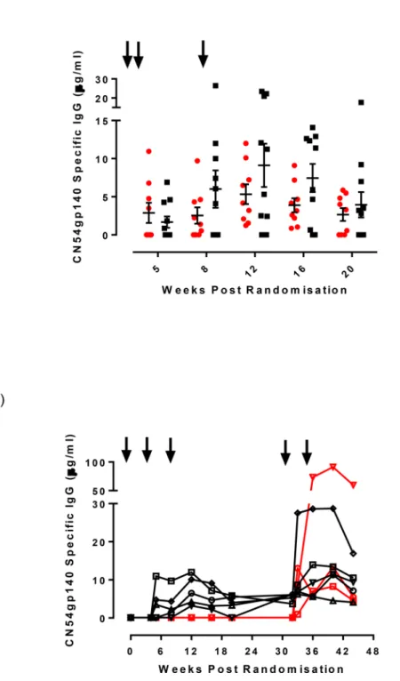
Fig 3. Magnitude of serum CN54gp140 IgG antibody responses for the two intramuscular vaccination groups by timepoint from randomisation.Panel a) Serum CN54gp140 IgG levels measured by ELISA expressed asμg/ml in either IM20 group (red circles) or IM100 group (black squares) at weeks 5, 8 12, 16 and 20. The black arrows indicate immunizations administered at weeks 0, 4 and 8 weeks. Panel b) Serum IgG responses measured in the 8 participants that received boost immunizations at weeks 32 and 34 (shown as arrows). Black symbols/lines indicate the 5 participants from the IM100 group, red symbols/lines indicate 3 participants from the IN prime group.
Only 1/36 trial participants, an individual in the IVAG group, developed a cervico-vaginal IgA response post vaccination, detected in the Softcup sample taken at Week 20 (0.157μg/ml),
but not detectable at any other time-point. CN54gp140-specific IgG was also detected in the Softcup sample at this time-point and at Week 8 in the absence of a serum IgG response. In addition, two other participants had low but detectable IgA binding antibodies prior to CN54gp140 vaccination thought to be polyreactive. In one of these, a response was also detected at Weeks 2, 16, and 20. Total IgG and IgA were reliably and reproducibly detected in softcup samples across all visits (S1A Fig), where the ratio of IgG:IgA was somewhere in between cervical os and vaginal Weck-Cel samples (S1B and S1C FigandS2 Table).
In the majority of participants, detection of specific IgG antibodies in cervico-vaginal secre-tions corresponded with detection of specific IgG in serum (S2 Table). At Week 12, the 8 par-ticipants with vaginal specific IgG (Softcup or Weck-Cel) had significantly higher serum IgG titres than participants without detectable specific vaginal IgG (n = 8: median serum CN54gp140 IgG 10.67μg [1.23–23.47μg/ml] versus n = 11 median 2.47μg/ml [0.01–20.96μg/
ml];p = 0.02 Mann Whitney test). Detectable specific vaginal IgG (Softcup) was significantly correlated with serum IgG titer (Spearman’s rank rs= 0.79, p = 0.036). At Week 16 and Week 20, median serum IgG titres were also greater in those with cervico-vaginal IgG, but this was not significantly different. However, the systemic and mucosal levels were not necessarily cor-related as there were individuals who had systemic responses of a similar magnitude in the absence of any corresponding mucosal antibodies.
CN54gp140-specific IgG responses after additional boosting. Eight participants, 5 in the IM100 and 3 in the IN group, received two further boosts delivered by IM immunization with 100μg of CN54gp140 adjuvanted with GLA-AF, 4 weeks apart, within 12–24 weeks of the
last vaccination. IM boosting elicited systemic IgG responses in all 3 participants in the IN group, which were not previously apparent. These were detectable after the first IM vaccination and were present at all 4 scheduled sampling time-points (Fig 3B). In addition, specific IgG antibodies were also detected in the cervico-vaginal secretions from 1/3 participants, albeit only at 2 Weeks post boost. Of the five subjects in the IM100 group who received the additional two IM boosts, augmented IgG levels were observed after the first IM boost which although sustained by the second boost did not show an apparent additional benefit (Fig 3B). 3/5 had detectable cervico-vaginal specific IgG after the boost immunizations. Two of these individuals had previously shown mucosal IgG antibodies during the main phase of the trial.
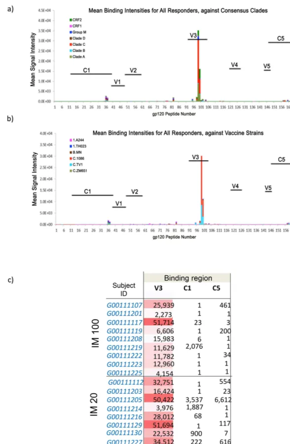
Serum peptide microarray analysis. The specificity of induced serum binding antibodies was assessed by peptide microarray analysis. All responding subjects displayed predominant responses to the V3 loop (Fig 4A and 4B). A subset of vaccinees also developed low levels of C5 or C1 gp120 Env IgG responses (Fig 4C). There was no significant binding to gp41 Env (data not shown). The V3 gp120 IgG responses were focused on Clade C, with evidence of cross-clade reactivity (mainly against Clade A, CRF02, and Group M). Among the 3 cross-clade C peptide sequences in the vaccine strain panel, the V3 response was targeted to 1086C and TV1
sequences. There was little correlation between the serum antibody titer and the number of epi-topes recognized in the peptide array (Spearman’s correlation 0.42).
(0.92, Spearman’s correlation) and weakly for MW965.26 (0.50). There was no detectable ADCC activity in any of the samples exhibiting anti-Env antibodies four weeks after the third gp140 vaccination (data not shown).
Cell-mediated immune responses. T-cell mediated immunity was assessed by intracellu-lar cytokine staining (ICS). There were no detectable cytokine responses in the CD8 T-cell pop-ulation in any group during the study (data not shown). Interestingly, modest responses to Env were primarily observed in the CD4 cells from participants that received IN immunizations, despite mounting no detectable antibody response. Responses were greatest in this cohort one week post third immunization (Week 9), with 80% of individuals positive for CD154, 60% for IL-2 and 60% for TNF-αexpression (Fig 6A–6C). By contrast, responses were infrequent, and of lower magnitude, in the IM20 parenteral group, and poorer still in the IM100 group. ICS responsiveness was not significantly associated with IgG antibody titer or neutralizing titer. Cytokine profiles were also measured by cytokine multiplex bead array (MBA) on supernatants of stimulated PBMC with whole CN54gp140 or matched peptides. Again, responses were pre-dominantly detected in participants that received IN immunizations with poor or no responses detected in all other vaccination groups. In subjects where cells were available, the most consis-tent responses to whole CN54gp140 in the IN group were seen in 75% of participants at Week 9 for IL-2, IL-5 and IL-13 (S2A–S2C Fig), with weaker and less frequent responses for GM-CSF (2/4), IL-4 (2/4), IL-10 (2/4), and IL-17A (3/4 participants, data not depicted). Similar
responses were seen for the IN group when PBMC were stimulated with matched peptides (two pools) in a Luminex assay. Here modest responses to most cytokines were seen in the IN group at Weeks 9 and 12, specifically IL-2, IL-15 and TNF-β, with weak responses across the other groups (S3A and S3B Fig).
Discussion
We conducted a novel randomized phase I trial of an HIV-1 Clade C gp140 envelope protein comparing the safety and immunogenicity of IM immunization with two different mucosal routes: IN and IVAG. All the participants completed their immunizations and there were no severe adverse reactions reported (Table 1).
Serum CN54gp140 IgG responses were detected in the majority of participants in the two parenteral groups. There was no statistical difference between the groups, suggesting the lower dose of gp140 (20μg) was similar to the standard dose (100μg) when administered with
adju-vant. There were no detectable serum IgA responses. This may reflect the adjuvant properties of GLA-AF. Given the potential negative correlation of specific systemic IgA in the RV144 trial [6,7] this may be a positive characteristic of the GLA formulation. Detectable specific vaginal IgG (softcup) was significantly correlated with serum IgG titer, likely reflecting transfer of sys-temic antibody into the vagina by passive transudation [13,14]. It is likely that the frequency of detectable vaginal antibody would be increased using strategies that elicit higher systemic responses. However, the systemic and mucosal antibodies were not necessarily correlated as there were some individuals who had systemic responses of a similar magnitude in the absence of any specific mucosal IgG, suggesting factors in addition to serum titer may influence IgG transfer to the vaginal lumen. The only previous clinical study of an HIV-1 subtype D gp140 envelope protein formulated in alum also showed antibody responses in all nine of the trial participants. It is not possible to compare data between two studies as only very limited
consensus clades; Panel b) shows mean binding intensities against a variety of HIV-1 vaccine strains. Panel c) Details the maximum binding intensities are shown according to participant at peak serum IgG response for the two groups that received intramuscular immunizations.
immunological analysis of serum antibody response was reported using a clinical diagnostic ELISA with no indication of serum dilutions. Mucosal and cellular responses were not assessed [15].
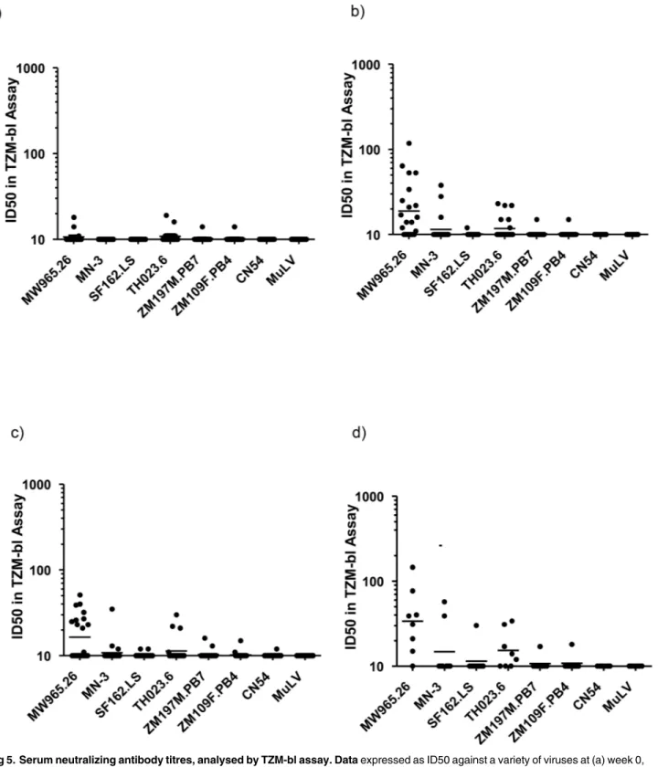
Fig 5. Serum neutralizing antibody titres, analysed by TZM-bl assay. Dataexpressed as ID50 against a variety of viruses at (a) week 0, b) week 12, c) week 20, and following additional boost in panel d).
Serum IgG responses were highly focused on the immunodominant V3 loop. This is reflec-tive of a number of gp140 immunogens recently evaluated in non-human primate models [30]. V3 dominated responses were mirrored by the low level of detectable neutralization to closely matched tier 1 isolates. Binding intensity to the V3 region positively correlated to the neutrali-zation of TH023.6 and only modestly with neutralineutrali-zation of MW965.26. Interestingly, as in rabbits and macaques [16,17], serum antibodies did not recognize the gp41-immunodominant domain (residues 598–609) [31] and showed no significant binding to any part of gp41. This suggests that the gp41 region of the molecule may be occluded from immune recognition, potentially avoiding induction of diverting responses associated with gp41-cross reacting microbiota [32]. The lack of any detectable ADCC activity likely reflects lack of response to dominant epitopes associated with ADCC, typically V1/V2, C1 and gp41 [24,33]. CN54 gp140 is naturally cleavage resistant constraining its ability to adopt a native-like conformation. It is anticipated that next generation stabilized cleaved soluble trimers may provide significant gains in neutralization breadth and potency [34].
Antibody responses to HIV-1 gp140 are thought to be dependent upon CD4 T- follicular helper (Tfh) cells. The paucity of detectable systemic CD4 T-cell responses in the two IM groups, despite robust antibody responses, suggests that required Tfh responses are likely restricted to the germinal centers of localized lymph nodes, and/or do not circulate in sufficient numbers at sam-pled time points (1 and 4 weeks post third immunization) to be detected. The observed paucity in CD4 response following parenteral immunization is in marked contrast to the IN group where 80% of participants had detectable circulating CD4 T-cell responses as assessed by CD154 expression (CD40L, a marker of T cell activation) in the apparent absence of detectable serum or vaginal antibody responses. It is unclear why we saw no effect of IN chitosan on humoral immu-nity, given that previous clinical studies have shown that chitosan promoted potent antibody responses to other immunogens [23,35,36], but it may be that these were intrinsically more anti-genic. However, the induction of CD4 T-cell responses suggests effective uptake of CN54gp140 by local antigen presenting cells, and subsequent presentation to CD4+ helper T cells. These data contrast with early preclinical studies from our group in mice, which suggested that chitosan efficiently promoted both humoral systemic and mucosal responses to CN54gp140 [9,37]. Inter-estingly, all participants in the IN group that agreed to the boosting protocol immediately responded to the first IM boost with serum antibody responses equivalent to those in the paren-teral groups after three immunizations. Thus IN administration appeared to act as a mucosal prime for subsequent systemic immunization. It is unclear whether priming was restricted to induction of CD4 T cell help or included priming of antibody responses that were too low to be detected. However the rapid anamnestic response on IM immunization is supportive of a boost-ing of primed antibodies rather than de novo primboost-ing at inductive sites. The possible dependence on potential adjuvant properties of chitosan cannot be inferred in the absence of a control arm. It is likely that higher doses, and/or the inclusion of a more potent mucosal adjuvant, could provide more positive immunogenicity data, even during the priming phase [38].
Vaginal CN54gp140-specific IgG antibody was detected in 36% and 44% of volunteers in the parenteral IM20 and IM100 groups, and at low intermittent levels in one individual in the IVAG group. There was no evidence for induction of specific IgA responses in any group,
Fig 6. T cell ICS analysis.The graphs display background corrected magnitude of T cell response for each cytokine and T cell subset by visit as percentage staining shown as box plots. Here, cohort 1 = IM20, cohort 2 = IM100, cohort 3 = IN and cohort 4 = IVAG. Responders are colored red and non-responders blue. Box plots based upon data from responders only are superimposed on the distributions, mid-line denotes median,ends of the box denote 25thand 75thpercentiles and where whiskers that extend from the top and
bottom are the extreme data points. Panel a) shows staining of CN54gp140 specific live CD3+/CD4+ CD154 + cells, b) CD3+/CD4+ IL-2+, c) CD3+/CD4+ TNF-α+ according to vaccination group at weeks 0, 12 and 24.
including the IVAG group. We did however observe evidence of pre-existent, polyreactive CN54gp140-binding IgA in two individuals prior to vaccination, as previously described [39]. The lack of IVAG boosting following a single IM prime contrasts to our previous studies in macaques where a single IM immunization primed animals for induced antibody following IVAG immuni-zation [13]. However, animals received 9 IVAG immunizations (100μg each) over one menstrual
cycle, as opposed to two higher doses (500μg) used in this human study. Lack of responsiveness
following IVAG administration may reflect the absence of mucosal adjuvant, prompted by our desire to minimize any local immune activation that might enhance HIV susceptibility [40]. How-ever, in a different human study using CN54gp140 and HSP70 as an adjuvant, IVAG administra-tion also failed to induce detectable systemic or mucosal antibody responses [18]. Furthermore, in recent studies a wider range of adjuvants failed to induce vaginal antibodies in NHP [38].
In summary, this study comparing different routes of administration demonstrates that IM administration of CN54gp140 elicited potent systemic IgG responses in the majority of sub-jects, and detectable vaginal IgG in just over a third. The observation that the lower dose of CN54gp140 (20μg) was similar to the standard dose (100μg) when formulated with GLA-AF
adjuvant, provides opportunities for dose sparing regimes. Interestingly, IN administration induced detectable CD4 T cell responses and primed subsequent intramuscular boosting, while IVAG administration in the absence of adjuvant had no detectable impact on systemic or mucosal responses in spite of IM priming.
Supporting Information
S1 CONSORT Checklist. CONSORT 2010 Checklist MUCOVAC2. (DOC)
S1 Fig. Measurement of total IgG (black symbols) and total IgA (red symbols) mucosal secretions.Data shown in either soft cup samples at weeks 0, 5, 8, 12, 16 and 20 (a) or cervical-os Weck-cel samples, (b) or vaginal vault Weck-Cel cel samples (c) at weeks 0, 5 and 12. (TIF)
S2 Fig. Cytokine Multiplex Bead Array (MBA) analysis measured in participants at weeks 0, 9 &12.Panel a) IL-2 responses, b) IL-5 responders are shown as red symbols and blue sym-bols non-responders. Here, cohort 1 = IM20, cohort 2 = IM100, cohort 3 = IN and cohort 4 = IVAG. c) IL-13 responders are shown as red symbols and blue symbols non-responders. Here, cohort 1 = IM20, cohort 2 = IM100, cohort 3 = IN and cohort 4 = IVAG.
(TIF)
S3 Fig. Luminex analysis shown as Trellis Plots.Analysis was performed on samples at weeks 0, 9, 12 and during the boost phase at weeks 28 or 40 in samples stimulated with either peptide pool 1 panel a) covering half the sequence of CN54gp140 peptides 1–78. Responders are shown as red symbols and non-responders as blue symbols. Here, cohort 1 = IM20, cohort 2 = IM100, cohort 3 = IN and cohort. b) peptide pool 2 covering latter half the sequence of CN54gp140 peptides 79–169. Responders are shown as red symbols and non-responders as blue symbols. Here, cohort 1 = IM20, cohort 2 = IM100, cohort 3 = IN and cohort.
(TIF)
S1 Study Protocol. MUCOVAC2 Clinical Study Protocol. (PDF)
S1 Table. Cervico-vaginal specific IgG responses at Week 12 and corresponding serum responses.
S2 Table. Total IgG and IgA levels (combined data from all groups) detected in cervico-vag-inal secretions collected by Softcup or Weck-Cel sampling.
(DOCX)
S1 Text. Supporting Methods. (DOCX)
Acknowledgments
We thank all the volunteers and trial participants for their assistance. We thank Professors Ralf Wagner and Hans Wolf, University of Regensburg and GENEART AG for the p97CN54-expressing plasmid. We thank Hongmei Gao and Kelli Greene of the CAVD for program man-agement. We gratefully acknowledge Dormeur Investment Service Ltd for providing funds to purchase equipment used in these studies. Recombinant CN54gp140 was manufactured to GMP specification and provided by Polymiun Scienfitc, Austria by Dietmar Kattinger.
Author Contributions
Conceived and designed the experiments: RS SM. Performed the experiments: CC CL AVC AB GM CY SB TC EB XS SD GF MS DM NF GT. Analyzed the data: WS LP XY AVC SJ. Contrib-uted reagents/materials/analysis tools: DC. Wrote the paper: RS AVC CC CL SM SJ.
References
1. Moog C, Dereuddre-Bosquet N, Teillaud JL, Biedma ME, Holl V, Van Ham G, et al. Protective effect of vaginal application of neutralizing and nonneutralizing inhibitory antibodies against vaginal SHIV chal-lenge in macaques. Mucosal Immunol 2014; 7:46–56. doi:10.1038/mi.2013.23PMID:23591718 2. Veazey RS, Shattock RJ, Pope M, Kirijan JC, Jones J, Hu Q, et al. Prevention of virus transmission to
macaque monkeys by a vaginally applied monoclonal antibody to HIV-1 gp120. Nat Med. 2003; 9:343– 46. PMID:12579198
3. Klein K, Veazey RS, Warrier R, Hraber P, Doyle-Meyers LA, Buffa V, et al. Neutralizing IgG at the portal of infection mediates protection against vaginal simian/human immunodeficiency virus challenge. J Virol. 2013; 87:11604–16. doi:10.1128/JVI.01361-13PMID:23966410
4. Mascola JR, Stiegler G, VanCott TC, Katinger H, Carpenter CB, Hanson CE, Beary H, et al. Protection of macaques against vaginal transmission of a pathogenic HIV-1/SIV chimeric virus by passive infusion of neutralizing antibodies. Nat Med. 2000; 6:207–10. PMID:10655111
5. Burton DR, Hessell AJ, Keele BF, Klasse PJ, Ketas TA, Moldt B, et al. Limited or no protection by weakly or nonneutralizing antibodies against vaginal SHIV challenge of macaques compared with a strongly neutralizing antibody. Proc Natl Acad Sci U SA 2011; 108:11181–86.
6. Kim JH, Excler JL, Michael NL. Lessons from the RV144 Thai phase III HIV-1 vaccine trial and the search for correlates of protection. Annu Rev Med 2015; 66:423–37. doi: 10.1146/annurev-med-052912-123749PMID:25341006
7. Tomaras GD, Ferrari G, Shen X, Alam SM, Liao HX, Pollara J, et al. Vaccine-induced plasma IgA spe-cific for the C1 region of the HIV-1 envelope blocks binding and effector function of IgG. Proc Natl Acad Sci U S A. 2013; 110:9019–24. doi:10.1073/pnas.1301456110PMID:23661056
8. Holmgren J, Czerkinsky C. Mucosal immunity and vaccines. Nat Med 2005; 11(4 Suppl):S45–53. PMID:15812489
9. Buffa V, Klein K, Fischetti L, Shattock RJ. Evaluation of TLR agonists as potential mucosal adjuvants for HIV gp140 and tetanus toxoid in mice. PLoS One 2012; 7:e50529. doi:10.1371/journal.pone. 0050529PMID:23272062
10. Arias MA, Van Roey GA, Tregoning JS, Moutaftsi M, Coler RN, Windish HP. Glucopyranosyl Lipid Adju-vant (GLA), a Synthetic TLR4 agonist, promotes potent systemic and mucosal responses to intranasal immunization with HIVgp140. PLoS One 2012; 7:e41144. doi:10.1371/journal.pone.0041144PMID: 22829921
12. Schiller JT, Castellsagué X, Garland SM. A review of clinical trials of human papillomavirus prophylactic vaccines. Vaccine 2012; 30 Suppl 5:F123–38. doi:10.1016/j.vaccine.2012.04.108PMID:23199956 13. Schwarz TF, Kocken M, Petaja T, Einstein MH, Spaczynski M, Louwers JA, et al. Correlation between
levels of human papillomavirus (HPV)-16 and 18 antibodies in serum and cervicovaginal secretions in girls and women vaccinated with the HPV-16/18 AS04-adjuvanted vaccine. Hum Vaccin 2010; 6:1054– 61. PMID:21157180
14. Scherpenisse M, Mollers M, Schepp RM, Meijer CJ, de Melker HE, Berbers GA, et al. Detection of sys-temic and mucosal HPV-specific IgG and IgA antibodies in adolescent girls one and two years after HPV vaccination. Hum Vaccin Immunother. 2013; 9:314–21. PMID:23149693
15. Hurwitz JL, Lockey TD, Jones B, Freiden P, Sealy R, Coleman J, Howlett N, Branum K, Slobod KS. First phase I clinical trial of an HIV-1 subtype D gp140 envelope protein vaccine: immune activity induced in all study participants. AIDS. 2008 Jan 2; 22(1):149–51 PMID:18090404
16. Cranage MP, Fraser CA, Stevens Z, Huting J, Chang M, Jeffs SA, et al. Repeated vaginal administra-tion of trimeric HIV-1 clade C gp140 induces serum and mucosal antibody responses. Mucosal Immu-nol 2010; 3:57–68. doi:10.1038/mi.2009.110PMID:19741600
17. Cranage MP, Fraser CA, Cope A, McKay PF, Seaman MS, Cole T, et al Antibody responses after intra-vaginal immunisation with trimeric HIV-1 CN54 clade C gp140 in Carbopol gel are augmented by sys-temic priming or boosting with an adjuvanted formulation. Vaccine. 2011; 29:1421–30. doi:10.1016/j. vaccine.2010.12.034PMID:21187177
18. Lewis DJ, Wang Y, Huo Z, Giemza R, Babaahmady K, Rahman D, et al. Effect of vaginal immunization with HIVgp140 and HSP70 on HIV-1 replication and innate and T cell adaptive immunity in women. J Virol. 2014; 88:11648–57. doi:10.1128/JVI.01621-14PMID:25008917
19. Lewis DJ, Fraser CA, Mahmoud AN, Wiggins RC, Woodrow M, Cope A, et al. Phase I randomised clini-cal trial of an HIV-1(CN54), clade C, trimeric envelope vaccine candidate delivered vaginally. PLoS One. 2011; 6:e25165. doi:10.1371/journal.pone.0025165PMID:21984924
20. Su L, Graf M, Zhang Y, von Briesen H, Xing H, Köstler J, et al. Characterization of a virtually full-length human immunodeficiency virus type 1 genome of a prevalent intersubtype (C/B') recombinant strain in China. J Virol 2000; 74:11367–76. PMID:11070037
21. Pabst M, Chang M, Stadlmann J, Altmann F. Glycan profiles of the 27 N-glycosylation sites of the HIV envelope protein CN54gp140. Biol Chem. 2012; 393:719–30. doi:10.1515/hsz-2012-0148PMID: 22944675
22. Clegg CH, Roque R, Perrone LA, Rininger JA, Bowen R, Reed SG. GLA-AF, an emulsion-free vaccine adjuvant for pandemic influenza. PLoS One. 2014; 9:e88979. doi:10.1371/journal.pone.0088979 PMID:24551202
23. Huo Z, Sinha R, McNeela EA, Borrow R, Giemza R, et al. Induction of protective serum meningococcal bactericidal and diphtheria-neutralizing antibodies and mucosal immunoglobulin A in volunteers by nasal insufflations of the Neisseria meningitidis serogroup C polysaccharide-CRM197 conjugate vac-cine mixed with chitosan. Infect Immun 2005; 73: 8256–65. PMID:16299322
24. Gottardo R, Bailer RT, Korber BT, Gnanakaran S, Phillips J, Shen X, et al. Plasma IgG to linear epi-topes in the V2 and V3 regions of HIV-1 gp120 correlate with a reduced risk of infection in the RV144 vaccine efficacy trial. PLoS One 2013; 8:e75665. doi:10.1371/journal.pone.0075665PMID:24086607 25. Sarzotti-Kelsoe M, Bailer RT, Turk E, Lin CL, Bilska M, Greene KM, et al. Optimization and validation of
the TZM-bl assay for standardized assessments of neutralizing antibodies against HIV-1. J Immunol Methods 2014; 409:131–46. doi:10.1016/j.jim.2013.11.022PMID:24291345
26. Pollara J, Bonsignori M, Moody MA, Pazgier M, Haynes BF, Ferrari G. Epitope specificity of human immunodeficiency virus-1 antibody dependent cellular cytotoxicity [ADCC] responses. Curr HIV Res 2013; 11:378–87. PMID:24191939
27. Horton H, Thomas EP, Stucky JA, Frank I, Moodie Z, et al. Optimization and validation of an 8-color intracellular cytokine staining (ICS) assay to quantify antigen-specific T cells induced by vaccination. J Immunol Methods 2007; 323:39–54. PMID:17451739
28. Finak G, McDavid A, Chattopadhyay P, Dominguez M, De Rosa S, Roederer M, et al. Mixture models for single-cell assays with applications to vaccine studies. Biostatistics 2014; 15:87–101. doi:10.1093/ biostatistics/kxt024PMID:23887981
29. Defawe OD, Fong Y, Vasilyeva E, Pickett M, Carter DK, Gabriel E, et al. Optimization and qualification of a multiplex bead array to assess cytokine and chemokine production by vaccine-specific cells. J Immunol Methods 2012; 382:117–28. doi:10.1016/j.jim.2012.05.011PMID:22626638
31. Gnann JW Jr, Nelson JA, Oldstone MB. Fine mapping of an immunodominant domain in the transmem-brane glycoprotein of human immunodeficiency virus. J Virol 1987; 61:2639–41. PMID:2439707 32. Williams WB, Liao HX, Moody MA, Kepler TB, Alam SM, Gao F, et al. HIV-1 VACCINES. Diversion of
HIV-1 vaccine-induced immunity by gp41-microbiota cross-reactive antibodies. Science. 2015; 349: aab1253. doi:10.1126/science.aab1253PMID:26229114
33. Pollara J, Bonsignori M, Moody MA, Liu P, Alam SM, Hwang KK, et al. HIV-1 vaccine-induced C1 and V2 Env-specific antibodies synergize for increased antiviral activities. J Virol. 2014; 88:7715–26. doi: 10.1128/JVI.00156-14PMID:24807721
34. Ringe RP, Sanders RW, Yasmeen A, Kim HJ, Lee JH, Cupo A, et al. Cleavage strongly influences whether soluble HIV-1 envelope glycoprotein trimers adopt a native-like conformation. Proc Natl Acad Sci U S A. 2013; 110:18256–61. doi:10.1073/pnas.1314351110PMID:24145402
35. McNeela EA, Jabbal-Gill I, Illum L, Pizza M, Rappuoli R, et al. Intranasal immunization with genetically detoxified diphtheria toxin induces T cell responses in humans: enhancement of Th2 responses and toxin-neutralizing antibodies by formulation with chitosan. Vaccine 2005; 22:909–14.
36. El-Kamary SS, Pasetti MF, Mendelman PM, Frey SE, Bernstein DI, Treanor JJ, et al. Adjuvanted intra-nasal Norwalk virus-like particle vaccine elicits antibodies and antibody-secreting cells that express homing receptors for mucosal and peripheral lymphoid tissues. J Infect Dis 2010; 202:1649–58. doi: 10.1086/657087PMID:20979455
37. Klein K, Mann JF, Rogers P, Shattock RJ. Polymeric penetration enhancers promote humoral immune responses to mucosal vaccines. J Control Release.2014; 183:43–50. doi:10.1016/j.jconrel.2014.03. 018PMID:24657807
38. Veazey R, Siddiqui A, Klein K, Buffa V, Fischetti L, Doyle-Meyers L, et al. Evaluation of mucosal adju-vants and immunization routes for the induction of systemic and mucosal humoral immune responses in macaques. Hum Vaccin ImmunotherIn press
39. Liao HX, Chen X, Munshaw S, Zhang R, Marshall DJ, Vandergrift N, et al. Initial antibodies binding to HIV-1 gp41 in acutely infected subjects are polyreactive and highly mutated. J Exp Med 2011; 208:2237–49. doi:10.1084/jem.20110363PMID:21987658