Vol.53, n. 1: pp. 105-114, January-February 2010
ISSN 1516-8913 Printed in Brazil BRAZILIAN ARCHIVES OF
BIOLOGY AND TECHNOLOGY
A N I N T E R N A T I O N A L J O U R N A L
Production and Characterization of Monoclonal Antibodies
Anti Fragment Fc of Bovine IgG
Tânia Regina Penha
1*, Ernesto Renato Krüger
2, Vanete Thomaz-Soccol
3, Jorge Victor
Bacila Agottani
1, Flávio Hiroshi Itano
1, Ludmilla Della Coletta Troiano
1and Josiane
Brodzinski
11Instituto de Tecnologia do Paraná; Rua João Américo de Oliveira, 330; Curitiba - PR - Brasil. 2Centro de
Diagnóstico Marcos Enrietti; Curitiba - PR - Brasil. 3Universidade Federal do Paraná; Curitiba - PR - Brasil
ABSTRACT
The aim of this work was to produce and characterize monoclonal antibodies anti bovine immunoglobulin G (IgG). Out of seven hybridomas, two were chosen based on the ELISA’S absorbance values and were labeled B4F11 and B3H12. These monoclonals were analyzed through Western Blot for IgG fragments obtained by proteolysis with papain, separated by electrophoresis in polyacrylamide gel electrophoresis with β-mercaptoetanol as reducing agent. This revealed that, possibly, the B4F11 was directed to a conformational antigen, and that B3H12 reacted in a specific fashion with Fc (Bovine IgG crystallizable fragment). This antibody could be used in the development of reagents to immunoassays relevant for research and diagnosis.
Key words: Monoclonal antibodies; bovine immunoglobulin G; Fragment Fc
*Author for correspondence: [email protected] INTRODUCTION
Biotechnology is currently considered the newest advance in the life sciences. This field became widely known at the end of the twentieth century, affecting various areas of research in human life and the environment. Recent progress in molecular biology and cell knowledge has made it possible to produce recombining DNA molecules with different applications in the production of protein of interest. Areas of animal health that have been greatly improved by biotechnology are: the development of diagnostic test for disease control, vaccine production, nutrition and parasite control. The production of antibodies for specific immunogens fostered by precise protocols of immunization has allowed the development of
highly specific reagents to be used as diagnostic tools. These antibodies may be of polyclonal or monoclonal origin, the latter having some advantages over the former due to their high specificity and production by immortalized cells. Monoclonal antibodies are used in immunoassays, which are extremely specific and sensitive to a great variety of viruses, bacteria, protozoa, and macro-parasitic components. The specificity of the test overcomes any restriction concerning the nature of the target material under evaluation. The remarkable sensitivity allows the identification of extremely small quantities of the target component, as small as 10 -12.
and quantify the immunoglobulins, which are the main components of the system that detects invaders in solutions of biological fluids or associated cells of this same medium. The bovine immunoglobulins consist of four classes: IgM, IgG, IgA, and IgE. The IgGs contain three subclasses: IgG1, IgG2, and IgG3 as well as two light ג (lambda) and K (Kappa) chains (Knight et al., 1988; Symons et al., 1989; Kacskovics et al., 1996; Rabbani et al., 1997). The three genes that codify the heavy chains of the IgGs (γ1, γ2, γ3) are located in the chromosome 21q24 (Gu et al., 1992). The production of monoclonal antibodies allows the development of specific laboratory tests for each class as well as each subclass. So far, no protein or mRNA has been identified for the IgD class. For the light chains, more than 90% are ג
(lambda); however, as no specific reagents to either kappa or lambda exist, future studies would have to be carried out in order to establish the relationship between ג and Ќ chains (Pastoret, 1998).
The immunoglobulins make up the largest portion of the humoral immunity, of which IgG is the main class of antibodies present in the blood, lymph, peritoneal, and cerebro-spinal fluids. They also constitute 75% of serum immunoglobulins. Subclasses of IgG exist in different species and their biochemical composition offers the basis for the biological effectors’ functions. The molecular and serological characteristics of the bovine IgG subclasses show the existence of allotype variation, providing polymorphic genetic markers for immunoglobulins with functional consequences (Blakeslee et al., 1971; Rapacz et al., 1972; Wegrezyn et al., 1978; Symons et al., 1989; Kacskovics et al., 1996; Rabbani et al., 1997).
The IgG1 displays unique characteristics: a) it constitutes 90% of the colostral immunoglobulins, providing neonatal protection as well as instructions for the action of the immune system. (Butler et al., 1972; Butler et al., 1974; Guidry et al., 1980); b) it is selectively transported by the FcRn, a receptor which is found in the lung alveolar and mammary gland epithelium (Dyxon et al., 1961; Kemler et al., 1965; Brandon et al., 1971); c) it binds to neutrophils such as IgG2 (Mcguire et al., 1979; Butler et al., 1985); d) it is synthesized locally and released as a secretory immunoglobulin on the mucosa (Brandon et al., 1971; Butler et al., 1985). Different allotypes among animals of the same species correspond to
the capacity of influencing the immune system and/or creating resistance to viral and bacterial infections. Allotype determinants are found in IgG1 and IgG2 (Saini et al., 2007). The most commonly studied, A1 and A2 are found in the IgG2 subclass and denominated IgG2a and IgG2b. The IgG2a allotype manifests before the IgG2b during the development of calves, which is efficient in the activation of complement, an important mechanism in the control of infections by pyogenic bacteria. Proteins synthesized by these bacteria bind to the allotype (b) and not to the allotype (a) (Corbeil et al., 1997; Bastida-Corcuera et al., 1999 (b).
The heterogeneous characteristic of IgG2 was proven additionally by polyclonal and monoclonal antibodies. Human IgG was used in hybridization tests showing that there were three subclasses of bovine IgG. Out of the three bovine genes that code for IgG, the first two ones code for IgG1 and IgG2, respectively, while the third gene codes for IgG3, which is not recognized by IgG1 and IgG2 antiserum. According to the immunoglobulin workshop promoted by the Veterinarian Immunology Committee, the three bovine IgG subclasses are labeled: IgG1, IgG2 and IgG3. It must be noted that the use of the same nomenclature for the subclasses (IgG1, IgG2, and IgG3) among different species frequently causes the misunderstanding that the subclasses are homologous and have the same function. This is true for IgM, IgG, IgA, IgD, and IgE but does not apply to the subclasses. Currently, the prevailing hypothesis is that of the derivation of the species preceded the subclasses evolution (Kehoe et al., 1974).
Thus, the present work was developed aiming at producing a monoclonal antibody against bovine IgG and improving the quality of cattle disease diagnoses.
MATERIAL AND METHODS
The methodologies used by Hallow et al., (1988) were followed in this work with the modifications and adaptations mentioned in the appropriate sections.
Blood serum
sent for examination at the Centro de Diagnóstico Marcos Enrietti, Curitiba-PR-Brazil. A serum pool was made with collected samples, which showed a clear aspect and absence of hemolysis. Later, it was centrifuged at 14,800g at 4ºC for 30 minutes. The supernatant was removed and samples were separated for immunodiffusion and electrophoresis. The rest was stored under -20ºC until further use.
Serum lipoproteins removal
Two procedures were used: the first one was labeled 1, in which 3.0g Polyvinylpyrolidone (PVP) was gradually mixed in 100ml of serum under stirring at 4ºC for 4 h. At the end, the serum was centrifuged at 14,800g at 4ºC for 30 minutes. The supernatant was collected and stored at 4ºC for subsequent stages. In the second procedure, labeled 2, the solution used was 5% dextran sulfate (5g of sodium dextran sulfate in 100mL of distilled water) added to a proportion of 1.25mL for every 25mL of serum, adding dropwise under stirring. After that, it was left at room temperature for 30 minutes, being constantly stirred on a 10-minute interval basis. Then, 2.25 mL of 11.1% calcium chloride solution was taken and added to 25mL of serum (dropwise under stirring). The suspension was left on ice bath for 30 minutes and then centrifuged at 14,800g at 4ºC for 30 minutes. After collecting the supernatant, the samples were separated for immunodiffusion and electrophoresis and the rest was stored at 4ºC.
Immunoglobulins precipitation
After the quantity of serum pre-set had been checked, its volume was doubled with PBS buffer (phosphate buffered saline).To the final volume, serum plus PBS, the same quantity of saturated ammonium sulfate solution was added dropwise (761g of ammonium sulfate in 1000mL of distilled water; pH 7.2) under stirring at room temperature, which precipitated all the immunoglobulin classes at 50% concentration. The suspension was homogenized for 30 minutes on a magnetic stirrer, after that it was centrifuged at 14,800g at 4ºC for 30 minutes. The precipitate was recovered and re-suspended in PBS to the original serum volume.
Immunoglobulins G precipitation
Saturated ammonium sulfate solution was added to achieve 33% concentration in order to get the IgG precipitation. This saturation was calculated from the immunoglobulin suspension volume, according the protocol presented above. Then, stirring was
maintained for 30 minutes at 4ºC and, after that, centrifuged at 14,800g for 30 minutes. The supernatant was kept between 2 and 8ºC; the precipitated IgG was re-suspended in a 5.0mL volume of PBS and dialyzed against PBS for 72 h with changes each 12 h at 4ºC.
Dialysis
The dialysis was run for three days in a flask containing 2-L of PBS, with changes every 12 h. After this, the G immunoglobulins were centrifuged at 3,700g for 15 minutes to remove the impurities. From the dialyzed IgG solution, samples were collected to measure the protein content and to perform the immunodiffusion and electrophoresis, and the rest was stored at -80ºC.
Quantification of proteins
A physical method employing metrolab 1700 spectrophotometer, wavelenght set at 260 and 280nm was used. The results were obtained according to the following formula:
Y= (1.55 x 280nm readings) – (0.76 x 260nm readings)
Y value x used dilution = protein concentration in mg/mL.
IgG Purification
Affinity chromatography column sensitized with protein G from Staphylococcus aureus, at draining speed of 1mL/min (Hitrap G HT, Amershan) was used. The column was balanced with 10 volumes of adsorption buffer (20mM of sodium phosphate, pH 7.0). After that, the sample was applied to a 1.0mL volume. Then, the column was washed with 10 column volumes of adsorption buffer, until there was no more protein detected at 280 nm. The material was eluted by the use of a five- column volume of elution buffer, (0.1M glycine-HCl, pH 2.7). From every 1.0mL fraction, a 50µl sample was submitted to a test with 10% trichloroacetic acid. Highly concentrated samples, with intense turbidity, were saved. To each fraction, 100µL of neutral buffer was added (1M of TRIS-HCl, pH 9.0). The column was washed with 10 volumes of adsorption buffer and conserved with ethanol at 20% (20mL of ethanol in 80mL of distilled water) at 4ºC.
Mice immunization protocol
100µL. The emulsion, antigen plus adjuvant, was homogenized until there was no water drop formation during the rest, or until there was no drop dissolution when it was poured over the water surface in the recipient. The emulsions containing bovine IgG were inoculated in a group of 10 mice via intra-peritoneal. The second and third doses were given at a 15-day interval and were processed like the first one. The adjuvant was replaced by Freund’s incomplete. At the end of the immunizations, total blood was collected from one mouse from each allotment through cardiac puncture and the serum was separated from the blood clot in order to evaluate the humoral immune response, through an immunodiffusion test. All mice were kept in constant surveillance during the execution of the immunization protocol.
Humoral immunity evaluation through
immunodiffusion
A six-centimeter diameter Petry dish, containing 7.0mL of 0.9% agar in PBS was used. The diameter of each well and the distance between them was 3 and 4mm, respectively. Then the purified bovine IgG (25µL) was added in the central well. In the peripheral wells, 25µL of bovine IgG immunized and non-immunized mouse serum was added alternately. The dishes were incubated in a humidity chamber at 28ºC for 48 h. The reading and interpretation obeyed the criteria of precipitation lines between the purified bovine IgG and immunized animal serum and no lines formed with negative serum.
Peritoneal macrophages
Twenty-four hours before fusion, peritoneal macrophages from five non-immunized mice were harvested through washing the peritoneal cavity with 0.34M sucrose in order to maintain the isotonicity. Then the skin from the mice abdomen was opened without drilling the peritoneal membrane. Five milliliters of sucrose solution were injected into the peritoneal cavity and the abdomen was massaged to release the macrophages; then the solution was removed. The cell suspension was centrifuged at 800g for 15 minutes at 4ºC; the supernatant was discarded and the cell pellet was re-suspended with 10mL of RPMI culture medium. A sample of the macrophage suspension was diluted to 1:20 proportion and counted in a Fuchs-Rosenthal chamber. The cell pool was diluted to obtain approximately 5 x 10⁵ cells/ ml. The macrophage
suspension was distributed in a volume of 100µl/well, through five 96 flat bottom multi-well plates, incubated at 37ºC into a CO2 incubator for
24h.
Spleen lymphocytes from immunized animals
The animals were sacrificed through inhalation of ethyl ether. After the asepsis with alcohol 70GL, the spleen, removed in a chirurgical procedure, was put into a Petri dish with 5mL of RPMI culture medium, without fetal serum. The spleen had the capsule displaced; the pulp was triturated into small pieces and then filtered through a thin mesh metal bolter. The lymphocytes released were transferred to a centrifuge tube with the same culture medium and left still for two minutes in order for the biggest spleen tissue blocks to precipitate. The supernatant was transferred to a new centrifuge tube and was centrifuged at 800g at 4ºC for 10 minutes. The supernatant was separated and the pellet containing the lymphocytes was re-suspended in a 10mL volume of RPMI culture medium without bovine fetal serum. The cells were counted in a Fuchs-Rosenthal chamber and diluted to approximately 108/mL lymphocytes (splenocytes).
Fusion procedure
A suspension of 108/mL of lymphocytes was added to another one containing 107/mL of myeloma cells SP2-0, keeping a proportion of 10:1. The lymphocytes-myeloma mixture was centrifuged at 800g for 10 minutes. After that, the supernatant was removed and the precipitated cells were dissolved with soft hand beats. In the sequence, 1.0mL of polyethylene glycol (PEG 1450 (50% w/v), previously warmed at 37ºC, was added dropwise under continuous stirring for one minute. Then the RPMI culture medium (1mL) without bovine fetal serum was added, left still for one minute and, after that, 2.0mL was again added after one minute, making a total of 3.0mL of cells culture medium without serum at 37ºC. Another 7.0mL of medium was added for 2 minutes. Finally, 10mL was added, resulting in a 20mL final volume.
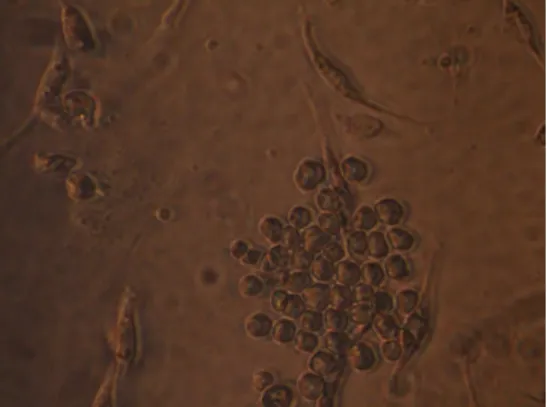
The plates were daily observed on an inverted microscope, replacing 50% of the culture medium by the 5th day, when it was possible to observe the first hybridomal colony formation. From this
stage, the medium changes took place every 48 h until the cellular confluence was approximately 70 to 80% (Fig 1).
Figure 1 - Hybridoma colony formation after an incubation of seven days.
ELISA screening of antibodies production
The method used was an indirect one to detect the monoclonal antibodies anti IgG in hybridoma culture medium. Purified bovine IgG had the concentration adjusted to 1 mg/ml, from which 10µL was added to 1000µL of adsorption buffer (0.2M carbonate-bicarbonate, pH 9.6). The final concentration, 500ng of proteins/50µL, was added to each well of an ELISA plate and incubated at 4°C overnight. The plates were washed with 200µL washing buffer, 8.5g sodium chlorine (NaCl), 0.32g sodium phosphate monobasic (NaH2PO4), 110g sodium phosphate dibasic
(Na2HPO4), and 1000mL purified water; pH 7.2).
After that, 50µL blocking buffer was added (3% bovine serum albumin BSA, and 0.05% Tween 20; pH 7.4). 50µL supernatant from each hybridoma was diluted to 1:2 in blocking buffer and added to each well. The plates were incubated at 37ºC for 90 minutes. Then, the plates were washed as described before and 50µL peroxydase conjugated anti mice IgG, diluted 1:3.200, was added to each well and incubated at 37ºC for 90 minutes. The standard washing procedure was followed and then 50µL substrate/chromogen solution was added (400mg of OPD, 100mL citrate buffer and 5µl hydrogen peroxide). After 15 minutes, the reaction was stopped by the addition of 50µL 1M sulfuric acid. The plates were read in a spectrophotometer at 490nm. H9 and H10 wells were used as negative control whereas H11 and H12 wells were used as positive (hyper immune serum). A check board titration of antigen and
conjugate was performed before the test by diluting the antigen (IgG). This was performed from an initial concentration of 2mg/50µL, column 1 through 11, and conjugate initial dilution 1:400, from row A to H, base two dilutions.
Fractionation of bovine immunoglobulin G
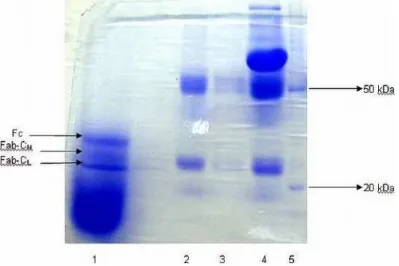
Figure 2 - SDS-PAGE gel at 10 % after being fractioned with papain. 1) Fragmented sample of CL and CH Fab and Fc of purified bovine IgG; 2) Purified bovine IgG; 3) Semi purified bovine IgG; 4) Bovine serum; 5) Molecular mass marker.
Bovine IgG characterization through SDS-PAGE and western blot
The samples of the bovine serum, unpurified IgG and purified IgG (by chromatography) were submitted to electrophoresis SDS-PAGE and Western Blot. Samples had the concentration of the proteins initially adjusted in order to get 150µg into 25µl sample buffer (4.0mL of 1M Tris pH 8.0; 4.0mL glycerol 100%; 0.2mL 0.5M EDTA; 0.4mL bromophenol blue 1%; 11.4mL purified water; 5% mercaptoetanol). The samples were boiled for 5 minutes and applied to the gel. Also, the molecular mass markers Bench Marck and Magic Marck, Amershan brand, with molecular masses ranging from 20 to 220KDa, were applied without having been boiled.
To perform the SDS-PAGE, the standard procedure was used with continuous system and 15% acrilamide bis-acrilamide concentration in the separating gel (7.5mL acrilamide bis-acrilamide 30:0.8; 2.1mL 1M Tris-HCl, pH 8.8, 150µL 10% SDS, 37µL 20% ammonium persulfate , 7.5µL Temed; 5.2mL purified water). The 5% stacking gel was made of 2.5mL acrilamide bis-acrilamide 30:0.8; 1M Tris-HCl, pH 6.8; 150µL SDS 10%; 37µL 20% ammonium persulfate; 7.5µL Temed; 11.3mL purified water. The running buffer used was tris-glycine (stock solution : 30.3g tris-base; 144g glycine; 5g SDS; 1000mL purified water; pH 8.6). Use solution:
100mL stock, 900mL purified water) and the migration occurred at 70V per minute. Finally, the gel was stained with coomassie blue R250 (0.1mL coomassie blue; 45mL methanol; 10mL acetic acid; 45mL purified water) for 60 minutes and destained (4mL methanol; 7.5mL acetic acid; 3mL purified water) for 60 minutes (Fig.3). From a second gel, proteins were transferred to the nitrocellulose membrane applying 20 to 30V, overnight at 4ºC. In the next step, the membranes were blocked (blocking solution: 0.02g bovine albumin fraction V, 100 purified water), under stirring for 90 minutes. Monoclonal antibody anti bovine IgG was diluted to 1:2, added to nitrocellulose stripes and incubated for 90 minutes at 37ºC. The membranes were washed 5 times with the washing buffer and the peroxidase conjugated anti mice IgG added at a dilution of 1:1000 and incubated for 90 minutes at 37ºC. The membranes were washed 5 times with the washing buffer and the chromogen-substrate was added (0.67 mL 4-chloro-1-naftol, 10mL 0.1M sodium acetate, 10ul hydrogen peroxide, pH 5.2) and remained for 15 minutes (Fig 4).
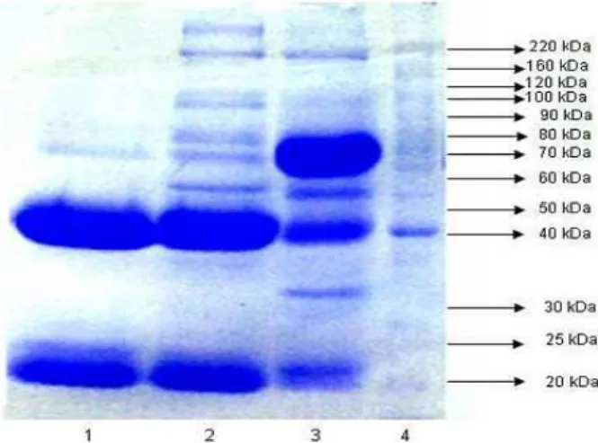
Figure 3 – SDS-PAGE Gel at 10 %. 1) Purified bovine IgG; 2) Semi purified bovine IgG (33% SAS); 3) Bovine serum; 4) Molecular mass marker, with the obtaining of the heavy chains bands at the 50KDa band and the light chains at the band with 25 KDa.
Figure 4 - Result of the Western Blot test. 1 and 2) Fc fragments of bovine IgG after 16 hours with papain digestion 3) Molecular mass marker.
RESULTS AND DISCUSSION
The samples of bovine IgG purified by affinity chromatography and quantified by spectrophotometer with absorption of ultra-violet rays showed concentrations that oscillated between 2.2 to 3.6mg/mL. Considering the fixed retaining capacity of the columns (25mg/mL), around which the IgG concentration in bovine serum oscillated, and the standard protocol followed in all the purification procedures, the variations registered in the concentration could be attributed to different affinity levels of the protein G for different subclasses of IgG, since the used serum
constituted in a pool of harvests from different animals.
possible to determine the purity grade from the obtained samples. Despite the staining method uses coomassie blue as dye, which allowed the detection of proteins in concentrations from 1.0 to 2.2µg/mL, there were not any extra bands, except for those pertaining to the subject under study. The purity grade of samples has relative importance to the immunization process of animals, because from the generated hybridomas it is possible to screen those that secrete anti bovine IgG. However, considering that the bovine IgGs used for the immunization would be the same used in the ELISA plates sensitization, which would be used in the selection processes of the anti IgG clones, special caution was taken to make the samples as pure as possible. We did consider the possibility of mice immunizations with pure Fc fragment, but taking in account the changes in the conformation and epitope destruction, we decided in favor of the whole molecule.
At the end of the immunization protocol, the mice were evaluated for their humoral response for bovine IgG. The chosen evaluation method was the immunodiffusion in agar gel. Blood serum samples obtained by cardiac punction of the mice were evaluated against purified IgG in the immunodiffusion tests. After an incubation period of 48h, it was possible to see the immunoprecipitation lines between the immunized mice serum and purified IgG, and absence of lines when the serum of non-immune mice was used. That was a reaction which made it possible to understand that intense and clear precipitation lines were convincing evidence that the mice spleen had significant quantities of clones of sensitized B lymphocytes. Inherent to the immunodiffusion methodology is its high specificity and low sensitivity, since it only detects the large protein quantities (3 to 20µg/mL). The present work revealed intense precipitation lines, which showed that the animals were ready for fusion.
Out of a total of five 96-well plates, 125 wells had the presence of hybridomas colonies in which, when 70 to 80% of the well surface was reached, the supernatants were removed and submitted to ELISA test to evaluate the antibodies secretion anti bovine IgG. Seven clones with expressive optical density were expanded and kept into liquid nitrogen. The 25% of yeld was not significant, suggesting that the fusion protocol needed improving.
produced by B3H12 clone within the Fc region of bovine IgG. Regarding the second clone (B4F11), the same was not possible, probably due to the nature of the target antigen, which was conformation dependent.
The fragments generated by the proteolysis and submitted to electrophoresis, with reducing agent, produced a pattern of bands that were ordinated by small molecular mass differences. In such order, proteins with the lowest molecular masses are placed close to the end of the migratory path. In the present case, the immunoglobulin light chains were preceded by a band of heavy chain and at the end by the fragment Fc band. Considering the lack of molecular mass markers that would produce bands with smaller weight differences, and due to the fact that two batches of markers used here were inefficient regarding the nitrocellulose transfer (though they had been previously chosen due to their specifications for Western Blot), there was no other alternative but established an equivalence in mm, the distance between the band position and the gel final extremity. The stained band on the Blot membrane was compared to those from the gel. Concerning the monoclonal antibody B4F11, there was no noticeable reaction with any of the fragments, which showed that this kind of antibody was taken to an antigen epitope, which was disarranged when there was chain separation by the breaking of intra and intermolecular disulfide bridges. It is relevant to state that this hybridoma presents excellent growth performance and has a good antibody production level, as evidenced by the optical density level in ELISA.
This work tried to expose the relevance of good quality diagnostic reagent production for the infectious and parasitic diseases of animals. To the best of our knowledge, this kind of reagents has never been produced before in this country. This would set the basis to develop a series of tagged antibodies for diagnostic purpose. Ideally, one should pursuit immunizations to produce subclass specific monoclonal antibodies, which would demand different protocols of purifications and characterizations. It is also important to consider the possibility of generating anti-ruminant monoclonal antibodies from immunization with bovine IgG.
The chosen clone for reagent elaboration was the B3H12, which should be evaluated for the real possibility to react with IgG from others ruminant species in the next researches. The characterization
of which segment of the heavy chain the produced antibody interacted must be given equal importance, which could be used to study the isotopes, IgG quantification, Fc binding of bacterial proteins implicated in the evasion of the immune system, and the possibility of this antibody to be specific for glycoproteic epitopes.
RESUMO
No Brasil, anticorpos anti-espécie específica usados em métodos de diagnóstico geralmente são importados, aumentando o custo das análises. Visando produzir insumos para métodos diagnóstico por enzimaimunoensaio, o presente trabalho teve como objetivo produzir e caracterizar anticorpos monoclonais anti imunoglobulina G (IgG) bovina. De sete hibridomas obtidos e que apresentaram valores relevantes de absorbância em teste imunoenzimático (ELISA indireta), dois clones com melhor performance foram selecionados e designados B4F11 e B3H12. Estes anticorpos monoclonais foram analisados em Western Blot para reatividade com fragmentos de IgG bovina, obtidos por proteólise com papaína e separados por eletroforese em gel de poliacrilamida, com presença do agente redutor beta-mercaptoetanol. A eletroforese mostrou que o anticorpo B4F11, foi direcionado para um antígeno conformacional, e que o monoclonal B3H12 reagiu especificamente com a porção Fc da IgG bovina (fragmento cristalizável). Este anticorpo será utilizado no desenvolvimento de reagentes para imunoensaios de interesse à pesquisa e diagnósticos.
REFERENCES
Purification of Specific Groups of Molecules, Affinity Chromatography, Principle and Methods. Amersham Pharmacia Biotechnology, Edition AB 18-1020-90, Chapter 3, 32-39.
Bastida-Corkuera, F.D.; Butler, J.E.; Yahiro, S. and Corbeil, L.B. (1999a), Differential complement activation by bovine IgG2 allotypes. Veterinary Immunology and Immunophatology, 71, 115-123. Bastida-Corkuera, F.D.; Nielsen, K.H. and Corbeil,
L.B. (1999b), Binding of bovine IgG2a and IgG2b allotypes to protein A, protein G, and Haemophilus
somnus IgBPs. Veterinary Immunology and
Blakeslee D.; Rapcz J. and Butler JE. (1971), Bovine immunoglobulin allotypes, Journal of Dairy Science, 54, 1319-1320.
Brandon M.R.; Watson D.L. and Lascelles A.K. (1971). The mechanism of transfer of immunoglobulin into mammary secretions of cows. Australian Journal of Experimental Biology and Medical Science, 49, 613-623.
Butler, J.E; Maxwel, C.F.; Pierce, C.S.; Hylton, M.B.; Asofsky, R. and Kiddy, C.A. (1972), Studies on the relative synthesis on distribuition of IgA and IgG1 in various tissue and body fluds of the cow. Journal of Immunology, 109, 38-46.
Butler, J.E. (1974), Immunoglobulins of the mammary secretions. In-Larson B.L. and Smith, V. eds. Lactation, A Comprehensive Treatise, Chapter V.
New York, Academic Press, 217-55.
Butler, J.E, (1986), Biochemistry and Biology of Ruminant Immunoglobulins. In-Pandey R., eds. Progress in Veterinary Microbiology and Immunology, 2. Basel: Karger, 1-53. Microbiology and Immunology, 2, 1-53.
Corbeil, L.B.; Gogolewski, R.P.; Kacskovics, I.; Nielsen, K.H.; Corbeil, R.R.; Morrill, J.L.; Greenwood, R. and Butler, J.E. (1997), Bovine IgG2a antibodies to Haemophilus somnus and allotype expression. Canadian Journal Veterinary Medice, 61, 207-213.
Dyxon, F.J.; Weigle, W.O. and Vasques, J.J. (1961), Metabolism and mammary secretions of serum protein in the cow. Laboratory Investigation, 10, 216-237.
Guidry, A.J.; Butler, J.E.; Person, R.B. and Weinland, B.T. (1980), IgA, IgG1, IgG2, IgM and BSA in serum and mammary secretion throughout lactation.
Veterinary Immunology and Immunopathology, 1, 329-341.
Gu, F.; Chowdhary, B.P.; Andersson, L.; Harbitz, I. and Gustavysson, I. (1992), Assignment of the bovine immunoglobulin gamma heavy chain (IGHG) gene to chromosome 21q24 by in situ hybridization.
Hereditas, 117, 237-240.
Harlow, E. D. and Lane, D. (1988), Antibodies. A Laboratory Manual, NY: Cold Spring Harbor Laboratory Press.
Jerne, N.K. (1984), The generative grammar of the immune system. Nobelprize Organization, Nobel lecture, Physiology or Medicine.
Kackskovics, I. and Butler, J.E. (1996), The heterogeneity of bovine IgG2. The complete cDNA sequence of bovine IgG2a (A2) and IgG1. Molecular Immunology, 33, 2, 189-195.
Kackskovics, I. (2004), Fc receptors in liverstock species, mini-revew. Veterinary Immunology and Immunophatology, 102, 351-362.
Kehoe, J. M. and Capra, J.D. (1974), Nature and significance of immunoglobulin subclasses. New York State Journal of Medicine, 74, 489-491.
Kemler, R.; Mossmam, H.; Strohmaier, U.; Kickhöten, B. and Hammer, D.K. (1975), In vitro studies on the selective binding of IgG from different species to tissue sections of the bovine mammary gland.
European Journal of Immunology, 5, 603-608. Knight, K.I.; Suter, M. and Becker, R.S. (1988),
Genetic engineering of cattle Ig. Construction and characterization of hapten-binding cattle/murine chimeric IgE, IgA, IgG1, IgG2 and IgG3 molecules.
Journal of Immunology, 140, 3654-9.
Letesson, J.J.; Lostrie-Trussant, N. and Depelchin, A. (1985), Production d’anticorps monoclonaux specifiques d’isotypes d’ immunoglulines bovines.
Annales Médécine Vétérinaire, 129, 131-141. Martinez, J.; Tomás, G.; Merino, S.; Arriero, E. and
Moreno, J. (2003), Detection of serum immunoglobulins in wild birds by direct ELISA: a methodological study to validate the technique in different species using antichicken antibodies, Funcional Ecology, 17, 700-706.
Mcguire, T.; Musoke, A. J. and Kurtti T. (1979), Functional properties of bovine IgG1 and IgG2: interaction with complement macrophages, neutrophils and skin. Immunology, 39, 249-256. Milstein, C. (1984), From the structure of antibodies to
the diversification of the immune response. Nobelprize, Nobel Lecture, Physiology or Medicine. Nakane, P.K. and Kawaoi, A. (1974),
Peroxidase-labeled antibody a new method of conjugation. The Journal of Histochemistry and Cytochemistry, 22, 12, 1084-1091.
Pastoret, P.P. (1998), Immunology of Catle, chapter VIII. In-Handbook of Vertebrate Immunology Academic Press.
Rabbani, H.; Brown, W.R.; Butler, J.E. and Hammarström L. (1997), Polymorphism of the IGHG3 gene in cattle. Immunogenetics, 46, 326-331. Rapcz, J. and Hasler-Rapacz, J. (1978) γ
1-globulin-allotype C1 in cattle. Animal Blood Groups
Biochemistry and Genetics, 9, 59-63.
Saini, S.S.; Farrugia, W.; Muthusamy, N.; Ramsland, P.A. and Kaushik, A.K. (2007), Structural evidence for a new IgG1 antibody sequence allele of cattle.
Scandinavian Journal of Immunology, 65, 1, p.32-38. Symons, D.B.A.; Clarkson, C.A. and Beale, D. (1989),
Structure of bovine immunoglobulin constant region heavy chain gamma 1 and gamma 2 genes. Molecular Immunology, 26, 9, 841-850.
Wegrezyn, J.A. and Wegrezyn, Z. (1978), Allelism of genes determining two IgG1 allotypes in cattle. Animal Blood Group Biochemistry and Genetics, 9, 59-63.