Leishmania donovani
Infection Induces Anemia in
Hamsters by Differentially Altering Erythropoiesis in
Bone Marrow and Spleen
William P. Lafuse1,2, Ryan Story3, Jocelyn Mahylis3, Gaurav Gupta4, Sanjay Varikuti4, Heidi Steinkamp4, Steve Oghumu4, Abhay R. Satoskar2,4*
1Department of Microbial Infection and Immunity, Wexner Medical Center at the Ohio State University, Columbus, Ohio, United States of America,2Center for Microbial Interface Biology, Wexner Medical Center at the Ohio State University, Columbus, Ohio, United States of America,3Medical School, Wexner Medical Center at the Ohio State University, Columbus, Ohio, United States of America,4Department of Pathology, Wexner Medical Center at the Ohio State University, Columbus, Ohio, United States of America
Abstract
Leishmania donovaniis a parasite that causes visceral leishmaniasis by infecting and replicating in macrophages of the bone marrow, spleen, and liver. Severe anemia and leucopenia is associated with the disease. Although immune defense
mechanisms against the parasite have been studied, we have a limited understanding of how L. donovani alters
hematopoiesis. In this study, we used Syrian golden hamsters to investigate effects of L. donovani infection on
erythropoiesis. Infection resulted in severe anemia and leucopenia by 8 weeks post-infection. Anemia was associated with increased levels of serum erythropoietin, which indicates the hamsters respond to the anemia by producing erythropoietin. We found that infection also increased numbers of BFU-E and CFU-E progenitor populations in the spleen and bone marrow and differentially altered erythroid gene expression in these organs. In the bone marrow, the mRNA expression of erythroid differentiation genes (a-globin,b-globin, ALAS2) were inhibited by 50%, but mRNA levels of erythroid receptor (c-kit, EpoR) and transcription factors (GATA1, GATA2, FOG1) were not affected by the infection. This suggests that infection has a negative effect on differentiation of erythroblasts. In the spleen, erythroid gene expression was enhanced by infection, indicating that the anemia activates a stress erythropoiesis response in the spleen. Analysis of cytokine mRNA levels in
spleen and bone marrow found that IFN-cmRNA is highly increased by L. donovaniinfection. Expression of the IFN-c
inducible cytokine, TNF-related apoptosis-inducing ligand (TRAIL), was also up-regulated. Since TRAIL induces erythroblasts apoptosis, apoptosis of bone marrow erythroblasts from infected hamsters was examined by flow cytometry. Percentage of erythroblasts that were apoptotic was significantly increased byL. donovaniinfection. Together, our results suggest thatL. donovaniinfection inhibits erythropoiesis in the bone marrow by cytokine-mediated apoptosis of erythroblasts.
Citation:Lafuse WP, Story R, Mahylis J, Gupta G, Varikuti S, et al. (2013)Leishmania donovaniInfection Induces Anemia in Hamsters by Differentially Altering Erythropoiesis in Bone Marrow and Spleen. PLoS ONE 8(3): e59509. doi:10.1371/journal.pone.0059509
Editor:Ger van Zandbergen, Federal Institute for Vaccines and Biomedicines, Germany ReceivedSeptember 17, 2012;AcceptedFebruary 15, 2013;PublishedMarch 22, 2013
Copyright:ß2013 Lafuse et al. This is an open-access article distributed under the terms of the Creative Commons Attribution License, which permits unrestricted use, distribution, and reproduction in any medium, provided the original author and source are credited.
Funding:These studies were funded in part through NIH grants AI092624 and AT004160 to ARS. The funders had no role in study design, data collection and analysis, decision to publish, or preparation of the manuscript. No additional external funding was received for this study
Competing Interests:The authors have declared that no competing interests exist.
* E-mail: abhay.satoskar@osumc.edu
Introduction
Visceral leishmaniasis (VL) is a parasitic disease caused by
Leishmania donovaniand related species (L. infantum, L. chagasi)which widely disseminate in the body by infecting and growing in phagocytes of the bone marrow, spleen, and liver [1–3]. Visceral leishmaniasis is characterized by massive hepatosplenomegaly, fever, anemia, and leucopenia [4–6]. The disease is usually fatal without drug treatment and even with drug treatment patients may die from the pathology or opportunistic bacterial infections [7,8]. Visceral leishmaniasis caused by L. donovaniis endemic in India, Bangladesh, Nepal, and Sudan [3]. L. chagasi causes leishmaniasis in South America and L. chagasicauses the disease in Southern Europe [3], where co-infection with HIV is prevalent [9,10]. It is estimated that there are 500,000 reported new cases of VL each year [11].
Infection of mice withL. donovanihas been widely used to study the immunology of VL. However, the course of the disease in mice
marrow of infected Syrian hamsters. We report that the numbers of erythroid progenitors are enhanced by infection in spleen and bone marrow, but erythroblast differentiation is differentially affected by the infection. In the bone marrow, erythroblast differentiation is inhibited with increased numbers of apoptotic erythroblasts, while in the spleen mRNA expression of erythroid-specific genes is enhanced, suggesting induction of a stress erythropoiesis response to the anemia, similar to that described in mice [31,32].
Results
Infection of Hamsters withL. donovaniResults in Severe Anemia
Hamsters infected withL. donovanidevelop a progressive disease in which uncontrolled parasite growth in the spleen and liver results in hepatosplenomegaly, anemia, leucopenia, and eventually death [21–24]. In the current study, hamsters were infected with 109amastigotes by intraperitoneal injection and disease assessed at 8 weeks post infection. Severe splenomegaly was present with a 20-fold increase in spleen weight (2.2336.326 g N = 4) compared to control non-infected hamsters (0.1056.0022 g N = 4). Hamsters were anemic with significantly decreased hemoglobin levels and hemocrit (Figure 1). Leucopenia was present with decreased numbers of red and white blood cells in the blood (Figure 1).
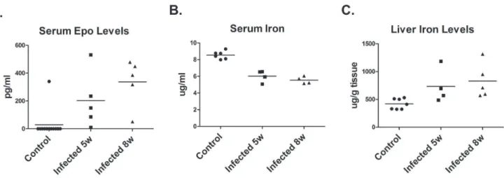
Acute anemia results in a systemic stress response in which tissue hypoxia triggers production of erythropoietin by the kidney [33]. Therefore, circulating levels of Epo were determined by ELISA. Epo was detected in the serum of hamsters infected for 5–6 weeks and eight weeks, while Epo was only detected in the serum of one control non-infected hamster (Figure 2).
Since iron deficiency resulting from chronic infections can lead to anemia associated with chronic disease and inflammation, iron levels were measured in the blood and liver. Iron levels were decreased in the serum of infected hamsters and accumulated in the liver (Figure 2). The decrease in serum iron levels while statistically significant was relatively small and is not sufficient to account for the decreased numbers of circulating red blood cells. Further, there were no significant differences in MCV and MCHC between infected and control hamsters (data not shown), which would be expected to be decreased if iron deficiency is disrupting hemoglobin biosynthesis.
L. donovaniInfection Increases the Frequency of Erythroid Progenitors
To determine whether anemia associated with VL is due to reduction in erythroid progenitors, we determined the frequency of erythroid progenitors in the bone marrow and spleen by the
Erythroid Gene Expression in the Bone Marrow and Spleen is Differentially Altered byL. donovaniInfection
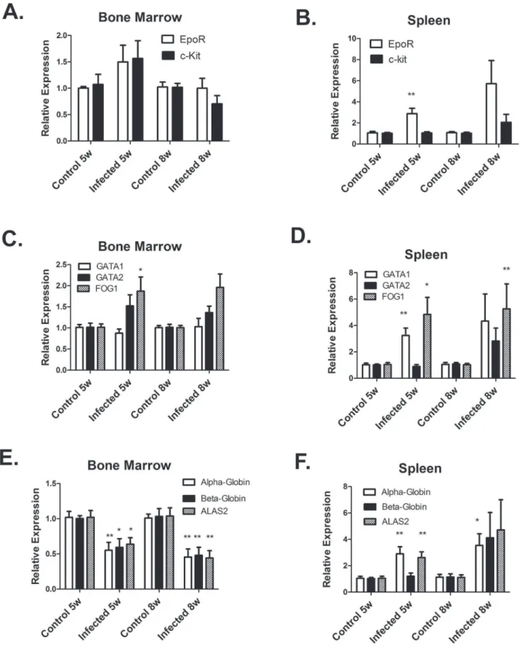
We next examined the expression of erythroid genes in the bone marrow and spleen by real-time RT-PCR. We first cloned and sequenced cDNA segments of erythroid receptors c-kit and Epo-R; key transcription factors GATA1, GATA2, and FOG1; and erythroid differentiation genes b-globin and ALAS2. Real-time PCR primers were then designed from the sequences. We also designed primers for a-globin using sequence information in GenBank. In the bone marrow, expression of erythroid receptors and transcription factors mRNA were unchanged byLeishmania
infection (Figure 4). However, erythroid differentiation genesa -globin,b-globin, and ALAS2 were inhibited by 50% in the bone marrow ofLeishmaniainfected hamsters. In the spleen, expression of EpoR, GATA1, and FOG1 mRNA were increased by
Leishmania infection at 5–6 weeks p.i (Figure 4). Furthermore, levels ofa-globin and ALAS2 transcripts were also increased by
Leishmaniainfection. At 8 weeks p.i, expression of these genes also tended to be increased, but there was more variability in expression between individual infected hamsters (Figure 4). We interpret these results to indicate that in the bone marrow a block in erythroid differentiation inLeishmaniainfected hamsters inhibits erythrocyte production. In the spleen, increased erythroid gene expression inLeishmaniainfected hamsters suggests that the spleen is responding to the anemia by activation of the stress erythro-poiesis response that has been described in mice [31,32].
IFN-cmRNA Levels in the Bone Marrow and Spleen are Highly Increased byL. donovaniInfection
Since pro-inflammatory cytokines have an inhibitory effect on erythroid differentiation [34], we measured the expression of cytokine mRNA by real-time RT-PCR in control (non-infected) hamsters and hamster infected with L. donovani for 5–6 and 8 weeks. The most highly elevated cytokine was IFN-c. IFN-c
IL-12p40 mRNA. Surprisingly, despite the increased expression of IFN-c mRNA, IL-12 p40 mRNA expression levels between control and infected hamsters differed by less than 2-fold in both the spleen and bone marrow (data not shown).
Expression of TRAIL mRNA is Increased byL. donovani
TRAIL (TNF-related apoptosis-inducing ligand) is a member of the TNF-family of cytokines that is induced by IFN-c and is a negative regulator of erythropoiesis [35–37]. Trail mRNA expression was examined by real-time RT-PCR. To obtain sequence information to design real-time PCR primers, we first cloned and sequenced a cDNA segment of the hamster TRAIL gene. As shown in Figure. 5B, expression levels of TRAIL mRNA in the bone marrow and spleen were increased 3–6 fold by infection withL. donovani.
Flow Cytometry Analysis of Erythroblasts
Because TRAIL is a negative regulator of erythropoiesis and inducer of apoptosis, we hypothesized that the block in erythroid
differentiation inLeishmaniainfected hamster results from apoptosis of erythroblasts during differentiation. To test this hypothesis, we determined if Leishmania infection increases the frequency of apoptotic erythroblasts by flow cytometry, using annexin-V binding and DNA fragmentation by the TUNEL assay. To detect hamster erythroblasts, a rat-monoclonal antibody (DECMA-1) that recognizes mouse, human, and canine E-cadherin (CD324) was used. E-cadherin is an epithelial cellular adhesion molecule that is also expressed on erythroblasts but not erythrocytes [38,39]. It is also absent on all other hematopoietic cells. Bone marrow cells from hamsters infected withLeishmania donovanifor 6–7 weeks and control non-infected hamsters were analyzed by flow cytometry for E-cadherin positive erythroblasts (Figure 7). There were no significant differences in the % percentage of E-cadherin positive erythroblasts, 33% of bone marrow cells were E-cadherin positive for control hamsters and 31% for infected hamsters (data not shown). However, the percentage of apoptotic erythroblasts that were annexin–V positive (Figure 7A and C) and TUNEL-positive (Figure 7B and D)were significantly higher in infected Figure 1.L. donovaniinfected hamsters develop severe anemia and leukopenia.Hematological analysis of blood from control hamsters and hamsters infected withL. donovanifor 8 weeks. Data shown are from individual hamsters and the mean. Hemogloblin levels (A) and hematocrit (B) were statistically significant at p,0.01. Red blood cell (C) and white blood cell counts (D) were significant at p,0.05, Student’s t test.
doi:10.1371/journal.pone.0059509.g001
hamsters than control hamsters. Thus,Leishmania donovaniinfection in hamsters increases apoptosis of erythroblasts.
Discussion
Visceral leishmaniasis (VL) in humans is a progressive disease that upon diagnosis can present a range of symptoms from asymptomatic infections, to sub-clinical infections, and active VL [40–42]. Experimental visceral leishmaniasis induced in Syrian Golden Hamsters by infection withL. donovani also progresses at different rates in individual hamsters. This is evident by the range in hemoglobin levels we observed in individual hamsters that ranged from levels near control hamsters to extremely low levels (below 5 g/dL)(Fig. 1). Similar variability is also present in the HCT, red blood cells counts, and white blood cell counts. Syrian Golden Hamsters are out-bred, so genetic differences could be involved in the variability. Genetic factors may also influence progression of human infections with L. donovaniorL. chagasi as
there are large differences in the ratio of asymptomatic to symptomatic infection among different racial groups [43,44].
L. donovaniestablishes infections by infecting and replicating in macrophages of bone marrow, spleen, and liver [1–3]. The progressive parasite burden in these organs is due in part to the ability of the parasite to alter hematopoiesis.L. donovaniinfection of mice results in expansion of progenitor cells and proliferative activity in the spleen and bone marrow [29]. This increase is highly selective for myleopoiesis. In the spleen, numbers of colony-forming unit-granulocyte, monocyte (CFU-GM) increased 20–30-fold at 28–56 days post-infection. Erythroid progenitors, burst-forming unit-erythrocyte (BFU-E), also increased, but to a lesser extent. Overall, splenomegaly was present, with 2–4 fold increase in total spleen cell numbers. An even greater selective expansion of hematopoiesis likely occurs in infected hamsters. For example, spleen weights inL. donovani infected hamsters increased 20-fold compared to control non-infected hamsters. Although we did not measure CFU-GM progenitor numbers, CFU-E progenitors in the Figure 2. Erythropoietin and Iron levels in infected hamsters.(A) Serum erythropoietin (Epo) levels in control hamsters and hamsters infected withL. donovanifor 5 and 8 were determined by ELISA. EPO levels were significantly increased by infection, p,0.005, one-way ANOVA. (B) Serum iron levels in control and infected hamsters were determined by ferrozone assay. Serum iron levels were significantly decreased by infection, p,0.0005, one-way ANOVA. (C). Liver iron levels in control and infected hamsters were determined by ferrozone assay. Liver iron levels were significantly increased by infection p,0.05, one-way ANOVA.
doi:10.1371/journal.pone.0059509.g002
Figure 3.L. donovaniinfection increases BFU-E and CFU-E progenitor numbers in the bone marrow and spleen.(A) Frequency of BFU-E progenitors in the bone marrow of control hamsters and hamsters infected withL. donovanifor 8 weeks was determined by the semi-solid colony assay. Data represent mean+/2SEM from hamsters. *p,0.05, Student’s t test. (B) and (C) Frequency of CFU-E progenitors in bone marrow and spleen from control and infected hamsters. Data represent mean+/2SEM from 3–5 hamsters. *p,0.05; *p,0.01, student’s t test.
Figure 4. Erythroid gene expression is differentially altered byL. donovaniinfection in the bone marrow and spleen.mRNA levels of erythroid genes were determined by real-time RT-PCR in bone marrow (A,C,E) and spleen (B, D, F). mRNA levels were normalized tob-actin and expressed relative to control non-infected hamsters. Data represent mean+/2SEM of 6–8 hamsters in each group. *p,0.05; **p,0.01, Student’s t test.
doi:10.1371/journal.pone.0059509.g004
spleen of infected hamsters were increased only 4-fold. A similar 4–5 fold increase in BFU-E and CFU-E progenitors were observed in bone marrow cells from infected hamsters.
The selective expansion of myleopoiesis during VL occurs together with alternations in erythropoiesis. Erythropoiesis in the bone marrow maintains the steady-state levels of circulating erythrocytes [45]. Thus, alternations in erythropoiesis in L. Donovani infected individuals contributes to a progressive decline in circulating erythrocytes and eventually anemia, which occurs in almost all VL patients. Analysis of bone marrow biopsies of VL patients indicate cellular hyperplasia with many abnormal erythroblasts, which include large erythroblasts containing giant lysosomes and nuclei with little condensed chromatin and multinuclear erythroblasts with irregular nuclei, and nuclear budding [46–48]. This dysplasia is similar to myelodysplasia observed in Myelodysplastic syndromes (MDS), which are stem cell disorders in which abnormal proliferation and differentiation of hematopoietic progenitor cells results in ineffective erythropoi-esis [49,50]. Erythrocytes that are produced during VL are also more susceptible to oxidative damage and have shortened lifespan [51–53].
The present study examined for the first time the expression of erythroid genes in the bone marrow during VL. Proliferation and differentiation of erythroid progenitors requires interactions of stem cell factor with its receptor (c-Kit) and eyrthropoietin with its receptor (EpoR) [54,55]. Erythropoietin and EpoR are also required for survival and differentiation of erythroblasts [55]. Differentiation of erythroblasts requires expression of GATA1, GATA2 and FOG1 transcription factors [56–59]. We found that mRNA levels of c-Kit, EpoR-R, GATA1, GATA2, and FOG1 were not affected by L. donovaniinfection of hamsters. Thus, the expression of receptors and transcription factors required for erythropoiesis is not altered by VL. However, we did observe decreased expression of erythroid differentiation genes (alpha-globin, beta-(alpha-globin, and ALAS2), which are target genes of GATA1. These results indicate a block in erythroblast differen-tiation could be due suppression of GATA1-induced expression of erythroid differentiation gene and/or through decreased numbers of erythroblasts.
Homeostasis of circulating erythrocytes is a balance between erythrocyte production by the bone marrow and red blood cell destruction. Apoptosis of immature erythroblasts is a key pathway by which bone marrow production of erythrocytes is regulated [reviewed in 45]. During normal physiological conditions, apoptosis acts as a negative control of the rate of maturation of immature erythroblasts. This occurs through the interaction of FasL and membrane TRAIL on mature erythroblasts with Fas and TRAIL receptors on immature erythroblasts, which activates the caspase cascade that cleaves GATA1 and triggers apoptosis [45]. As the erythroblasts mature, the erythroblasts become resistant to apoptosis induced by Fas cross-linking and lose expression of Trail receptors. During pathological conditions, up-regulation of Fas and FasL expression can exacerbate Fas-control of erythropoiesis. For example, Fas and FasL expression is increased in the bone marrow of MDS patients compared to controls [60]. Also, the death receptor ligand TRAIL and its receptors TRAILR1 and TRAILR2 are increased in bone marrow of MDS patients and hemopoietic progenitor cells from MDS patients are more sensitive to TRAIL-mediated apoptosis [61,62]. Therefore, in the current study we examined apoptosis of bone marrow erythroblasts duringL. donovaniinfection of hamsters by flow cytometry. To detect hamster erythroblasts, we used a rat monoclonal antibody to E-cadherin, which we found to cross-react with hamster. E-cadherin is expressed primarily by early immature erythroblasts and declines in expression as the erythroblast matures [38,39]. Other antibodies used to detect erythroblasts, such as antibodies to mouse TER119 and human glycophorin A did not cross-react with hamster. We were also unable to find a cross-reactive antibody to CD71, which is used in flow cytometry in combination with antibodies to TER119 or glycophorin A to distinguish erythroblast stages. Our results do show that E-cadherin+ erythroblasts from L. donovani-infected hamsters have higher percentage of apoptotic cells, as detected by Annexin-V binding and the TUNEL assay, than erythroblasts from normal control hamsters. Thus,L. donovaniinfection of the bone marrow increases apoptosis during erythropoiesis.
In this study, we also examined erythroid gene expression in the spleen. In mice, erythropoiesis in the spleen occurs in response to Figure 5. IFN-cand TRAIL mRNA are highly expressed in L. Donovaniinfected hamsters.mRNA levels of IFN-c (A) and TNF-related apoptosis inducing ligand (TRAIL) (B) were determined by real-time RT-PCR in the bone marrow and spleen of control non-infected hamsters andL. donovaniinfected hamsters. mRNA levels are expressed relative to control non-infected hamsters. Data represent mean+/2SEM of 6–8 hamsters in each group. **p,0.01; ***p,0.001, Student’s t test.
anemia [32,32]. The response is regulated by high levels of circulating erythropoietin, which induces proliferation and differ-entiation of stress BFU-E progenitors into erythroblasts. Our data show that mRNA expression of erythroid genes is enhanced in
L.donovani-infected hamsters. Expression levels tended to be higher at 8 weeks post-infection compared to 5 weeks, but there was also more variability in expression among individual hamsters, which likely reflects the variability in VL disease progression. We also observed higher levels of serum erythropoietin in L. donovani
infected hamsters compared to control hamsters, which also tended to be higher at 8 weeks post-infection. The results are consistent with a stress erythropoiesis in infected hamsters induced by the anemia. Whether this stress erythropoiesis is countered by increased apoptosis of erythroblasts in the spleen of infected hamsters needs to be examined. However, erythropoietin has been shown in mice to inhibit apoptosis during the spleen stress erythropoiesis response by suppressing Fas-FasL expression [63].
The few studies that have examined mechanisms responsible for alterations in hematopoiesis focused on examining effects of cytokines and growth factors on the enhanced myelopoiesis during VL. Enhanced myelopoiesis in mice is associated with increased mRNA for growth factors GM-CSF, M-CSF, and G-CSF [29].L. donovani infection of mouse bone marrow derived macrophages
in vitro and co-incubation with spleen cells increased CFU-GM colony formation through the production of GM-CSF and
TNF-a[30]. Mouse studies with TNF-aknockout mice have shown that TNF-ais required to control intracellular growth ofL. donovaniand promote granuloma formation [64], but is also responsible for remodeling the splenic marginal zone [18]. TNF-amay also alter erythropoiesis during VL, since TNF-a has been reported to inhibit erythropoiesisin vitro[34, 65, and 66]. However, we did not observe increased mRNA expression of TNF-a in the spleen or bone marrow of infected hamsters at 5 and 8 wks post-infection compared to control hamsters. This was not unexpected, since prior studies [27,28] with hamsters have shown TNF-aexpression Figure 6. T cell and anti-inflammatory cytokine mRNA are increased byL. donovaniinfection.mRNA levels of T cell cytokines IL-2 and IL-4 (A and B) and anti- inflammatory cytokines IL-10 and TGF-b(C and D) in the spleen and bone marrow of control non-infected hamsters andL. donovaniinfected hamsters were determined by real-time RT-PCR, mRNA levels are expressed relative to control non-infected hamsters. Data represent mean+/2SEM of 6–8 hamsters in each group. *p,0.05; **p,0.01; ***p,.001, Student’s t test.
doi:10.1371/journal.pone.0059509.g006
in the spleen is induced early at 3–10 days post-infection and then declines. Whether TNF-a alters erythropoiesis early in the infection needs to be examined. However, its absence at 5 and 8 weeks post-infection suggest TNF-a is not involved in inhibiting erythropoiesis at these time points.
Our data show that mRNA for IL-10, TGF-b, and IFN-c
mRNA is up-regulated in bone marrow of L. donovaniinfection. Both IL-10 and TGF-b have been shown in vitro to inhibit erythropoiesis [67,68]. IL-10 inhibits BFU-E growth by suppres-sing T cell production of GM-CSF [67]. Since we observed increased BFU-E and CFU-E progenitor numbers in infected hamster, this effect of IL-10 does not appear to be involved in the inhibiting erythropoiesis in infected hamsters. TGF-b acts by inducing cell cycle arrest of immature erythroblasts and acceler-ating differentiation [68]. TGF-b is also involved in T cell immunosuppression during VL in infected hamsters and induces lymphocyte apoptosis [69,70]. Interestingly, while TGF-binduces lymphocyte apoptosis in hamsters, it does not appear to induce apoptosis in human erythroblasts. [68]. Whether TGF-b is a contributing factor to inhibition of erythropoiesis during VL needs further study.
The cytokine that we found to be induced in high amounts inL. donovaniinfected hamsters is IFN-c. IFN-cmRNA is produced in both the spleen and bone marrow throughout the course of infection in hamsters, with induction as early as 7–10 day post-infection [27,28]. IFN-c is also present in high amounts in the
serum of human patients with active VL [71–73]. IFN-cinvitro
bone marrow studies suppresses proliferation and differentiation of erythroblasts [34, 74, and 75] and induces apoptosis in maturing erythroblasts [76]. Studies by Feli et al. [37] have shown that
IFN-csuppresses erythropoiesis by inducing expression of members of the TNF super family, including TRAIL, FASL (CD95L), and TWEAK, which interact with their receptors on target cells to induce apoptosis. Other studies have identified TRAIL as negative regulator of erythropoiesis [35, 36, 61, and 62]. In the current study, we showed that TRAIL mRNA expression in bone marrow and spleen is up-regulated by L. donovani infection. Thus, our studies are consistent with L. donovani infection inhibiting erythropoiesis by inducing macrophage expression of TRAIL, which then interacts with its receptors on immature erythroblasts to induce apoptosis. IFN-c may also be involved in enhanced myelopoiesis, since IFN-c has been show to expand early hematopoietic progenitor cell populations, resulting in a bias toward the myeloid lineage [77,78].
Materials and Methods
Hamsters
Six 28 week old outbred Syrian golden hamsters (Mesocricetus auratus) were obtained from Charles River (Wilmington, DE). This study was carried out in strict accordance with the recommenda-tions of Office of Laboratory Animal Welfare, National Institutes Figure 7.L. donovani infection increases apoptosis of erythroblasts.Apoptosis of bone marrow erythroblasts was examined by flow cytometry from control non-infected hamsters andL. donovaniinfected hamsters. Apoptosis was determined by Annexin-V binding (A) and the TUNEL Assay (B). Representative flow cytometry plots are from a control non-infected hamster and a hamster infected withL. donovanifor 7 weeks. (A) Bone marrow cells were labeled with anti- E-cadherin antibody-e-Fluro 660 and FITC-Annexin V. Dead cells were excluded with 7-amino actinomycin (7-ADD). (B) BrdUTP incorporation by the TUNEL technique in erythroblasts was detected using Alexa-488 anti-BrdUTP and anti-E-cadherin-e-Fluro 660. (C) Percentage of E-cadherin positive erythroblasts that are Annexin-V positive. (D) Percentage of E-cadherin positive erythroblasts that are TUNEL positive. Data represent mean+/2SEM of 4 hamsters in each group. *p,0.05, Student’s t test.
doi:10.1371/journal.pone.0059509.g007
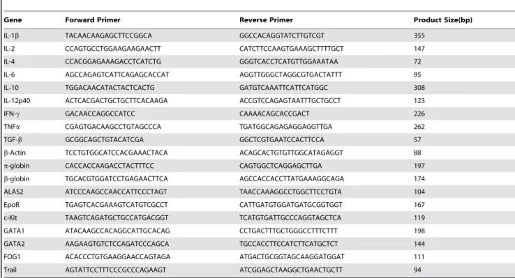
Table 1.List of primers used for RT-PCR.
Gene Forward Primer Reverse Primer Product Size(bp)
IL-1b TACAACAAGAGCTTCCGGCA GGCCACAGGTATCTTGTCGT 355
IL-2 CCAGTGCCTGGAAGAAGAACTT CATCTTCCAAGTGAAAGCTTTTGCT 147
IL-4 CCACGGAGAAAGACCTCATCTG GGGTCACCTCATGTTGGAAATAA 72
IL-6 AGCCAGAGTCATTCAGAGCACCAT AGGTTGGGCTAGGCGTGACTATTT 95
IL-10 TGGACAACATACTACTCACTG GATGTCAAATTCATTCATGGC 308
IL-12p40 ACTCACGACTGCTGCTTCACAAGA ACCGTCCAGAGTAATTTGCTGCCT 123
IFN-c GACAACCAGGCCATCC CAAAACAGCACCGACT 226
TNFa CGAGTGACAAGCCTGTAGCCCA TGATGGCAGAGAGGAGGTTGA 262
TGF-b GCGGCAGCTGTACATCGA GGCTCGTGAATCCACTTCCA 57
b-Actin TCCTGTGGCATCCACGAAACTACA ACAGCACTGTGTTGGCATAGAGGT 88
a-globin CACCACCAAGACCTACTTTCC CAGTGGCTCAGGAGCTTGA 197
b-globin TGCACGTGGATCCTGAGAACTTCA AGCCACCACCTTATGAAAGGCAGA 174
ALAS2 ATCCCAAGCCAACCATTCCCTAGT TAACCAAAGGCCTGGCTTCCTGTA 104
EpoR TGAGTCACGAAAGTCATGTCGCCT CATTGATGTGGATGATGCGGTGGT 167
c-Kit TAAGTCAGATGCTGCCATGACGGT TCATGTGATTGCCCAGGTAGCTCA 119
GATA1 ATACAAGCCACAGGCATTGCACAG CCTGACTTTGCTGGGCCTTTCTTT 198
GATA2 AAGAAGTGTCTCCAGATCCCAGCA TGCCACCTTCCATCTTCATGCTCT 144
FOG1 ACACCCTGTGAAGGAACCAGTAGA ATGACTGCGGTAGCAAGGATGGAT 111
Trail AGTATTCCTTTCCCGCCCAGAAGT ATCGGAGCTAAGGCTGAACTGCTT 94
doi:10.1371/journal.pone.0059509.t001
from marrow cavity with Iscove’s modified Dulbecco’s medium (IMDM). Livers were snap frozen and stored280uC.
Hematological Analysis
Blood was collected from euthanized hamsters by cardiac puncture in heparinized tubes (Sigma) and submitted to the Hematology Lab of the Ohio State University Veterinary Hospital for hematological analysis. Blood was also collected in non-heparinized tubes and allowed to clot overnight at 4uC. Serum was obtained and stored at 280uC until analyzed for serum erythropoietin and iron levels. Serum erythropoietin levels were determined by ELISA using a mouse erythropoietin ELISA kit (R&D Systems, Minneapolis, MN) and mouse erythropoietin as standard.
Serum and Liver Iron Analysis
Serum were de-proteinized by 1:1 dilution with 10% TCA in 1N HCL and incubation at 95ufor 1 hr in microcentrifuge tubes. Liver tissue was weighted and 75–150 mg of tissue homogenized in 1 ml of molecular grade water using a tissue homogenizer. Liver homogenates were de-proteinized by1:1 dilution with 10% TCA in 1N HCL and heating at 65uC for 20 hrs. Precipitated proteins were removed by centrifugation and iron content determined by the ferrozone assay [79].
Colony Assays for BFU-E and CFU-E
Single cell suspensions of splenocytes and bone marrow cells were incubated with RBC lysing buffer to lyse red blood cells, pelleted by centrifugation, resuspended in IMDM with 2% FBS and counted. For the CFU-E assay, 56105bone marrow cells and
86105splenocytes were plated in duplicate 35 mm culture plates containing methylcellulose media (Stem Cell Technologies) with 3 units/ml Epo. After 3 days of culture at 37u, the plates were stained with of 2, 7-diaminofluroene (DAF) to detect hemoglobin and hemoglobin positive colonies counted under an inverted microscope [80]. For the BFU-E assay, 36105bone marrow cells were plated in duplicate 35 mm culture plates containing methylcellulose media with 3 units/ml Epo, 150 ng/ml mouse SCF, and 10% pokeweed mitogen hamster spleen conditioned media. After 10 days of culture, colonies of least 30 red-colored cells were counted.
Flow Cytometry Analysis
Bone marrow cells were isolated from hamsters infected with
Leishmania donovaniand control non-infected hamsters. Cells were stained with E-cadherin (CD234) monoclonal antibody DECMA-1 conjugated with e-Fluro 660 antibody (e-Bioscience) washed in
E-cadherin antibody conjugated with e-Fluro 660 and Alexa 488 anti-BrdU antibody.
Cloning of Hamster cDNA and Real-time RT- PCR Primer Design
To obtain hamster gene sequences for primer design, we designed PCR primers from the mouse gene sequences that were conserved with rat and then used these primers to amplify hamster cDNA by PCR. PCR products 300–700 bp in size were isolated by agarose gel electrophoresis and purified on Qiagen spin columns (QIAquick gel extraction kit). PCR products were cloned into pGEM-T Easy Vector (Promega) and transformed into DH5a
E. coli.(Invitrogen). Plasmid DNA was isolated from 2–3 colonies using Qiagen QIAprep Miniprep Kit and sequenced. Identity of the hamster cDNA sequences were confirmed by NCBI Blast search and were 82–93% identical to the mouse and rat homologues DNA sequences for GATA1 (JX569328), GATA2 (JX569329), FOG1 (JX569330), EpoR (JX569331), c-Kit (JX569332), b-globin (JX569333), ALAS2 (JX569334), and TRAIL (JX569335) have been submitted to GenBank. Primers (Table 1) for real-time RT-PCR were designed from these sequences using PrimerQuest (Integrated DNA Technologies). Hamster gene sequences in GenBank were used to design primers for a-globin (X57029), IL-6 (AB028635), IL12p40 (AB085792), andb-actin (AJ312092). Primer sequences for IL-1b, IL-2, IL-4, IL-10, IFN-c, and TGF-bhave been previously reported [81–83].
Isolation of Hamster RNA and Real-time RT PCR Analysis
Spleen and bone marrow cells were lysed by homogenization with TRIzol reagent (Invitrogen) and RNA isolated using the RNeasy mini kit (Qiagen). Residual DNA was removed during RNA purification by on-column DNase digestion. RNA was reversed-transcribed using oligo (dT)15 primers by the Promega Reverse Transcription System. The mRNA expression was analyzed by real-time RT-PCR using FastStart DNA STBR Green 1 reaction mixture (Roche). The amplification conditions were 95uC for 10 minutes followed by 40 cycles of 95uC for 15 s, 57uC for 30 s, and 72uC for 30 s. Each measurement was carried out in duplicate. Relative expression was calculated by theDCT method usingb-actin expression as the normalize [84]. Results are expressed as relative gene expression to samples from uninfected hamsters. PCR products were electrophoresed on 2% agarose gels to confirm amplification of products with the correct size.
Statistical Analysis
for multiple comparisons. Pairwise comparisons were done using Student’s t-test.
Author Contributions
Conceived and designed the experiments: WL ARS SO GG. Performed the experiments: SV HS. Analyzed the data: WL ARS RS JM SO GG. Contributed reagents/materials/analysis tools: ARS WL. Wrote the paper: WL ARS.
References
1. Alexander J, Russell DG (1992) The interaction of Leishmania species with macrophages. Adv Parasitol 31: 175–254.
2. Liew FY, O’Donnell CA (1993) Immunology of leishmaniasis. Adv Parasitol 32: 161–258.
3. Murray HW, Berman JD, Davies CR, Saravia NG (2005) Advances in leishmaniasis. Lancet 366: 1561–1577.
4. Zijlstra EE, Ali MS, el-Hassan AM, el-Toum IA, Satti M, et al. (1992) Clinical aspects of kala-azar in children from the Sudan: a comparison with the disease in adults. J Trop Pediat 38: 17–21.
5. al-Jurayyan NA, Al-Naaer M, Al-Fawaz I, al-Herbish AS, al-Mazrou AM, et al. (1995) The haematological manifestations of visceral leishmaniasis in infancy and childhood. J Trop Pediat 41: 143–148.
6. Pearson RD, Sousa AQ (1996) Clinical Spectrum of leishmaniasis. Clin Infect Dis 22: 1–13.
7. Santos MA, Marques RC, Fairas CA, Vasconcelos DM, Stewart JM, et al. (2002) Predictors of an unsatisfactory responses to pentavalent antimony in the treatment of American visceral leishmaniasis. Rev Soc Bras Med Trop 35: 629– 633.
8. de Araujo VE, Morais MH, Reis IA, Rabello A, Carnerio M (2012) Early clinical manifestions associated with death from visceral leishmaniasis. Plos Neg Trop Dis 6: e1511.
9. Pasaquau F, Ena J, Sanchez R, Cuadrado JM, Amador C, et al. (2005) Leishmaniasis as an opportunistic infection in HIV-infected patients: determi-nants of relapse and mortality in a collaborative study of 228 episodes in a Mediterreanean region. Eur J Clin Microbiol Infect Dis 24: 411–418. 10. Alvar J. Aparicio P, Aseffa A, Den Boer M, Canavate C, et al. (2008) The
relationship between Leishmaniasis and AIDS: the second 10 years. Clin Microbiol Rev 21: 334–359.
11. Savioli L, Daumerie D, Crompton DWT, Annon. (2010) Leishmaniasis. In: Savioli L, Daumerie D, Crompton DWT, editors. Working to overcome the global impact of neglected tropical diseases. First WHO report on neglected tropical diseases. Geneva: World Health Organization. 91–96.
12. Miralles GD, Stoeckle MY, McDermott DF, Finkelman FD, Murray HW. (1994) Th1 and Th2 cell-associated cytokines in experimental visceral leishmaniasis. Infect Immun 62: 1058–1063.
13. Wilson ME, Sandor M, Blum AM, Young BM, Metwali A, et al. (1996) Local suppression of IFN-c in hematic granulomas correlates with tissue-specific replication ofLeishmania chagasi.J Immunol 156: 2231–2239.
14. Engwerda CR, Murphy M, Cotterell SE, Smelt SC, Kaye PM (1998) Neutralization of IL-12 demonstrates the existence of discrete organ-specific phases in the control ofLeishmania donovani. Eur J Immunol 28: 669–680. 15. Engwerda CR, Kaye PM (2000) Organ-specific immune responses associated
with infectious disease. Immunology Today 21: 73–78.
16. Smelt SC, Engwerda CR, McCrossen M, Kaye PM (1997) Destruction of follicular dendritic cells during chronic visceral leishmaniasis. J. Immunol. 158: 3813–3821.
17. Melby PC, Tabares A, Restrepo BJ, Cardona AE, McGuff HS, et al. (2001)
Leishmania donovani: evolution and architecture of the splenic cellular immune response related to control of infection. Exp Parasitol 99: 17–25.
18. Engwerda CR, Ato M., Cotterell SE, Mynott TL, Tschannerl A, et al. (2002) A role for tumor necrosis factor-ain remodeling the splenic marginal zone during
Leishmania donovaniinfection. Am J Path 161: 429–437.
19. Engwerda CR, Ato M, Kaye PM (2004) Macrophages, pathology, and parasite persistence in experimental visceral leishmaniasis. Trends Parasitol 20: 524–530. 20. Ott KJ, Hanson WL, Stauber L (1967) Course of infection ofLeishmania donovani.
J Parasitol 53: 641–645.
21. Farrell JP (1976)Leishmania donovani: acquired resistance to visceral leishmaniasis in the golden hamster. Exp Parasitol 40: 89–94.
22. Gifawesen C, Farrell JP (1989) Comparison of T-cell responses in self-limiting versus progressive visceralLeishmania donovaniinfections in golden hamsters.Infect Immun57: 3091–3096.
23. Evans TG, Smith D, Pearson RD (1990) Humoral factors and nonspecific immune suppression in Syrian hamsters infected with Leishmania donovani.
J Parasitol 76: 212–217.
24. Biswas T, Chakraborty M, Naskar K, Ghosh DK, Ghosal J (1992) Anemia in experimental visceral leishmaniasis in hamsters. J Parsitol 78: 140–142. 25. Requena JM, Soto M, Doria MD, Alonso C (2000) Immune and clinical
parameters associated withLeishmania infantumin the golden hamster model. Vet Immunol Immunopathol 76: 269–281.
26. Zivcec M, Safronetz D, Haddock E, Feldmann H, Ebihara H (2011) Validation of assays to monitor immune responses in the Syrian golden hamster (Mesocricetus auratus). J Immunol Methods 368: 24–35.
27. Melby PC, Tryon VV, Chandrasekar B, Freeman GL. (1998) Cloning of Syrian hamster (Mesocrietus auratus) cytokine cDNAs and analysis of cytokine mRNA in experimental visceral leishmaniasis. Infect Immun 66: 2135–2142.
28. Melby PC, Chandrasekar B, Zhao W, Coe JE (2001) The hamster as a model of human visceral leishmaniasis: progressive disease and impaired generation of nitric oxide in the face of a prominent TH1-like cytokine response. J Immunol 166: 1912–1920.
29. Cotterell SE, Engwerda CR, Kaye PM (2000) Enhanced hematopoietic activity accompanies parasite expansion in the spleen and bone marrow of mice infected withLeishmania donovani.Infect Immun 68: 1840–1848.
30. Cotterell SE, Engwerda CR, Kaye PM (2000)Leishmania donovaniinfection of bone marrow stromal macrophages selectively enhances myelopoiesis, by a mechanism involving GM-CSF and TNF-a. Blood 95: 1642–1651. 31. Perry J, Harandi OF, Paulson R (2007) BMP4, SCF, and Hypoxia cooperatively
regulate the expansion of murine stress erythroid progenitors. Blood 109: 4494– 4502.
32. Perry JM, Harandi OF, Porayette P, Hedge S, Kannan AK, et al. (2009) Maintenance of the BMP4 dependent stress erythropoiesis pathway in the murine spleen requires hedgehog signaling. Blood 113: 911–918.
33. Sasaki R, Masuda S, Nago M (2000) Erythropoietin: multiple physiological functions and regulation of biosynthesis. Biosci Biotechnol Biochem 64: 1775– 1793.
34. Morceau F, Dicato M, Diecherich M (2009) Pro-inflammatory cytokine-mediated anemia: Regarding molecular mechanisms of erythropoiesis. Media-tors Inflamm 2009: 405016.
35. Zauli G, Secchiero P (2006) The role of the TRAIL/TRAIL receptors system in hematopoiesis and endothelial cell biology. Cytokine Growth Factor Rev 17: 245–257.
36. Silvestris F,Cafforio P,Tucci M, Dammacco F (2002) Negative regulation of erythroblast maturation by Fas-L+/TRAIL+ highly malignant plasma cells: a major pathogenetic mechanism of anemia in multiple myeloma. Blood 99: 1305–1313.
37. Felli N, Pedini F, Zeuner A, Petrucci E, Testa U, et al. (2005) Multiple members of the TNF superfamily contribute to IFN-c-mediated inhibition of erythropoi-esis. J Immunol 175: 1464–1472.
38. Armeanu S, Buhring HJ, Reuss-Borst M, Muller CA, Klein G (1995) E-cadherin is functionally involved in the maturation of the erythroid lineage. J Cell Biol 131: 243–249.
39. Lammers R, Giesert C, Grunebach F, Marxer A, Vogel W, et al. (2002) Monoclonal antibody 9C4 recognizes epithelial cellular adhesion molecule, a cell surface antigen expressed in early steps of erythropoiesis. Exp Hematol 30: 537– 545.
40. Badaro R, Jones TC, Carvalho EM, Sampaio D, Reed SG, et al. (1986) New perspectives on a subclinical form of visceral leishmaniasis. J Infect Dis 154: 1003–1011.
41. Badaro R, Jones TC, Lorenco R, Cerf BJ, Sampaio D, et al. (1986) A prospective study of visceral leishmaniasis in an endemic area of Brazil. J Infect Dis 154: 639–649.
42. Wilson ME, Jeronimo SM, Pearson RD (2005) Immunopathogenesis of infection with the visceralizingLeishmaniaspecies. Microb Patho 38: 147–160. 43. E-l Hassan AM, Zijlstra EE, Ismael A, Ghalib HW (1995) Recent observations
on the epidemiology of kala-azar in the eastern and central states of Sudan. Trop Geogr Med 47: 151–157.
44. Ibrahim ME, Lambson B, Yousif AO, Deifalla N, Alnaiem DA, et al. (1999) Kala-azar in a high transmission foci: an ethnic and geographical dimension. AM J Trop Med Hyg 61: 941–944.
45. Testa U (2004) Apoptotic mechanisms in the control of erythropoiesis. Leukemia 18: 1176–1199.
46. Wickramasinghe SN, Abdalla SH, Kasili EG. (1987) Ultrastructure of bone marrow in patients with visceral leishmaniasis. J Clin Pathol 40: 267–275. 47. Yarali N, Fisgin T,Duru F, Kara A (2002) Myelodysplastic features in visceral
leishmaniasis. Am J Hematol 71: 191–205.
48. Kumar PV, Vasei M, Sadeghipour A, Sadeghi E, Soleimanpour H, et al. (2007) Visceral leishmaniasis: bone marrow biopsy findings. J Pediatr Hematol Oncol 29: 77–80.
49. Hasle H (1994) Myelodysplastic syndromes in childhood-classification, epide-miology, and treatment. Leuk Lymphoma 13: 11–16.
50. Hofmann WK, Ottmann OG, Ganser A, Hoelzer D (1996) Myelodysplastic syndromes: clinical features. Semin Hematol 33: 177–185.
51. Sen G, Mukhopadhyay R, Ghosal J, Biswas T (2001) Oxidative damage of erythrocytes: a possible mechanism for premature hemolysis in experimental visceral lesihmaniasis in hamsters. Ann Hematol 80: 32–37.
a paradigm for transcription factors in hematopoiesis. Mol Cell Biol 25: 1215– 1227.
60. Claessens YE, Bouscary D, Dupont JM, Picard F, Melle J, et al. (2002)In vitro
proliferation and differentiation of erythroid progenitors from patients with myelodysplastic syndromes: evidence for Fas-dependent apoptosis. Blood 99: 1594–1601,
61. Zang Y, Goodwin RG, Loken MR, Bryant E, Deeg HJ, et al. (2001) Expression of tumor necrosis factor-related apoptosis-inducing ligand, Apo2L, and its receptors in myelodysplastic syndrome: effects onin vitrohemopoiesis. Blood 98: 3058–3065.
62. Campioni D, Secchiero P, Corallini F, Melloni E, Capitani S, et al. (2005) Evidence for a role of TNF-related apoptosis-inducing ligand (TRAIL) in the anemia of myelodysplastic syndromes. Am J Pathol 166: 557–563.
63. Liu Y, Pop R, Sadegh C, Brugnara C, Haase VH, et al. (2006) Supression of Fas-FasL coexpression by erythropoietin mediates erythroblast expansion during the erythropoietic stress responsein vivo.Blood 108: 123–133.
64. Murray HW, Jungbluth A, Ritter E, Montelibano C, Marino MW (2000) Visceral leishmaniais in mice devoid of tumor necrosis factor and response to treatment. Infect Immun 68: 6289–6293.
65. Dufour C, Corcione A, Svahn J, Haupt R, Poggi V, et al. (2003) TNF-aand IFN-care overexpressed in the bone marrow of Fanconi anemia patients and TNFasuppresses erythropoiesisin vitro.Blood 102: 2053–2059.
66. Buck I, Morcau F, Cristofanon S, Heintz C, Chateauvieux S, et al. (2008) Tumor necrosis factorainhibits erythroid differentiation in human erythropoi-etin-dependent cells involving p38 MAPK pathway, GATA-1 and Fog-1 downregulaton and GATA-2 upregulation Biochem Pharm 76: 1229–1239. 67. Oehler L, Kollars M, Bohle B, Berer A, Reiter E, et al. (1999) Interleukin-10
inhibits burst-forming unit-erythroid growth by suppression of endogenous granulocyte-macrophage colony-stimulating factor production from T cells. Exp Hematol 27: 217–223.
lymphocytes. J Clin Invest 75: 1496–1503.
75. Selleri C, Maciejewski JP, Sato T, Young NS (1996) Interferon-gamma constitutively expressed in the stromal microenvironment of human marrow cultures mediates potent hematopoietic inhibition. Blood 87: 4149–4157. 76. Dai C, Krantz SB (1999) Interferon gamma induces upregulation and activation
of caspases 1,3 and 8 to produce apoptosis in human erythroid progenitor cells. Blood 93: 3309–3316.
77. Caux C, Moreau I, Saeland S, Bancherau J (1992) Interferon-cenhances factor-dependent myeloid proliferation of human CD34+hematopoietic cells. Blood 79: 2628–2635.
78. Zhao X, Ren G, Liang L, Ai PZ, Zheng B, et al. (2010) Brief Report: Interferon-cinduces expransion of Lin-Sca-1+
C-Kit+
cells. Stem Cells 28: 122–126. 79. Rebouche CJ, Wilcox CL, Widness JA (2004) Microanalysis of non-heme iron in
animal tissues. J Biochem Biophys Methods. 58: 239–51.
80. Worthington RE (1987) Quantitation of erythroid differentiation in vitro using a sensitive colorimetric assay for hemoglobin. Exp Hematol 15: 85–92. 81. Pacheco-Yepez J, Galvan-Moroyoqui JM, Meza I, Tsutsumi V, Shibayama M
(2011) Expression of cytokines and their regulation during amoebic liver abscess development. Parasite Immunol. 33: 56–64.
82. Rama Iniguez S, Dea-Ayuela MA, Sanchez-Brunete JA, Torrado JJ, Alunda JM, et al. (2006) Real-time reverse transcription-PCR quantification of cytokine mRNA expression in golden Syrian hamster infected withLeishmania infantumand treated with a new amphotericin B formulation. Antimicrob Agents Chemother 50: 1195–1201.
83. Li G, Duan T, Wu X, Tesh RB, Soong L, et al. (2008) Yellow fever virus infection in Syrian golden hamsters: relationship between cytokine expression and pathological changes. Int J Clin Exp Pathol 1: 169–179.
84. Livak KJ, Schmittgen DT (2001) Analysis of relative gene expression data using real-time quantitative PCR and the 22DDCt