Cytotoxic and genotoxic effects of Br-containing oxaphosphole
on
Allium cepa
L. root tip cells and mouse bone marrow cells
Vanya P. Kalcheva, Asya P. Dragoeva, Karamfil N. Kalchev and Dobromir D. Enchev
Faculty of Natural Sciences, University of Shumen, Shumen, Bulgaria
Abstract
The continuous production and release of chemicals into the environment has led to the need to assess their genotoxicity. Numerous organophosphorus compounds with different structures have been synthesized in recent years, and several oxaphosphole derivatives are known to possess biological activity. Such chemical compounds may influence proliferating cells and cause disturbances of the genetic material. In this study, we examined the cytotoxicity and genotoxicity of 4-bromo-N,N-diethyl-5,5-dimethyl-2,5-dihydro-1,2-oxaphosphol-2-amine 2-oxide (Br-oxph). InA. cepa cells, Br-oxph (10-9
M, 10-6
M and 10-3
M) reduced the mitotic index 48 h after treatment with the two highest concentrations, with no significant effect at earlier intervals. Mitotic cells showed abnormalities 24 h and 48 h after treatment with the two lowest concentrations but there were no consistent changes in interphase cells. Bone marrow cells from mice treated with Br-oxph (2.82 x 10⁻³
mg/kg) also showed a reduced mitotic index after 48 h and a greater percentage of cells with aberrations (principally chromatid and isochromatid breaks). These findings in-dicate the cytotoxicity and genotoxicity of Br-oxph in the two systems studied.
Key words: Allium cepaL. root tip cells, Br-containing oxaphosphole derivative, chromosome aberrations, ICR mouse bone marrow. Received: July 18, 2008; Accepted: December 5, 2008.
The chemistry of organophosphorus compounds is a subject of increasing interest, and a large number of com-pounds with different structures, properties and biological activites have been synthesized (Smee and Reist, 1996; Leblond et al., 2002). Heterocyclic organophosphorous compounds are an interesting group of molecules, espe-cially oxaphosphole derivatives that contain oxygen and phosphorus. Enchevet al.(1986) demonstrated that some oxaphospholes possess biological activity. Chemical com-pounds that possess biological activity may influence pro-liferating cells and cause disturbances of the genetic mate-rial. Since many organophosphorus compounds are known to be mutagenic (Lieberman et al., 1998; Blasiaket al., 1999), there is need to screen new organophosphorus com-pounds for possible genotoxicity.
To assess the potential genotoxicity of any com-pound, its ability to cause chromosomal damage needs to be evaluated in multiple tests (Repettoet al., 2001). Higher plants provide reliable bioassays for monitoring and testing genotoxins (Grant, 1999), with theAlliumtest being partic-ularly sensitive and reproducible (Fiskesjö, 1985). Small mammals are also useful models for testing genotoxicity (Topashka-Anchevaet al., 2003). The aim of this work was to investigate the cytotoxicity and genotoxicity of
4-bro- mo-N,N-diethyl-5,5-dimethyl-2,5-dihydro-1,2-oxaphos-phol-2-amine 2-oxide (Br-oxph) usingAllium cepaL. root tip cells and ICR mouse bone marrow cells.
Br-oxph was synthesized in the Laboratory of Or-ganic Chemistry of the University of Shumen (Bulgaria) (Angelov and Enchev, 1987). Since Enchevet al.(1986) showed that some oxaphospholes affected plant growth at concentrations of 10-9M, 10-6M and 10-3M, these concen-trations were also used in the Allium test. The solutions were prepared immediately before use and the cells were incubated with Br-oxph for 3 h and then for a further 24 h and 48 h in the absence of the compound. The 3 h incuba-tion was used since this period corresponded to the earliest appearance of DNA damage (Williams and Omoh, 1996; Miyamaeet al., 1997).
ThirtyAllium cepaL. cv. Stuttgarter Riesen seeds (2n = 16) purchased from a local market were placed on filter paper in Petri dishes containing 5 mL of distilled water and the dishes were then sealed and incubated at 25±1 °C for 72 h. Germinated seeds with roots of equal length (~1 cm) were used in three experiments. In the first experiment, 5 mL of Br-oxph (10-9M, 10-6M or 10-3M) was added to the dishes followed by incubation for 3 h at 25±1 °C. In the other two experiments, after the 3 h incubation described above, the seedlings were removed and placed on filter pa-per in Petri dishes containing 5 mL of distilled water and in-cubated for a further 24 h or 48 h at 25±1 °C in the absence
Send correspondence to Vanya P. Kalcheva. Faculty of Natural Sciences, University of Shumen, str. “Universitetska” 115, 9712 Shumen, Bulgaria. E-mail: [email protected].
of Br-oxph to assess their ability to recover from possible damage. Distilled water and methyl methanesulfonate (MMS, CAS 66-27-3; 10-4M for 24 h) were used as nega-tive and posinega-tive controls, respecnega-tively.
Chromosomal aberrations inAllium root cells were assessed by light microscopy (Rank, 2003). The roots were fixed in Clarke’s fixative (95% ethanol:acetic acid glacial, 3:1 v/v) for 90 min, hydrolyzed in 3 N HCl for 8 min and in 45% acetic acid for 30 min at room temperature, and stained for 30 min in 1% aceto-orcein. The terminal root tips (1-2 mm) were removed and squashed in 45% acetic acid. The microscopic analysis included calculation of the mitotic index and the scoring of aberrant cells. Each sample consisted of six root meristems with at least 600 cells ana-lyzed in each meristem. The mitotic index was calculated by counting the number of mitotic cells in 100 cells/root. The categories of aberrations scored included chromo-somal bridges and fragments, vagrant chromosomes, aber-rant metaphases and anaphases in dividing cells, micro-nuclei in interphase cells, and the presence of binucleate cells.
ICR mice (2n = 40) were obtained from the Base for Experimental Animals at Slivnitza (Bulgaria). All of the experiments were done under permission granted by the Faculty of Natural Sciences of the University of Shumen (Bulgaria) (permission no. 153/02). The mice were housed on a 12/12 h light/dark cycle at 24°±2 °C, with access to water and foodad libitum. Eleven experimental groups, each containing 10 mice (5 males and 5 females) were used. The mice were injected with Br-oxph (1 mL per 100 g/bw, i.p., of 10-3 M, 10-6 M and 10-9 M solutions that corre-sponded to doses of 2.82 x 10³mg/kg, 2.82mg/kg and 2.82 x 10⁻³ mg/kg, respectively). Saline (NaCl, 0.9% w/v) and
MMS (1.10 x 10²mg/kg bw) were used as negative and pos-itive controls, respectively. The mice were killed 3 h, 24 h and 48 h after the administration of Br-oxph, and the con-trols were killed 24 h after the administration of 0.9% NaCl or MMS.
Bone marrow cell preparations were prepared essen-tially as described by Prestonet al.(1987). The mice were injected with colchicine (4 mg/kg, i.p.) and 90 min later they were killed by cervical dislocation. The femurs were removed and the cells were flushed out with 0.075 M KCl and incubated in the same hypotonic solution for 25 min at 37 °C. The lysed cells were then fixed in methanol:acetic acid (3:1, v/v), air dried and stained with 5% Giemsa stain. The mitotic index was calculated by counting the number of mitotic cells in 1000 cells per mouse. Fifty well-spread metaphases per mouse were analyzed for chromosomal ab-errations (Prestonet al., 1987), using the following catego-ries: chromatid and isochromatid breaks, centromeric and telomeric fusions, and fragments. The number of tetraploid metaphases (as a result of spindle abnormalities), chromo-some gaps (defined as achromatic lesions; Ito and Ito, 2001) and apoptotic cells (identified by typical fragmented condensed nuclei; Gornevaet al., 2005) were also deter-mined.
The results were expressed as the mean±standard de-viation (SD) and statistical comparisons were done by us-ing Student’st-test, with p < 0.05 indicating significance.
Table 1 shows that there was no significant change in the mitotic index of Allium root cells immediately after (0 h) and 24 h after a 3 h incubation with Br-oxph. In con-trast, there was a significant reduction in the mitotic index 48 h after a 3 h incubation with the two highest concentra-tions (10-6M and 10-3M) of Br-oxph. Br-oxph induced a
Table 1- Mitotic index and abnormalities in mitotic and interphase cells in root tip meristems ofAllium cepaL. incubated with Br-oxph (10-9, 10-6or 10-3 M) for 3 h followed by no recovery interval (0 h) or by recovery for 24 h and 48 h.
Recovery time (h) Sample Mitotic index (%) Abnormalities in mitotic cells (% total) Abnormalities in interphase cells (% total)
0 NC 6.79±2.31 1.94±1.13 0.16±0.26
10-9M
6.54±1.31 12.64±8.06** 0.50±0.32
10-6M 7.72±2.00 4.71±3.64 0.62±0.55
10-3M 7.56±2.35 4.24±3.02 0.11±0.14
24 NC 7.83±2.60 2.01±2.61 0.09±0.09
10-9M
6.10±1.29 9.45±5.66* 0.25±0.39
10-6M
6.24±2.18 10.34±4.25** 0.43±0.63
10-3M 5.63±2.67 4.25±4.37 0.54±0.48*
PC 3.86±1.49** 20.30±11.27** 1.11±0.73**
48 NC 6.61±1.08 1.46±1.74 0.22±0.29
10-9M 6.03±0.99 12.39±0.55*** 0.45±0.54
10-6M
4.14±2.41* 13.24±4.23*** 0.19±0.48
10-3M
3.51±1.64** 11.11±12.87 0.06±0.09
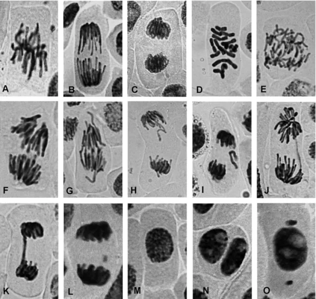
variety of chromosomal aberrations in mitotic cells of
A. cepaL. root tips (Figure 1) in which anaphases with spindle abnormalities and anaphases and telophases with vagrant chromosomes were the most frequent alterations; anaphase/telophase fragments and bridges and C-mitoses were less frequent.
The treatment with 10-9M Br-oxph significantly in-creased the percentage of chromosomal aberrations in mi-totic cells (by ~7-fold) compared to the controls (Table 1), but there were no significant changes with the two higher concentrations. During the 24 h recovery period after the 3 h incubation with 10-9M and 10-6M Br-oxph, the percent-age of chromosomal aberrations was ~5-fold higher than in the control cells; the highest concentration (10-3M) did not significantly affect the number of these aberrations. Similar responses were seen after the 48 h recovery period, al-though the increases were greater. Abnormalities (the pres-ence of micronuclei and binucleate cells) were also seen in
interphase cells (Figure 1). There were no significant changes in the percentage of abnormal interphase cells im-mediately after the 3 h incubation or during the 24 h and 48 recovery periods, except for an increase with 10-3 M Br-oxph after 24 h.
Br-oxph triggered apoptosis in bone marrow cells 3 h after administration. Light microscopy revealed typical signs of apoptosis,i.e., nuclear fragmentation and conden-sation (Figure 2), and this apoptotic effect persisted up to 48 h after treatment. Chromosomal aberrations were scored only when the frequency of mitotic cells was enough for de-termination of at least 50 well spread metaphases per ani-mal. When apoptosis was very extensive, it was impossible to determine the mitotic index and chromosomal aberra-tions. For this reason, Table 2 shows only the effect of the lowest dose of Br-oxph tested (2.82 x 10⁻³mg/kg; 24 h and 48 h after the treatment). This dose significantly reduced the mitotic index by 55% 48 h after the treatment, and there
was a significant increase (2-fold) in the level of chromo-somal aberrations in mitotic cells 24 h and 48 h after the in-jection of Br-oxph (Table 2). Chromatid breaks were the most frequent aberrations, but isochromatid breaks and centromeric and telomeric fusions were also observed; fragmentation was seen only 48 h after treatment. There was no change in the percentage of cells with gaps or in the number of tetraploid cells.
These results show that in both systems Br-oxph de-pressed cellular proliferation (mitosis) 24 h and 48 h after treatment. Interestingly, Br-oxph (10-6M and 10-3M) ap-peared to stimulate cell division inA. ceparoot tips imme-diately after a 3 h treatment, but this effect was transitory and was not seen after 24 h and 48 h. The decrease in the mitotic index indicates that Br-oxph can arrest cell growth. The suppression of mitotic activity is often used to assess cytotoxicity (Smaka-Kincl et al., 1996). The ability of Br-oxph to induce chromosomal aberrations inA. ceparoot tips and bone marrow cells after treatment for 3 h and dur-ing 24 h and 48 h of recovery agrees with the finddur-ings of Williams and Omoh (1996) and Miyamaeet al. (1997), who observed DNA damage after a 3 h exposure to other compounds.
Br-oxph was generally less genotoxic than the posi-tive control (MMS). The abnormalities caused by Br-oxph showed little concentration- or time-dependence. Interest-ingly, 10-3M Br-oxph caused fewer aberrations inA. cepa
immediately after the treatment and during the 24 h recov-ery than did concentrations of 10-9M and 10-6M. A lack of
concentration-dependence in the effects of other oxaphos-pholes has also been reported (Enchevet al., 1986). In the case of Br-oxph, the nonlinear relationship may reflect the influence of this compound on cell division. The increase in the percentage of chromosomal aberrations inA. cepa mi-totic cells 48 h after treatment with 10-6 M and 10-3 M Br-oxph correlated with the inhibition of cell division. A number of factors, such as compound solubility, rate of transport and biodistribution, and concentration at the tar-get site (which is influenced by time and cellular perme-ability), can modulate the time of occurrence of chemi-cally-induced aberrations (McFee and Tice, 1990). In addition, there was marked individual variation in the re-sponses to Br-oxph, which meant that in some experiments the changes observed were not significant.
There were differences in the chromosomal aberra-tions caused by Br-oxph in the two test systems. The occur-rence of abnormal anaphases and C-mitosis in A. cepa
indicated that spindle formation was adversely affected (El-Ghameryet al., 2000). According to Rank (2003), va-grant chromosomes are also indicators of spindle poison-ing. In bone marrow cells, chromatid breaks were the most frequent aberrations, whereas the number of tetraploid metaphases in bone marrow cells was unaffected by the treatment. These findings suggest a plant-specific action of Br-oxph on spindle formation.
Fusion between chromatids can be initiated by the si-multaneous breakage of two chromatids or by the loss of telomere capping (Gilleyet al., 2005). The bridges seen in
A. cepacells were also probably formed by breakage and fusion of chromosomes and chromatids (Türkoglu, 2007). The relatively low percentage of bridges and fragments in
A. ceparoot tips agreed with the relatively low percentage of cells with micronuclei (Krishna and Hayashi, 2000). The detection of a binucleate condition inAlliumindicated that Br-oxph solutions inhibited cytokinesis.
Apoptosis is an energy-dependent, genetically con-trolled process by which unnecessary or damaged cells die (Martin, 1993; Earnshaw, 1995). DNA damage can induce cell death and thus, the occurrence of apoptosis in mouse bone marrow cells was another indication of the
cytoto-Figure 2- Bone marrow nuclei of ICR mice treated with Br-oxph showed a typical apoptotic morphology that included fragmentation and condensa-tion: A - normal nucleus, B - condensed nucleus, and C - fragmented nu-cleus.
Table 2- Cytogenetic analysis of mouse bone marrow cells 24 h and 48 h after treatment with Br-oxph (2.82 x 10⁻³mg/kg).
Dose (mg/kg)
Time after treatment (h)
Mitotic index (%)
Metaphases scored
Type of aberration Cells with aberrations (%)#
Cells with gaps (%)
Tetraploid metaphases (%)
ICB CB c/c t/t Fr
NC 24 1.97±1.03 500 1 21 1 1 0 4.40±3.10 3.00±2.71 1.20±1.69
2.82 x 10⁻³ 24
1.48±1.13 350 5 28 2 1 0 9.14±3.44* 1.71±2.14 0.00±0.00*
48 0.89±0.57* 350 4 31 1 0 4 10.80±6.57* 2.00±1.15 0.29±0.76
PC 24 0.67±0.29** 400 28 106 1 2 0 20.44±8.99*** 2.25±1.98 0.44±1.33
The results are expressed as the mean±SD. *p£0.05, **p£0.01, and ***p£0.001 compared with the corresponding negative control (NC; 0.9% NaCl).
#
xicity of Br-oxph. Light microscopy showed the presence of apoptotic nuclei with an altered morphology with typical nuclear fragmentation and condensation, as described by others (Kam and Ferch, 2000; Gornevaet al., 2005). Ac-cording to Grishinet al.(2001), genotoxic stresses activate intracellular signaling molecules, which lead to growth ar-rest, DNA repair, and/or apoptosis. Although several of the pathways linking DNA damage to mitochondria-dependent and -independent mechanisms of death have been eluci-dated, the connectivity of these pathways is subject to regu-lation by various other poorly understood factors (Borgeset al., 2008).
In conclusion, the results of this study indicate cyto-oxicity and genotcyto-oxicity of Br-oxph inA. ceparoot tip cells and ICR mouse bone marrow cells, with the effects being observed up to 48 h after treatment for 3 h. Chromosomal aberrations provide a sensitive endpoint for assessing the genotoxicity of chemicals (Topashka-Anchevaet al., 2003) and, as shown here,A. cepamay be a sensitive biosensor for screening the genotoxicity of oxaphospholes. On the other hand, our data are in accordance with observation that rodent bioassays are useful for investigating the pharma-cokinetics, mechanisms of action, and differential toxicity of various chemicals (Roldan-Arjonaet al., 1991).
References
Angelov CM and Enchev DD (1987) 1,2-Alkadienephosphonic amidoesters and their cyclization with electrophilic re-agents. Phosphorus Sulfur Relat Elem 34:163-168. Blasiak J, Jaloszynski P, Trzeciak A and Szyfter K (1999)In vitro
studies on the genotoxicity of the organophosphorus insecti-cide malathion and its two analogues. Mutat Res 445:275-283.
Borges HL, Linden R and Wang JYJ (2008) DNA damage-induced cell death: Lessons from the central nervous system. Cell Res 18:17-26.
Earnshaw WC (1995) Nuclear changes in apoptosis. Curr Opin Cell Biol 7:337-343.
El-Ghamery AA, El-Nahas AI and Mansour MM (2000) The ac-tion of atrazine herbicide as an inhibitor of cell division on chromosomes and nucleic acids content in root meristems of
Allium cepaandVicia faba. Cytologia 65:277-287. Enchev D, Nicolov N and Angelov C (1986) Growth-regulating
activity of 2-N,N -dialkylamino-2,5-dihydro-1,2-oxaphos-phole 2-oxides. In: Proceedings of the IV International Sym-posium on Plant Growth Regulators, Pamporovo, Bulgaria, I:365-368.
Fiskesjö G (1985) TheAlliumtest as a standard in environmental monitoring. Hereditas 102:99-112.
Gilley D, Tanaka H and Herbert BS (2005) Telomere dysfunction in aging and cancer. Int J Biochem Cell Biol 37:1000-1013. Gorneva G, Mateva R, Gugova R and Golovinsky E (2005) The
study of the apoptogenic effect of pyrimidine derivatives on murine leukemia cells. Arch Oncol 13:62-64.
Grant WF (1999) Higher plant assays for the detection of chromo-somal aberrations and gene mutations - A brief historical background on their use for screening and monitoring envi-ronmental chemicals. Mutat Res 426:107-112.
Grishin AV, Azhipa O, Semenov I and Corey SJ (2001) Interac-tion between growth arrest-DNA damage protein 34 and Src
kinase Lyn negatively regulates genotoxic apoptosis. Proc Natl Acad Sci USA 98:10172-10177.
Ito Y and Ito M (2001) Suppressive effect of (-)-epigallocatechin gallate on aflatoxin B1-induced chromosome aberrations in rat bone marrow cells. J Health Sci 47:248-257.
Kam PCA and Ferch NI (2000) Apoptosis: Mechanisms and clini-cal implications. Anaesthesia 55:1081-1093.
Krishna G and Hayashi M (2000)In vivorodent micronucleus as-say: Protocol, conduct and data interpretation. Mutat Res 455:155-166.
Leblond L, Attardo G, Hamelin B, Bouffard DY, Nguyen-Ba N and Gourdeau H (2002) BCH-1868 [(-)-2-R-dihydroxy-phosphinoyl-5-(S) - (guanine-9’-yl-methyl) tetrahydrofu-ran]: A cyclic nucleoside phosphonate with antitumor activ-ity. Mol Cancer Ther 1:737-746.
Lieberman AD, Craven MR, Lewis MR and Nemenzo JH (1998) Genotoxicity from domestic use of organophosphate pesti-cides. J Occup Environ Med 40:954-957.
Martin SJ (1993) Apoptosis: Suicide, execution or murder? Trends Cell Biol 3:141-145.
McFee AF and Tice RR (1990) Influence of treatment to sacrifice time and the presence of BrdUrd on chemically-induced ab-erration rates in mouse marrow cells. Mutat Res 241:95-108. Miyamae Y, Zaizen K, Ohara K, Mine Y and Sasaki YF (1997) Detection of DNA lesions induced by chemical mutagens by the single cell gel electrophoresis (Comet) assay. I. Rela-tionship between the onset of DNA damage and the charac-teristics of mutagens. Mutat Res 393:99-106.
Preston RJ, Dean BJ, Galloway S, Holden H, McFee AF and Shelby M (1987) Mammalianin vivo cytogenetic assays. Analysis of chromosome aberrations in bone marrow cells. Mutat Res 189:157-165.
Rank J (2003) The method ofAlliumanaphase-telophase chromo-some aberration assay. Ekologija (Vilnius) 1:38-42. Repetto G, Jos A, Hazen MJ, Molero ML, del Peso A, Salguero
M, del Castillo P, Rodriguez-Vicente MC and Repetto M (2001) A test battery for the ecotoxicological evaluation of pentachlorophenol. Toxicolin Vitro15:503-509.
Roldan-Arjona T, Garcia-Pedrajas D, Luque-Romero F, Hera C and Pueyo C (1991) An association between mutagenicity of the Ara test ofSalmonella typhimuriumand carcinogenicity in rodents for halogenated aliphatic hydrocarbons. Muta-genesis 6:199-205.
Smaka-Kincl V, Stegnar P, Lovka M and Toman MJ (1996) The evaluation of waste, surface and ground water quality using theAlliumtest procedure. Mutat Res 368:171-179. Smee DF and Reist EJ (1996) Potent anti-murine cytomegalovirus
activity and reduced nephrotoxicity of ganciclovir cyclic phosphonate. Antimicrob Agents Chemother 40:1964-1966. Topashka-Ancheva M, Metcheva R and Teodorova S (2003) A comparative analysis of the heavy metal loading of small mammals in different regions of Bulgaria II: Chromosomal aberrations and blood pathology. Ecotoxicol Environ Saf 54:188-193.
Türkoglu S (2007) Genotoxicity of five food preservatives tested on root tips ofAllium cepaL. Mutat Res 626:4-14. Williams GO and Omoh LE (1996) Mitotic effects of the aqueous
leaf extract ofCymbopogon citratusinAllium ceparoot tips. Cytobios 87:161-168.
Associate Editor: Catarina S. Takahashi