GUILHERME MIRANDA TAVARES
β
-LACTOGLOBULIN AND LACTOFERRIN COMPLEX
COACERVATES: CHARACTERIZATION AND PUTATIVE
APPLICATIONS AS ENCAPSULATION DEVICE
Dissertation thesis presented to the Universidade Federal de Viçosa as part of the requirements of the PostGraduate Progam in Food Science and Technology, to obtain the title of Doctor Scientiae.
RENNES
FRANÇA
Ficha catalográfica preparada pela Seção de Catalogação e Classificação da Biblioteca Central da UFV
T
Tavares, Guilherme Miranda, 1988-
T231b β-lactoglobulin and lactoferrin complex coacervates : characterization and 2015 putative applications as encapsulation device / Guilherme Miranda Tavares. - Rennes, FR, 2015.
xxviii,176f. : il. (algumas color.) ; 29 cm. Orientador: Antônio Fernandes de Carvalho.
Tese (doutorado) – Universidade Federal de Viçosa. Referências bibliográficas: f.161-176.
1. Leite - Proteínas. 2. Beta-lactoglobulina. 3. Lactoferrina. 4.
Biomoléculas. 5. Encapsulação. I. Universidade Federal de Viçosa.
Departamento de Tecnologia de Alimentos. Programa de Pós-graduação em Ciência e Tecnologia de Alimentos. II. Título.
GUILHERME MIRANDA TAVARES
β
-LACTOGLOBULIN AND LACTOFERRIN COMPLEX
COACERVATES: CHARACTERIZATION AND PUTATIVE
APPLICATIONS AS ENCAPSULATION DEVICE
Dissertation thesis presented to the Universidade Federal de Viçosa as part of the requirements of the PostGraduate Progam in Food Science and Technology, to obtain the title of Doctor Scientiae.
APROVED: October 8th, 2015
André Matagne Christian Sanchez
Francis Canon Michel Desmadril
Saïd Bouhallab Thomas Croguennec
ii “The only source of knowledge is experience”
iii REMERCIEMENTS
Ce n’est vraiment pas facile pour moi d’écrire ces remerciements, c’est probablement la partie
de la thèse la plus compliquée à rédiger. Ces trois années de thèse ont été définitivement intenses. Quotidiennement, je me suis rendu compte de mon évolution personnelle et scientifique grâce à tous les moments partagés avec les gens extraordinaires que j’ai eu la
chance d’avoir à mes côtés.
La réalisation de ce projet de thèse n’aurait pas pu être possible sans le support financier du
Ministère Brésilien de Science et Technologie (au travers du programme Science sans Frontières du Conselho Nacional do Desenvolvimento Científico e Tecnológico – CNPq), de
l’INRA, de l’Université Européenne de Bretagne (Collège Doctoral International), du Conseil
Régional de Bretagne et de l’Université Fédérale de Viçosa. Merci beaucoup. Je remercie
également Joëlle Léonil de m’avoir accueilli au sein de son laboratoire, le STLO.
Tout au long de mon parcours académique j’ai eu de la chance d’être encadré par des
personnes que j’admire, pour la thèse ce n’était pas différent. Dans l’ordre dans lequel ils sont
« rentrés » dans ma vie : Antônio, Thomas et Saïd, MERCI.
Professor Antônio, merci beaucoup pour toute ton aide, tes encouragements et pour avoir cru
dans mon potentiel depuis 2006 quand j’ai rejoint ton équipe. Je n’aurais pas pu arriver où je
suis aujourd’hui sans toi.
Thomas, merci pour ta présence et pour ta patience, même quand j’arrivais avec mes
manuscrits pleins de fautes d’anglais et mes énormes phrases. Ce serait impossible de ne pas
vouloir travailler sur les protéines laitières après avoir suivi tes cours passionnés/passionnants sur la physico-chimie du lait.
Saïd, j’ai beaucoup appris avec toi pendant ces trois années. Même si tu n’as pas eu l’occasion
iv déterminantes pour mon avenir professionnel. Merci également d’avoir toujours priorisé ma formation en tant que chercheur par rapport à l’obtention des résultats.
J’espère sincèrement que vous avez apprécié autant que moi ces trois ans de travail.
Un grand merci également à Ken Mok de m’avoir accueilli et encadré pendant les trois mois
au sein de son labo à Dublin. Merci pour toutes nos discussions et ta curiosité sur mon travail.
J’espère que l’on se recroisera dans un futur proche.
Je ne peux pas continuer mes remerciements sans remercier Pascaline. C’est facile de mener
un projet de thèse lorsque l’on sait que l’on peut compter sur quelqu’un d’aussi compétent que
toi. Merci pour tous les moments partagés devant l’HPLC et l’ITC ou à purifier des protéines.
Merci pour tout ce que tu m’as appris et pour tout ce qu’on a pu apprendre ensemble.
Je remercie aussi Olivia Lerideau pour son engagement lors de son stage au sein de l’équipe
et Anne-Laure pour toutes nos discussions scientifiques. Anne-Laure, je te souhaite bonne continuation pour la suite de ta thèse.
Merci à André Matagne, Christian Sanchez, Francis Canon et Michel Desmadril d’avoir accepté de faire partie de mon jury de thèse.
Merci à Taco Nicolai, Denis Poncelet et à Sylvain Guyot pour tous leurs précieux conseils lors de mes comités de thèse. Merci à tous les membres de l’équipe ISF-PL Power (Coralie,
j’essaie aussi d’éterniser le nom) pour votre grande sympathie et pour toutes nos discussions
lors des réunions d’équipe. Un merci spécial à Stéphane, Marie-Hélène et Fanny.
v
Egalement, cette thèse n’aurait jamais pu être la même sans l’aide des personnes responsables
de tâches parallèles à la recherche qui sont tellement importantes. Un grand merci à Paulette, Jessica, Laurence, Danielle, Nathalie, Anne Giboulot, Marie-Claude, Dominique, Michel, Laurent, Christophe et Rachel.
Même si à priori ces remerciements sont pour la thèse, pour moi c’est impossible de dissocier la thèse du temps que j’ai passé en master. Pour moi, la thèse représente l’aboutissement de
ces quatre ans passés à Rennes. La fin d’une très belle partie de ma vie.
Je remercie donc le team poudre du STLO (Romain, Pierre et Anne Dolivet) pour tout que j’ai pu apprendre avec eux pendant mon master. Merci à tous mes collègues et voisins de bureau : Marielle, Samira, Kéra, Solène, Céline, Melanie, Claire, Christelle, Naaman... pour la bonne humeur et pour avoir su me supporter pendant tout ce temps (pas facile, j’en suis conscient). Merci encore à Naaman pour les longues discussions du vendredi après-midi, même pour celles dont la conclusion était : il faut refaire le monde. Ce sera toujours un grand plaisir pour moi de parler de science (pas que) avec toi.
Merci à tous les doctorants de l’unité pour les moments de détente lors des repas du midi et
pauses chez Paulette. D’ailleurs, merci encore Paulette de nous offrir tous les jours des bons
moments pendant les pauses…
Je remercie également tous ceux qui ont activement participé à ma vie pendant mon séjour en Irlande. Merci à Anna, Ji Yoon, Nadia, Nadine, Sara, Marcelo, Vanessa, Moacir et tous les
membres de l’IFSC Force, mon séjour était aussi agréable grâce à vous.
Une grande partie de mon temps « libre » pendant ces quatre ans de Rennes a été consacré aux activités du Jeong Tong Taekwondo dojang. Merci à tous les membres du club, avec vous
vi Tibo, Etienne, Vincent, Gaëtan, Lydie, Didier, Blandine, Judith, Mathilde, Kévin, Emilien, Nathalie, Kévin, Phuc-loi...PATCHOOO !
Après le travail et le sport, le troisième point clé de ma vie à Rennes : Merci énormément à tous les membres (et ex-membres) de la Goutte Pendante, du Square de la Rance et de
l’Alcoolocation pour tous les moments de détente. Merci à Samira pour la meta 10. Merci aux
Carinhosos pour avoir renforcé ma certitude sur l’effet papillon. Merci à Gaëtan pour ton
grand cœur. Merci à tous les grands amis que j’ai pu avoir pendant ce séjour à Rennes :
Samira, Arlan, Marilia, Lélia, Juliana, Gaëtan, Anne-C, Xaxa, Féfé, Carlos, Clarisse, Elise, Flavia, Guillaume, Livia, Rachid, Song, Andreza (meu monstro), Perrine, Andreas, Adèle, Priscilla, Naaman, Rosangela, Renam, Rozenn....
Finalement je tiens à remercier ma famille (pai, mãe, mã et Iago). Même loin vous avez
essayé d’être proches. Eu dedico a vocês esse diploma, sem vocês eu não teria chegado aqui.
C’est une très belle étape de ma vie qui finit. Je rentre au Brésil, prêt pour de nouvelles
vii
Table of Contents
TABLE OF FIGURES ... xiii
TABLE OF TABLES ... xix
LIST OF ABBREVIATIONS ... xx
RESUMO ... xxiv
RESUME ... xxv
ABSTRACT ... xxvi
THESIS OUTPUTS ... xxvii
1 GENERAL INTRODUCTION ... 1
2 REVIEW OF LITERATURE ... 5
2.1 Whey Proteins ... 6
2.1.1 β-lactoglobulin (β-LG) ... 7
2.1.2 α-lactalbumin (α-LA) ... 9
2.1.3 Bovine Serum Albumin (BSA) ... 10
2.1.4 Lactoferrin (LF) ... 10
2.2 Whey proteins as encapsulation devices ... 11
2.2.1 Binding properties of whey proteins ... 16
2.2.2 Encapsulation devices obtained through the “bottom-up” approach ... 18
2.2.3 Encapsulation devices obtained from a “top-down” approach ... 21
2.2.4 Supra-molecular structures with putative encapsulation properties ... 24
viii
2.3.1 Complex coacervation: principle... 27
2.3.2 Complex coacervation of globular proteins ... 29
2.3.3 Specific case of complex coacervation between β-LG and LF ... 35
RESULTS AND DISCUSSION ... 37
3 CHAPTER 1: Multiscale characterization of Lactoferrin and β-lactoglobulin complex coacervation ... 38
PART 1: SELECTIVE COACERVATION BETWEEN LACTOFERRIN AND THE TWO ISOFORMS OF β-LACTOGLOBULIN ... 39
3.1 Introduction ... 42
3.2 Materials and Methods ... 45
3.2.1 Reagents and stock solutions ... 45
3.2.2 Turbidity measurements ... 46
3.2.3 Phase boundaries of coacervation ... 46
3.2.4 Isothermal titration calorimetry (ITC) ... 47
3.3 Results ... 48
3.3.1 Optimal pH for coacervation ... 48
3.3.2 Formation of coacervates as a function of protein concentration ... 50
3.3.3 Protein recovery and stoichiometry in formed coacervates ... 52
3.3.4 βLG and LF interaction at molecular level ... 56
3.4 Discussion ... 58
3.4.1 Selective interaction of LF with β-LG isoforms ... 58
ix
3.4.3 Proposed mechanism ... 63
3.5 Conclusion ... 64
3.6 Acknowledgements ... 65
PART 2: MOLECULAR MECHANISM OF β-LACTOGLOBULIN AND LACTOFERRIN COMPLEX COACERVATION ... 66
3.7 Introduction ... 69
3.8 Experimental section ... 71
3.8.1 Sample preparation: Reagents and Solutions ... 71
3.8.2 Preparation of β-LG-LF coacervates ... 71
3.8.3 Evaluation of β-LG and LF heterocomplex by rigid docking ... 72
3.8.4 FRAP: analysis of β-LG binding to LF ... 73
3.8.5 NMR: Estimation of hydrodynamic radius and relative abundance of the different β-LG/LF complexes ... 76
3.9 Results ... 77
3.9.1 β-LG and LF complexes: rigid Docking ... 77
3.10 β-LG binding within coacervate phase: FRAP analysis ... 81
3.10.1 Rotation diffusion coefficients show the presence of three different complexes: NMR analysis ... 83
3.11 Discussion ... 87
3.11.1 Building blocks of the coacervates ... 88
x
3.11.3 Specific thermodynamic equilibrium governs the stability of the coacervates .. 91
3.12 Acknowledgements ... 92
3.13 Additional Data: Characterization of the coacervates thermal-stability ... 93
4 CHAPTER 2: How the presence of a small molecule affects the complex coacervation between lactoferrin and -Lactoglobulin ... 95
PART 1: HOW THE PRESENCE OF ANS AFFECTS THE COMPLEX COACERVATION BETWEEN LACTOFERRIN AND β-LACTOGLOBULIN ... 96
4.1 Introduction ... 99
4.2 Materials and Methods ... 101
4.2.1 Reagents and Solutions ... 101
4.2.2 Preparation of the coacervates ... 102
4.2.3 Isothermal titration calorimetry (ITC) ... 102
4.2.4 Hydrodynamic diameter (Dh) measurement ... 103
4.2.5 ζ-potential measurement ... 104
4.2.6 Turbidity measurements ... 104
4.2.7 Optical microscopy observation ... 105
4.2.8 Quantification of proteins and ANS in dense and coacervate phases ... 105
4.3 Results ... 105
4.3.1 Characterization of the ANS-β-LG and ANS-LF interactions ... 105
4.3.2 Coacervation process in the presence of ANS ... 108
4.3.3 Partition of proteins and ANS between dilute and coacervate phases ... 110
xi
4.4.1 ANS-LF binding versus β-LG-LF coacervation ... 112
4.5 Conclusion ... 116
4.6 Acknowledgements ... 117
PART 2: HOW THE PRESENCE OF FOLIC ACID AFFECTS THE COMPLEX COACERVATION BETWEEN LACTOFERRIN AND β-LACTOGLOBULIN ... 118
4.7 Introduction ... 120
4.8 Material and Methods ... 122
4.8.1 Reagents and Solutions ... 122
4.8.2 Isothermal titration calorimetry (ITC) ... 122
4.8.3 Quantification of free and bound FA ... 123
4.8.4 Measurement of hydrodynamic Diameter (Dh) ... 124
4.8.5 ζ-Potential measurement ... 124
4.9 Results ... 125
4.9.1 Isotherm of FA-LF interaction ... 125
4.9.2 Influence of physicochemical parameters on FA-LF interaction ... 127
4.9.3 Independent determination of the FA-LF binding parameters ... 128
4.9.4 Characterization of FA-induced LF nanoparticles ... 129
4.10 Model to characterize the energy of FA-LF interaction ... 131
4.10.1 Deduction of the theoretical FA-LF binding isotherm ... 133
4.10.2 Estimation of saturated LF concentration for each ITC injection ... 134
4.10.3 Deduction of the energy contribution of the self-association step ... 136
xii
4.11.1 Overview of the ITC isotherms and the proposed model ... 137
4.11.2 Specific features of FA-LF nanoparticles ... 139
4.12 Conclusion ... 140
4.13 Acknowledgements ... 141
4.14 Supporting Information ... 141
4.14.1 Determination of saturated LF concentration after each ITC injection... 142
4.14.2 Determination of the energy contribution of saturated LF self-association ... 144
4.15 Additional Data: Effect of FA on β-LG – LF complex coacervation ... 145
5 FINAL CONSIDERATIONS ... 146
5.1 General Discussion ... 147
5.1.1 Mechanism of -LG-LF coacervation ... 147
5.1.2 Coacervation in the presence of small ligands ... 152
5.2 General Conclusion & Perspectives ... 155
REFERENCES ... 161
xiii TABLE OF FIGURES
Figure 2.1. -LG dimer (RCSB PDB code: 2Q2M). The arrows show the dimer interface and
the position of the AA64, an important substitution between isoforms A and B. ... 9 Figure 2.2. Tertiary structure of (Alactalbumin (RCSB PDB code: 1ALC), (BBSA
(RCSB PDB code: 3V03) and (CLactoferrin (RCSB PDB code: 1BLF). ... 11 Figure 2.3. Illustration of encapsulation strategies applied to whey proteins (adapted from
Tavares et al. [4]). ... 12 Figure 2.4. SEM cross-section image (12,000 x magnification) of a whey protein-isolate gel
with encapsulated Lactobacillus rhamnosus (adapted from Reid et al. [87]) ... 23 Figure 2.5. Different supramolecular structures obtained from whey proteins. (A) TEM
micrograph of whey protein isolate (WPI) obtained from 2 % of WPI at pH 2 solution heated
at 80 °C for 16h (adapted from Loveday et al. [104]). (B) AFM micrograph of -LG ribbons
obtained after heating -LG (2 %wt.) at 90 °C and pH 2 (adapted from Lara et al. [95]). (C)
Polarised light microscopy micrograph of -LG spherulites (adapted from Krebs et al. [105]).
(D) TEM micrograph of hydrolysed a-LA nanotubes (adapted from Graveland-Bikker et al. [106]). (E) Confocal scanning laser micrograph of heteroprotein complex coacervates obtained from apo a-LA (0.2 mM) and LYS (0.2 mM) mixture (adapted from Nigen et al. [107]). ... 26 Figure 2.6. (A) Glass beads glued together using proteins complex coacervates produced by
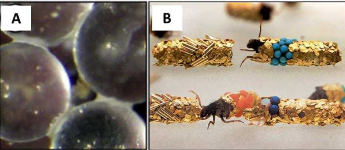
sandcastle worm (adapted from Stewart et al. [112]); and (B) Gold and gemstones jewelry built by Caddisfly Larvae (adapted from Matus [113]) ... 28 Figure 2.7. Summary of the interactions, supramolecular structure formation and phase
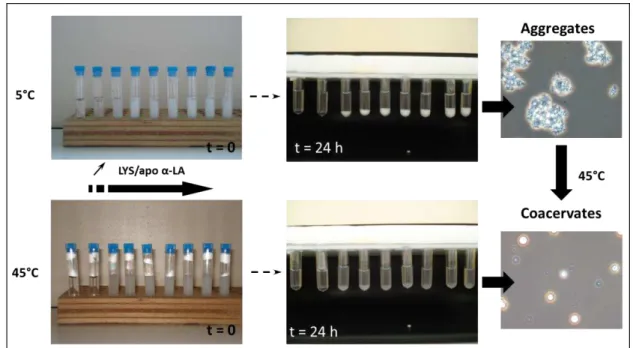
separation of apo -LA-LYS mixture at 5 and 45 °C ranging from 0.05 to 2 LYS-LA
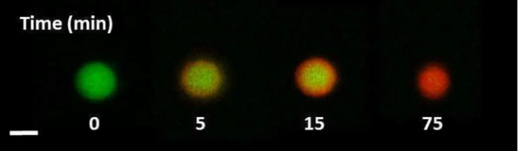
xiv Figure 2.8. Internal dynamic of apo -LA-LYS coacervates and time-dependent protein exchange between coacervates and dilute phase (adapted from Nigen et al. [107]). ... 32 Figure 2.9. Proposed mechanism explaining selective formation of coacervates following the
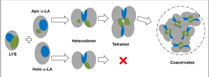
interaction of lysozyme with calcium depleted -LA (apo -LA - LYS) but not with holo
-LA (adapted from Salvatore et al. [131]). Blue zones indicate the binding sites leading to the heterodimer formation, green zones indicate the binding site required to the formation of tetramer. ... 33
Figure 3.1. Change in turbidity of mixtures containing 40 µM of LF and 500 µM of (■) βLG
A, (●)βLG B or (▲)βLG A+B in 10 mM MES buffer at different pHs. ... 49
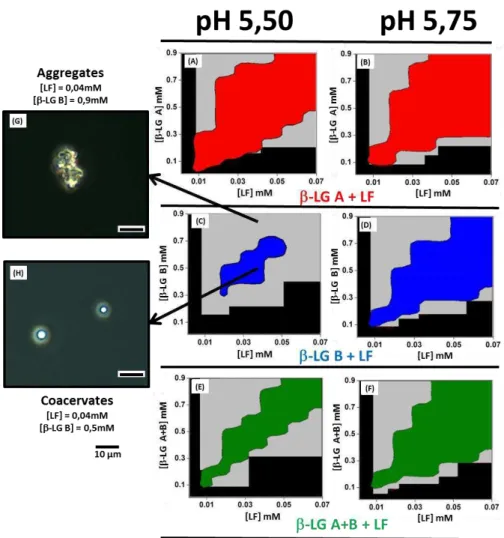
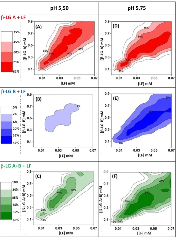
Figure 3.2. Phase boundaries of co-assembly of LF with LG isoforms at pH 5.50 and pH
5.75. (A,B): LF - LG A; (C,D): LF - LG B; (E,F) LF - LG AB. Black zones: domains
without detectable supramolecular structures; Gray zones: aggregation domains. Red, Blue and Green zones: coacervation domains. Optical microscopy of aggregates formed by mixing
for example 40 µM LF and 900 µM LG B at pH 5.50 (G) versus coacervates formed by
mixing for example LF 40 µM and LG B 500 µM at pH 5.50 (H)... 51
Figure 3.3. Superposition of the coacervation domains obtained by mixing LF and LG A
(red), LG B (blue) or LG A+B (green) at pH 5.50 (A) and pH 5.75 (B). Co-assembly
experiments were conducted at 25°C in MES buffer 10 mM. ... 52
Figure 3.4. Fraction of LF recovered in the coacervates (dense phase) formed with LG A
(A and D), LG B (B and E) or LG A+B (C and F) at pH 5.50 and pH 5.75 in 10 mM
MES buffer and at 25°C. ... 54 Figure 3.5. Average of the relative proportion of LG A and LG B quantified in the
xv Figure 3.6. Binding isotherms of LF with (■) LG A or (■) LG B and dissociation isotherms upon dilution of (□) LG A and (□) LG B dimers. LF 0.1 mM was titrated with
successive injections of -LG 2 mM. ITC experiments were conducted at 25 °C in 10 mM MES buffer at pH 5.50 (A) and pH 5.75. ... 57 Figure 3.7. Proposed steps for the coacervation process involving LF and β-LG co-assembly.64
Figure 3.8. Molecular docking between -LG2 and LF (A, B), and between -LG2 and
already formed LF-LG2 complex (C, D). ... 78
Figure 3.9. Molecular docking between trimeric and pentameric complexes of LF and -LG2.80
Figure 3.10. FRAP analysis of the FITC labeled -LG within the coacervate phase. ... 82
Figure 3.11. NMR signal Assignments. ... 84
Figure 3.12. Assignment of NMR signals to different complexes in the coacervates. ... 86
Figure 3.13. (A) DSC thermograms of the -LG (~300 g/L), LF (~ 300 g/L) and the
coacervate phase (-LG ~150 g/L + LF ~150 g/L). (B) Image of the coacervate phase. ... 94
Figure 4.1. Binding isotherms of the titration of LF 0.05 mM and -LG 0.05 mM with 4.25
mM ANS in MES buffer pH 5.5 at 25 °C. LF without () and with () 50 mM NaCl; -LG
without NaCl (●). Injection of ANS in the MES buffer without (□) and with 50 mM NaCl (Δ).107
Figure 4.2. Evolution of the hydrodynamic diameter Dh and -potential of the complexes as a
function of the initial ANS/LF molar ratio at 25°C without added NaCl (, □) and in the
presence of 50 mM of NaCl (). ... 108
Figure 4.3. Evolution of the turbidity of mixtures containing 0.5 mM of -LG, 0.05 mM of
LF and variable concentrations of ANS. Different ANS/LF molar ratios were tested: 0 (straight line), 10 (dashed line), 20 (dotted line) and 45 (dash-dotted line). These results
xvi Figure 4.4. Optical microscopy images in phase contrast mode (A, C, E) and epifluorescence
mode (B, C, E) for mixtures containing 0.5 mM of -LG, 0.05 mM of LF and different
concentrations of ANS according to mixing order 2. Different ANS/LF molar ratios were tested: 25 (A, B), 30 (C, D) and 40 (E, F). ... 110 Figure 4.5. Evolution of recovered ANS (in %) and corresponding ANS/LF molar ratio in the
dense phase (A) and of Dh of the structures remaining in the dilute phase after centrifugation of the mixtures (B) as a function of the initial ANS/LF molar ratio. Protein concentrations
were 0.05 mM for LF and 0.5 mM for-LG. The squares, circles and triangles correspond to
mixing order 1, 2 and 3, respectively. ... 112 Figure 4.6. Proposed mechanism describing how increasing ANS concentration (represented
by the blue circle) affects -LG-LF coacervation process at constant protein concentration.115
Figure 4.7. Representative raw data (upper panel) and binding isotherm (bottom panel) of the
titration of 0.01 mM LF with successive injections of FA stock solution. The experiment was carried out at 20°C in 10 mM MES buffer, pH 5.5. ... 126 Figure 4.8. Experimental binding isotherms of the interaction of FA with LF at various: (A)
Temperatures, titration of LF 0.01 mM with 1 mM FA; (B) Cell LF concentrations, titration with 1 mM FA at 20°C; (C) ionic strength, titration of LF 0.01 mM with 1 mM FA at 20°C. All experiments were carried out in 10 mM MES buffer, pH 5.5. ... 128 Figure 4.9. Scatchard plot for the interaction between LF (10 µM) and FA (0-150 µM) in 10
mM MES buffer, pH 5.5. ... 129
Figure 4.10. (A) Evolution of the hydrodynamic diameter Dh (●) and -potential () as a function of the FA/LF molar ratio at 25°C. (B) Particle size in MES 10 mM pH 5.5, 25 °C for
xvii Figure 4.11. Algorithm used to develop the proposed model for FA binding to LF and
subsequent self-association of FA/LF complexes. ... 133 Figure 4.12. Superimposition of the experimental () and theoretical (●) FA/LF binding isotherms. Experimental titration of 0.01 mM LF was performed with FA in 10 mM MES buffer pH, 5.50, at 20°C. ... 134 Figure 4.13. (A) Evolution of [LF saturated] as a function of FA/LF molar ratio () and the sigmoidal curve fitted to the experimental data (Δ). (B) Concentration of LF which saturates
at each ITC injection as a function of FA/LF molar ratio (data obtained from the fitted sigmoidal curve). ... 135 Figure 4.14. Superimposition of experimental binding isotherm (), theoretical FA/LF
binding isotherm (●), theoretical energy contribution of the LF self-association () and the
theoretical global isotherm (binding + self-association) (♦). Results were obtained from the raw ITC data acquired at 20°C and (A) [LF] = 0.01mM and (B) [LF] = 0.02mM. ... 136 Figure 4.15. Proposed mechanism for the formation of folic acid/lactoferrin nanoparticles of
finite size. ... 140 Figure 4.16. Raw data (upper panel) and binding isotherm (bottom panel) of the titration of
0.06 mM LF with successive injections of sodium citrate stock solution. The experiment was carried out at 25°C in 10 mM MES buffer, pH 5.5. ... 141 Figure 4.17. Simulation program developed with software R (version 3.1.1) to determine the
fraction of saturated LF for specific FAbound/LF molar ratio. ... 143 Figure 4.18. Optical microscopy images in phase contrast mode for mixtures containing 0.16
mM of -LG, 0.016 mM of LF and different concentrations of FA according to mixing order
2. Different FA/LF molar ratios were tested: (A) 0, (B) 1, (C) 2 and (D) 10. Barre = 10 µm.145
Figure 5.1. Relative stability of different -LG2 - LF complexes according to the docking
xviii Figure 5.2. Hypothetic scheme of the self-assembly of trimers LF -LG2 formed by LF and
-LG A homodimers. Representation of the role of the electronegative region on the -LG A
homodimers interface promoting the self-assembly into coacervates... 152 Figure 5.3. Binding isotherms of the titration of LF 0.05 mM with 4.25 mM ANS (A) and LF
0.01 mM with 1.0 mM FA (B). LF with (and □) and without () 50 mM of NaCl. ... 153
Figure 5.4. Phase boundaries of complex coacervation of LF with -LG A at pH 5.5. (A)
xix TABLE OF TABLES
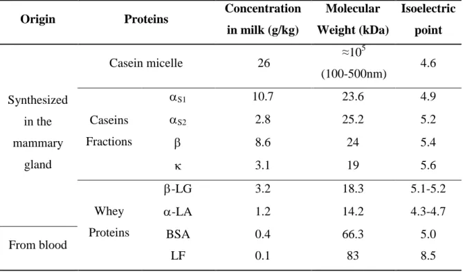
Table 2.1. Physical-chemical properties of bovine milk proteins ... 7
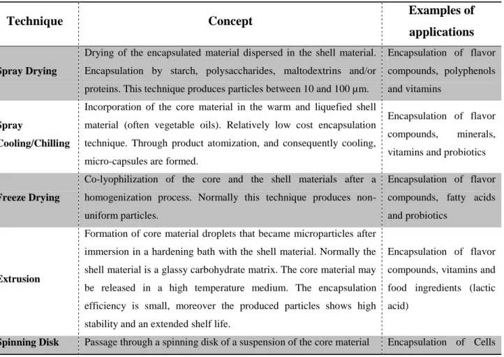
Table 2.2. Main conventional techniques used for the encapsulation of bioactive and food ingredients. Please refer to the following papers for more details: [34, 35, 40, 41]. ... 13
Table 2.3. Main studies concerning the spontaneous co-assembly of oppositely charged food proteins (adapted from Bouhallab and Croguennec [15]). ... 34
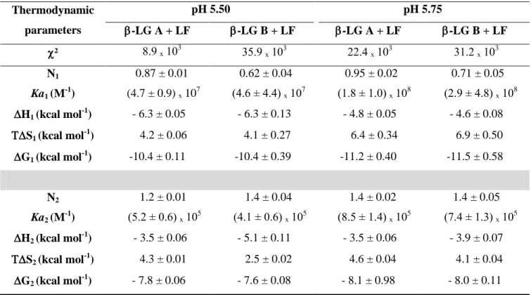
Table 3.1. Thermodynamic parameters of the interaction of LF with the two β-LG isoforms at 25°C in 10 mM MES buffer. ITC data were fitted with two independent binding sites. ... 57
Table 3.2. Comparison of the experimental conditions and the main results obtained here and by others on the coacervation of β-LG/ LF system. ... 62
Table 3.3. FRAP fitting parameters ... 82
Table 3.4. Abundance (w/w) and dynamics of complexes in the coacervate phase. ... 87
xx LIST OF ABBREVIATIONS
AA: amino acid
-CN: -casein
AFM: atomic force microscopy Ala: alanine
-LA: α-lactalbumin
ANS: 8-anilinonaphthalene-1-sulfonic acid
Apo--LA: apo -lactalbumin Asp: aspartic acid
-CN: -casein
-LG: β-lactoglobulin native
-LG A: β-lactoglobulin isoform A
-LG B: β-lactoglobulin isoform B
-LG (A+B): β-lactoglobulin equimolar mixture of isoforms A and B
BSA: bovine serum albumin
Ceq: concentration of bound fluorescent molecule
CNPq: Conselho Nacional de Desenvolvimento Científico e Tecnológico CP: cross polarization
Cys: cysteine
Deff: effective diffusion Df: pure diffusion
G: Gibbs free energy
H: change in enthalpy
xxi DMSO: dimethyl sulfoxide
S: change in entropy
DSC: differential scanning calorimetry EGCG: epigallocathechin-3-gallate FA: folic acid
Feq: concentration of unbound fluorescent molecule FITC: Fluorescein isothiocyanate
frap(t): fluorescence intensity at time t
FRAP: fluorescence recovery after photo bleaching FRET: Fluorescence resonance energy transfer Gly: glycine
HAMLET/BAMLET: human/bovine α-lactalbumin made lethal to tumor cells
Holo--LA: apo -lactalbumin
HPLC: high pressure liquid chromatography HSA: human serum albumin
INRA: Institut National de la Recherche Agronomique ITC: isotherm titration calorimetry
Ka: association constant / binding constant Kb: Boltzmann constant
-CN: -casein
Kd: dissociation constant
koff: dissociation rate
kon: association rate
l: light path length
xxii LYS: lysozyme
MAS: magic angle spinning
MES: 4-Morpholineethanesulfonic acid Mw: molecular weight
: solvent viscosity
n: stoichiometry
NMR: Nuclear Magnetic Resonance
PDADMAC: poly-dimethyldiallyammonium chloride RBITC: Rhodamine B isothiocyanate
Rh: hydrodynamic radius
RP-HPLC: reverse phase –HPLC SA: serum albumin
SANS: Small-angle neutron scattering SAXS: Small-angle X-ray scattering SDS: sodium dodecyl sulfate
T: temperature
: turbidity
T1 and T2: spin relaxation times
c: correlation time
TEM: transmission electronic microscopy Tmax: denaturation temperature
TTMA: cationic gold nanoparticle coupled to 3,6,9,12-tetraoxatricosan-1-aminium, 23-mercapto-N,N,N-trimethyl
Val: valine
xxiii WPC: whey protein concentrate
WPI: whey protein isolate
[FA bound]: bound folic acid concentration [FA free]: unbound folic acid concentration [FA]: initial folic acid concentration
xxiv RESUMO
TAVARES, Guilherme Miranda, D.Sc., Universidade Federal de Viçosa, outubro de 2015. Coacervados complexos de -lactoglobulina e lactoferrina: caracterização e aplicação potencial para a encapsulação de bioativos. Orientador: Antônio Fernandes de Carvalho; Coorientadores: Saïd Bouhallab, Thomas Croguennec, Eduardo Basílio e Ítalo Tuler Perrone.
xxv RESUME
TAVARES, Guilherme Miranda, D.Sc., Universidade Federal de Viçosa, Octobre 2015. Coacervats de -lactoglobuline et de lactoferrine : caractérisation et
application potentielle pour l’encapsulation de bioactifs. Encadrant: Antônio Fernandes de Carvalho; Co-encadrants: Saïd Bouhallab, Thomas Croguennec, Eduardo Basílio e Ítalo Tuler Perrone.
Le bénéfice de l’encapsulation des molécules bioactives a séduit les industries
agroalimentaires depuis plusieurs décennies et constitue toujours un levier de développement pour des produits innovants. Plus récemment des études ont montré la capacité de protéines alimentaires de charge opposée à s’assembler en microsphères par coacervation complexe. La compréhension des forces gouvernant
le processus de coacervation complexe entre protéines et l’influence exercée par la
présence de petits ligands (bioactifs) demeurent des prérequis pour l’utilisation des
coacervats complexes de protéines comme agent d’encapsulation. Dans ce contexte, l’objectif de mon projet de thèse a été de comprendre le mécanisme de coacervation
complexe entre une protéine chargée négativement, la -lactoglobuline (-LG), et une protéine chargée positivement, la lactoferrine (LF), issues du lactosérum en absence et en présence de petits ligands. Les conditions optimales de coacervation entre la -LG et la LF ont été définies entre pH 5.4 et 6.0 ainsi qu’en présence d’un excès de -LG. La LF a présenté une coacervation préférentielle avec le variant A de la -LG qui se distingue du variant B par la substitution de 2 acides aminés. Au niveau moléculaire, deux sites de fixation de la -LG sur la LF ont été identifiés. En
outre, par la mesure d’une part des coefficients de diffusion rotationnel et d’autre part
de la cinétique de diffusion des entités moléculaires constituant les coacervats, il est suggéré que ces derniers sont formés à partir de -LG libre, de pentamère, LF( -LG2)2, ainsi que des entités plus larges, (LF-LG2)n. Afin d’évaluer l’effet de la présence de petits ligands sur la coacervation complexe entre la -LG et la LF, des
ligands modèles, l’un hydrophobe (ANS), l’autre hydrophile (acide folique) ont été utilisés. Dans les conditions expérimentales testées ces deux ligands n’ont pas d’affinité pour la -LG, mais après interaction avec la LF ils sont capables d’induire son auto-association en nanoparticules. En concentrations élevées de ligands, la coacervation complexe entre la -LG et la LF est perturbée et une transition vers un
xxvi ABSTRACT
TAVARES, Guilherme Miranda, D.Sc., Universidade Federal de Viçosa, October, 2015. -lactoglobulin and lactoferrin complex coacervates: Characterization and putative applications as encapsulation device. Advisor: Antônio Fernandes de Carvalho; Co-advisor: Saïd Bouhallab, Thomas Croguennec, Eduardo Basílio e Ítalo Tuler Perrone.
Encapsulation of bioactives has been used by the food industries for decades and represents a great potential for the development of innovative products. Given their versatile functional properties, milk proteins in particular from whey have been used for encapsulation purposes using several encapsulation techniques. In parallel, recent studies showed the ability of oppositely charged food proteins to co-assemble into microspheres through complex coacervation. Understanding the driving forces governing heteroprotein coacervation process and how it is affected by the presence of ligands (bioactives) is a prerequisite to use heteroprotein coacervates as encapsulation device. In this context, the objective of my thesis work was to understand the mechanism of complex coacervation between -lactoglobulin (-LG) and lactoferrin (LF) in the absence and presence of small ligands. The conditions of optimal -LG - LF coacervation were found at pH range 5.4-6 with a molar excess of
xxvii THESIS OUTPUTS
Review
Tavares, G.M., Croguennec, T., Carvalho, A.F., Bouhallab, S. (2014). Milk proteins as encapsulation devices and delivery vehicles: Applications and trends. Trends in Food Science and Technology, 37(1), 5 – 20.
Published or Submitted papers
Tavares, G.M., Croguennec, T., Hamon, P., Carvalho, A.F., Bouhallab, S. (2015). Selective coacervation between lactoferrin and the two isoforms of beta-lactoglobulin. Food Hydrocolloids, 48, 238 – 247.
Tavares, G.M., Croguennec, T., Lê, S., Lerideau, O., Hamon, P., Carvalho, A.F., Bouhallab, S. Binding of folic acid induces specific self-association of lactoferrin into nanoparticles: thermodynamic characterization. Submitted to Langmuir.
Papers in preparation
Peixoto, P., Tavares, G.M., Croguennec, T., Nicolas, A., Hamon, P., Roiland, C., Bouhallab, S. Molecular mechanism of proteins complex coacervation: Case of β -lactoglobulin and lactoferrin. In preparation for submission to Soft Matter.
Tavares, G.M., Croguennec, T., Lerideau, O., Hamon, P., Carvalho, A.F., Bouhallab, S. How the presence of a small molecule affects the complex coacervation between lactoferrin
and β-lactoglobulin. In preparation for submission to Food Chemistry.
Oral presentations
Tavares, G.M., Croguennec, T., Hamon, P., Carvalho, A.F., Bouhallab, S.
Biopolymers 2013: Biopolymer assemblies for material design (December 2013). Specificity
xxviii Bouhallab, S., Tavares, G.M., Hamon, P., Croguennec, T. Minerals & Dairy Products
Symposium (February 2014). Specific binding of Minerals affects spontaneous co-assembly of
globular proteins. Auckland, New Zealand.
Tavares, G.M., Croguennec, T., Hamon, P., Carvalho, A.F., Bouhallab, S. Food
Structure and Functionality forum Symposium: From molecules to functionality (April 2014).
Particular spontaneous assembly of lactoferrin with the two variants A and B of beta-lactoglobulin. Amsterdam, Netherlands.
Tavares, G.M., Croguennec, T., Hamon, P., Carvalho, A.F., Bouhallab, S. 6es
Rencontres Biologie-Physique du Grand Ouest (June 2014). Co-assemblage de la
beta-lactoglobuline et de la Lactoferrine en microsphères. Le Mans, France.
Tavares, G.M., Croguennec, T., Carvalho, A.F., Bouhallab, S. VII Workshop de
laticinios (October 2014). Proteinas Lácteas como dispositivos de encapsulação: aplicações e
tendências. Viçosa, Brazil.
Tavares, G.M., Croguennec, T., Hamon, P., Peixoto, P., Carvalho, A.F., Bouhallab, S.
6th International Symposium on Delivery of Functionality in Complex Food-Systems (July
2015). Protein co-assembly for the encapsulation of bioactives. Paris, France. Poster presentations
Tavares, G.M., Croguennec, T., Peixoto, P., Hamon, P., Carvalho, A.F., Bouhallab, S.
6th International Symposium on Delivery of Functionality in Complex Food-Systems (July
2015). Amino acid substitution on -lactoglobulin changes its coacervation properties with
lactoferrin. Paris, France. Supervision
Co-supervision of undergraduate student (Biologie-Environnement/Université
1
2 Milk is a product secreted by the females of all mammals whose objective is to provide to the newborns all necessary elements for their survival and development during the first stages of life [1]. After the domestication of animals, milk became an important part of the human diet, even for adults. Nowadays, it is estimated that in USA, several European countries, Canada, Australia and New Zealand, 30% of the protein diet is supplied by the consumption of dairy products [2]. Thanks to the domestication of animals and the better control of milk production, different milk products could be developed aiming milk conservation or the diversification of sensory aspects. However, for centuries the knowledge associated with milk was empirical. A great example of how the development of scientific knowledge changed the dairy market and industry is the whey.
Smithers [3] well described in his review “Whey and whey proteins - From gutter-to-gold” how a set of factors (e.g. environmental, scientific and technological advances...) contributed to change the image and perception of whey by the consumers and industries. The whey which was considered as an industrial by-product is now recognized for its high biological value and the technical and functional properties of its proteins.
3 Several different encapsulation techniques using whey proteins, such as spray drying, has been applied at industrial scale for decades. It is estimated that the encapsulation applied to food represented in 2014 a market of around 3 billion of dollars and a turnover of more than 5 billion of dollars is expected in 2020 [5]. The use of new and innovative whey proteins based supramolecular structures as encapsulation devices is expected to be more interesting compared to the traditional encapsulation techniques. These supramolecular structures allows a better control of the assembly and disassembly process, essential for the delivery and control of release of bioactives. Special attention is given to coacervates (microspheres) which require very low energy to be formed, being durable potential encapsulation devices. All these factors match with the consumer demands for healthy and sustainable food systems.
In this context, two main objectives were assigned to the work of this thesis: (i) to understand
the mechanism of coacervation process between two oppositely charged whey proteins:
-lactoglobulin (LG) and Lactoferrin (LF); (ii) to determine how the presence of small
ligands (bioactive) affects the process of coacervation between these two proteins and therefore to extract some important parameters conditioning the use the heteroprotein coacervates as encapsulation devices.
This document is divided into 3 distinct sections:
4 - Results and Discussion section is divided into two chapters, each divided into two parts.
The first chapter presents the study of the complex coacervation between -LG and LF.
Different scales were explored; the first part focuses on the macro/mesoscopic study, identifying the optimal conditions for coacervation. This part was published in 2015 in Food Hydrocolloids [6]. The second part explores the interaction between these two proteins at
molecular level, a mechanism leading to LG - LF coacervation is proposed. These results
will be submitted soon to Soft Matter.
The second chapter explores the interaction of small ligands (models for bioactive molecules) with the proteins and how they affect the coacervation process between the two proteins. The first part focuses on the impact of ANS (chosen as model molecule to mimic a hydrophobic bioactive molecule) on the complex coacervation between -LG and LF. This first part is in
preparation for submission to Food Chemistry. The second part describes how a hydrophilic charged bioactive, i.e. folic acid, interacts and affects the properties of the two proteins. The obtained results were submitted for publication in Langmuir.
5
6 Encapsulation technologies that have been used for a long time in the pharmaceutical industry for drug delivery applications offer a real opportunity for the food industry. Encapsulation represents a means to develop innovative products to satisfy the growing demand of the consumer for foods with health and well-being benefits. For some food applications, encapsulation can be performed using relatively simple operations, such as emulsions, suspensions, gels and solid matrices. Due to their high abundance, nutritional value and high acceptance by consumers, whey proteins have largely been tested to design encapsulation devices, owing to their versatility and excellent functional properties. Special attention is given to the novel potential of heteroprotein complex coacervates, reversibly co-assembled protein supramolecular structures.
2.1 Whey Proteins
Proteins in milk are divided into two main groups: caseins and whey proteins (Table 2.1). Caseins are the fraction of proteins that precipitate at pH 4.6 and are often considered as intrinsically unstructured proteins associated as micelles with calcium phosphate in milk [7]. The caseins are synthesized exclusively in the mammary glands, suggesting that one of their functions is to provide amino acids (AA) and soluble calcium required for the development of the neonate [1]. Besides this function, caseins allow milk to be supersaturated in calcium phosphate, due to their capacity to bind divalent and multivalent ions [1, 8].
Whey proteins are typically globular proteins that exhibit various biological functions including source of essential amino acids, transport of molecules, co-factors for enzymes. β
7
Table 2.1. Physical-chemical properties of bovine milk proteins
Origin Proteins Concentration
in milk (g/kg)
Molecular Weight (kDa) Isoelectric point Synthesized in the mammary gland
Casein micelle 26 ≈10
5
(100-500nm) 4.6
Caseins Fractions
S1 10.7 23.6 4.9
S2 2.8 25.2 5.2
8.6 24 5.4
3.1 19 5.6
Whey Proteins
-LG 3.2 18.3 5.1-5.2
-LA 1.2 14.2 4.3-4.7
From blood BSA 0.4 66.3 5.0
LF 0.1 83 8.5
2.1.1 β-lactoglobulin (β-LG)
-LG, although absent in human milk, is the major whey protein in cow milk (~ 3.2 g/kg). It
belongs to the lipocalin family of proteins because of its ability to bind small hydrophobic molecules into its internal cavity [1]. It has 162 amino acids (AA), a molecular weight of 18.3 kDa and presents more than ten known genetic variants, although isoforms A and B are the most common [10]. -LG A and -LG B differ only by the substitution of two amino acids:
AA64 corresponds to an aspartic acid in -LG A and to a glycine -LG B, in addition, AA118
corresponds to a valine in -LG A and to an alanine -LG B. Thus, -LG A is slightly more
8
-LG is an acidic protein presenting an isoelectric point of 5.1/5.2 (respectively for isoforms
A and B) [12]. It has five cysteine (Cys) residues, four involved on two disulfide bridges and
the other thiol group is free but hidden in the center of the -LG structure [13]. The denaturation temperature (Tmax) of this protein is around 75 °C and the Cys residues presented in the structure are essential to insure the protein thermo-stability [14].
The structure of β-LG is similar to that of retinol-binding proteins, even though little milk
endogenous retinol is linked to β-lactoglobulin. Although β-LG has several binding sites for
hydrophobic ligands such as fatty acids and vitamins, the biological function of this protein is still not well defined despite numerous studies conducted over many years [1].
-LG is mainly in a monomeric state below pH 3.5 and above pH 7.5, and is able to form
stable noncovalent dimer in equilibrium with the monomeric form between the two pH values [15]. The residues 145 to 153 are implicated in the dimer interface [16], as illustrated in Figure 2.1. In the dimeric state of the protein, the residue 64 is near the dimer interface,
resulting in high electronegative zone close to the dimer interface for the -LG A homodimers
9
Figure 2.1. -LG dimer (RCSB PDB code: 2Q2M). The arrows show the dimer interface and the position of the AA64, an important substitution between isoforms A and B.
2.1.2 α-lactalbumin (α-LA)
-LA is the second most quantitatively important whey protein of cow milk (~1.2 g/kg).
Contrarily to -LG, -LA is present in the milk of all species of mammals [1]. It has 123 AA, a molecular weight of 14.2 kDa and a pI between 4.3 and 4.7. This protein has a high similarity to the hen egg white lysozyme (LYS), these two homologous proteins have 54
identical AA and 23 others are structurally similar [18]. -LA tertiary structure is shown in Figure 2.2 A.
No free thiol group is present in -LA, all the eight Cys residues are involved in disulfide
bounds conferring certain rigidity to the protein. -LA also presents a very specific calcium
10 2.1.3 Bovine Serum Albumin (BSA)
Although its concentration in milk is relatively low (~0.4 g/kg), BSA is an important protein in blood for the transport of molecules. It has 582 AA and a molecular weight of 66.4 kDa [15]. BSA is a monomeric protein presenting 17 disulfide bridges and only one free cysteine (Figure 2.2 B). This protein and human serum albumin (HSA) share 75% sequence identity [1].
BSA is also an acidic protein, with a pI around 4.9 [21]. Its denaturation temperature is around 60 °C, although its thermo-stability can be increased by binding of hydrophobic ligands [1, 22].
2.1.4 Lactoferrin (LF)
LF is an iron-binding glycoprotein composed by 689 AA distributed in two homologous lobes (around 40% of similarity) (Figure 2.2 C). Its molecular weight is around 83 kDa and it is the major basic protein in cow milk having a pI around 8.6 - 8.9 [15]. Its charges are distributed on its surface and important positive patches are located on the N lobe and on the inter-lobe region [23].
Lactoferrin tends to polymerize especially at high concentrations. It can exist in different polymeric forms ranging from monomers to tetramers. Usually lactoferrin is presented in a monomeric state at low ionic strengths, however increasing the ionic strength dimers/aggregates can be formed [24, 25].
11 pH, apo LF denaturation temperature is around 71 °C whereas holo LF has a denaturation temperature of 91 °C [27]. The native-LF presents two denaturation peaks, the major one corresponds to the apo form presenting a denaturation temperature around 62 °C while the small one corresponding to the holo form population, presents a denaturation temperature around 90 °C [27].
Behind its role as iron transporter, LF has several biological activities such as antimicrobial, immunomodulatory and anticarcinogenic [28]. In addition, LF is a protein with very high industrial interest, especially for enrichment and formulation of infant formula, a market in full growth [29].
Figure 2.2. Tertiary structure of (Alactalbumin (RCSB PDB code: 1ALC), (BBSA (RCSB PDB code: 3V03) and (CLactoferrin (RCSB PDB code: 1BLF).
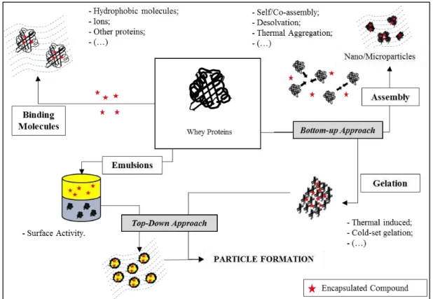
2.2 Whey proteins as encapsulation devices
12 because of their ability to bind different bioactives or to entrap them through the formation of supramolecular structures, emulsions or hydrogels. The main strategies applied for encapsulation that involve whey proteins are summarized in Figure 2.3. The different studies were classified according to the applied encapsulation strategy.
Figure 2.3. Illustration of encapsulation strategies applied to whey proteins (adapted from
Tavares et al. [4]).
13 storage of the products [36]. In addition, it is also important to ensure the production of encapsulation devices that can be easily incorporated into food products without altering texture and flavor [34].
The most widely used encapsulation techniques and some related applications are summarized in Table 2.2. Furthermore, some procedures have been specifically developed to optimize the potential of biopolymers such as whey proteins as encapsulating agents. Several articles have summarized the potential of whey proteins to transport and protect bioactives such as ions, fatty acids and vitamins [8, 37-39]. Additionally, two distinct and complementary approaches i.e. "top-down" and "bottom-up", have been explored to widen the use of whey proteins as encapsulating biomaterials.
Table 2.2. Main conventional techniques used for the encapsulation of bioactive and food
ingredients. Please refer to the following papers for more details: [34, 35, 40, 41].
Technique Concept Examples of
applications
Spray Drying
Drying of the encapsulated material dispersed in the shell material.
Encapsulation by starch, polysaccharides, maltodextrins and/or
proteins. This technique produces particles between 10 and 100 µm.
Encapsulation of flavor
compounds, polyphenols
and vitamins
Spray
Cooling/Chilling
Incorporation of the core material in the warm and liquefied shell
material (often vegetable oils). Relatively low cost encapsulation
technique. Through product atomization, and consequently cooling,
micro-capsules are formed.
Encapsulation of flavor
compounds, minerals,
vitamins and probiotics
Freeze Drying
Co-lyophilization of the core and the shell materials after a
homogenization process. Normally this technique produces
non-uniform particles.
Encapsulation of flavor
compounds, fatty acids
and probiotics
Extrusion
Formation of core material droplets that became microparticles after
immersion in a hardening bath with the shell material. Normally the
shell material is a glassy carbohydrate matrix. The core material may
be released in a high temperature medium. The encapsulation
efficiency is small, moreover the produced particles shows high
stability and an extended shelf life.
Encapsulation of flavor
compounds, vitamins and
food ingredients (lactic
acid)
14 in the shell material. During processing, the shell material forms a
thin film around the core material particles. Production of particles
from20 µm to few millimeters of diameter.
(yeast)
Supercritical
Fluid Extraction
This technique similar to spray drying, except that the shell material
and the core material are solubilized/dispersed in a supercritical fluid.
Encapsulation of
Heat-sensible cores as
vitamins and polyphenols
Fludized bed
This technique is applied for solid particle encapsulation (100µm to
few mm). The shell material is atomized onto core material fluidized
by an upward stream of air.
Encapsulation of
acidulates, vitamins and
cells
Cocrystallization
Spontaneous crystallization of a supersaturated solution of sucrose
simultaneously with the addition of the core material, forming a
crystalline irregular network, allowing the encapsulation into the
pores of the network.
Encapsulation of acids,
flavor compounds,
anti-oxidants and minerals
Coacervation
Phase separation of one or many polyelectrolytes from a solution and
deposition of the colloidal particles around the active ingredient
suspended or emulsified in the same reaction media. When
hydrocolloids are used, they can be cross-linked using appropriate
chemical or enzymatic agent.
Encapsulation of fatty
acids and flavonoids
Liposomes
Spherical particles consisting of a membranous system formed by
one or more concentric bi-layers of lipids (often phospholipids).
They can be used in the entrapment, delivery and released of
water-soluble, lipid-soluble and amphiphilic materials.
Encapsulation of
vitamins, enzymes and
peptides
Inclusion
This technique refers to the supra-molecular association through
non-covalent interactions of a ligand (“encapsulated” compound) into a cavity formed by a “shell” material (e.g. cyclodextrins).
Encapsulation of
vitamins, flavor
compounds and essential
oils
15 encapsulating heat-sensitive bioactives, including probiotics [38] and occurs in two steps; the first step involves an unfolding of the globular proteins (by a thermal treatment), resulting in the exposure of some reactive groups. After cooling and the addition of the heat-sensitive molecules of interest, the second step consists of a cold aggregation of the denatured proteins through a reduction in electrostatic repulsions, by decreasing pH or/and adding salts [45, 47]. Another advantage of this method is that reactive groups exposed during the heat treatment of the proteins might establish interactions with the bioactives [38].
In contrast, the “bottom-up” approach is based on the association of molecules or small
particles into larger and more complex supra-structures [38, 48]. This approach allows a better kinetic and thermodynamic control of the supramolecular structure formation, but it is often
more complex to implement. In addition, the ”bottom-up" approach usually requires less
energy input than the “top-down” approach [37]. The assembly of proteins into
16 solvents such as acetone or ethanol. The change in solvent properties strengthens hydrogen bonds and reduces hydrophobic associations between heat-denatured proteins [54, 56]. These nanoparticles, which can be further stabilized using a cross-linking reagent, are suitable for the encapsulation and transport of micronutrients [57] and bioactives [55]. Recently, milk proteins have been used to form nano- and microcapsules by electrospraying [58, 59]. Electrospraying is a versatile and low-cost technique that is easy to implement and allows the production of capsules or fibers (in this case the technique is called electrospinning) from polymer solutions [60]. Charges are induced on the surface of the liquid jet or droplet through the application of an electric field, which generates a repulsive force opposite to that of surface tension; while the jet or the droplet is exposed to the electric field, the solvent evaporates, generating fibers or capsules [61]. Usually, organic solvents are used for the dissolution of the polymers (polyethylene glycol, polyvinylchloride, polystyrene). The advantage of using biopolymers such as proteins, is that the electrospraying technique can be applied in aqueous solution [58]. This technique was used for the production of nano-, sub-micro- and microcapsules from solutions of whey protein concentrate (WPC) at different pH
values, to encapsulate -carotene in the presence of glycerol [58]. The authors reported an
encapsulation rate of about 90%, with a good stability of -carotene against photo-oxidation
after resolubilization of the capsules and exposure of the solution to UV radiation for 50 h.
2.2.1 Binding properties of whey proteins
β-LG has been widely studied for its ability to bind hydrophobic and amphiphilic compounds
such as flavor compounds, vitamins, fatty acids and polyphenols. Globally, the interactions
between β-lactoglobulin and bioactives are mainly driven by hydrophobic bonds, although
hydrogen bonds are also involved in the binding of polyphenols [62] and fatty acids [63]. It
17 internal calyx, but additional binding sites in the cavity near to the alpha-helix and the
external surface of the β-barrel have also been described [64].
The parameters of the interaction (localization and number of binding sites on the protein and affinity) depend on the chemical nature of the ligand, the physico-chemical conditions of the medium and the conformation of the protein. The binding affinity constant of curcumin with
native β-lactoglobulin was higher than that with heat-denatured protein [65]. This change was
attributed to a conformational change of the internal calyx of β-LG, resulting in a non-specific
binding of curcumin molecules. In contrast, Tavel et al. [66] showed that β-ionone and
guaiacol aroma exhibited a higher binding affinity for partially denatured β-LG molecules
(molten globule state) than for the native protein. This higher affinity was hypothesized to be due to the exposure of some internal hydrophobic regions on the surface of the partially denatured protein and an increased accessibility of the calyx [66]. In some cases, the ligand
binding might induce changes in the β-LG structure as reported by Le Maux et al. [67]. These
authors showed that linoleate binding to β-LG favored denaturation of the protein, with
subsequent formation of protein covalent dimers and trimers [67].
Whey proteins other than β-LG have also been studied for their ability to bind specific
ligands. Kuhn et al. [68] showed that BSA has two binding sites and has a higher affinity for 2-nonanone, a flavor compound, than β-LG or α-LA, which only possess one binding site. After binding to whey proteins, some ligand properties were improved, including, in a non-exhaustive manner: i) a reduction of UV radiation induced the photo-degradation of folic acid from 40% to 6% after 60 minutes of treatment [69]; ii) an increase in the photo-stability and solubility of resveratrol [70] and α-tocopherol [71]; iii) an increase in the solubility and half-life of curcumin [65]. In contrast, a decrease in the antioxidant activity of tea catechins was
observed when they formed complexes with β-LG [62] and BSA [72]. In some cases, the
18
isolated molecules. This was the case for the complex formed between apo α-LA
(calcium-free form) and oleic acid, known as HAMLET/BAMLET (human/bovine α-lactalbumin made
lethal to tumor cells), which induces apoptosis of tumor cells [73]. Heat-denatured α-LA was also able to form a complex with oleic acid that induced apoptosis in cancer cells [74]. In
addition, a complex formed by β-LG and oleate was shown to induce apoptosis in cancer cells
comparable to the activity of BAMLET [75]. The improvement in fatty acid solubility through its binding to these proteins is probably a driving mechanism behind the observed apoptotic effect.
2.2.2 Encapsulation devices obtained through the “bottom-up” approach
The ability of whey proteins, particularly β-LG, to form nanoparticles used as encapsulating
agents has also been extensively studied. Thermal aggregation and desolvation are the main strategies used for the production of these nanoparticles.
19 astringency of EGCG were significantly reduced. The release of EGCG was limited during simulated gastric digestion, which suggests that the nanoparticles could be used to protect EGCG in the stomach, allowing a possible release of the bioactive into the gut [78].
High-pressure homogenization was used to produce nanoparticles from heat-induced aggregates of whey proteins for α-tocopherol encapsulation [79]. The formed particles exhibited a diameter between 212 and 293 nm, depending on the pressures employed. Compared to homogenization at 300 bar, a pressure at 1,200 bar induced some protein structural changes that modified the zeta potential of the produced particles and improved the
stability of encapsulated α-tocopherol during storage [79]. Alternatively, whey protein
nanoparticles with a controlled size were also produced by pH-cycling treatment (acidification and neutralization). Particles with a diameter ranging from 100 to 300 nm were produced through the acidification of a low-concentrated solution of heat-denatured whey proteins [50]. The whey proteins were linked by covalent bonds in the nanoparticles after the neutralization step. The particle size varied depending on the pH of acidification (5.0–6.0), aggregation time (0–75 h) and calcium concentration (0–5.0 mM). Calcium concentration also influenced the voluminosity of the particles: increasing the concentration of calcium decreased the voluminosity of the particles [50]. This technique was used to produce particles for entrapping hydrophobic aroma [80]. The retention efficiency was maximum when the aroma molecules were added to the protein dispersion before the formation of the particles at pH 5.0 or 5.5 and without added calcium [80].
Nanoparticles of BSA were produced in the presence of the flavonoid quercetin by a desolvation process induced by the addition of 10% dimethyl sulfoxide (DMSO) [81]. These
nanoparticles showed a -potential of -12.5 mV and a diameter close to 10 nm, surprisingly
20 the authors explained their results by the highest compaction of the nanoparticles in complexes with the flavonoid [81]. The antioxidant activity of encapsulated flavonoid was not substantially changed, but its stability under intestinal conditions appeared to increase. The latter observation was attributed to the formation of both hydrophobic interactions and hydrogen bonds between quercetin and BSA during the encapsulation process [82].
Desolvation was also used to produce nanoparticles of β-LG for curcumin encapsulation at
21 2.2.3 Encapsulation devices obtained from a “top-down” approach
Top-down approaches for the encapsulation of bioactives using whey proteins are basically restricted to strategies of protein cold gelation, the formation of emulsions or a combination of these two strategies using extrusion techniques. Remondetto et al. [84] reported that cold-set
gels of β-LG induced by the addition of iron show different morphologies, depending on the
iron/protein ratio. For the lower ratios tested, filamentous gels were formed, whereas particulate gels were obtained at high ratios [84]. Particulate gels are mainly stabilized by van der Waals interactions; the large concentration of iron causes a rapid decrease in the repulsive forces, which generates random aggregation and leads to particulate gels. Moreover, low concentrations of iron mainly drive the formation of gels via hydrophobic interactions; the low iron concentration causes a decrease in the surface charge of the molecules and/or aggregates, facilitating interaction between exposed hydrophobic regions, which orients the growth of the assembly in only one direction, leading to filaments [85]. The microstructure of the gels affects the iron release properties. At acidic pH, iron was released more efficiently from particulate gels than from filamentous gels. In contrast, iron release was more efficient with filamentous gels at neutral pH, providing greater iron absorption under intestinal conditions, according to in vitro tests. This suggests that filamentous matrices are more efficient in the protection and transport of iron for nutritional purposes [86].
23 efficiency of the bacteria reached 96%. Indeed, the encapsulation device increased the viability of the cells during storage and their resistance against gastric digestion. Hence, whey proteins constitute an ideal biopolymer for the encapsulation and protection of probiotics and their challenging delivery to intestinal absorption sites [89].
Figure 2.4. SEM cross-section image (12,000 x magnification) of a whey protein-isolate gel with encapsulated Lactobacillus rhamnosus (adapted from Reid et al. [87])
24 retinol, the lipid droplets were extruded into a calcium chloride solution to induce gelation of interfacial proteins [90]. The thus-formed encapsulating devices were about 2.0 mm in
diameter and were stabilized by intermolecular β-sheet structures between protein molecules
[90]. With the same objective, Liang et al. [91] produced an emulsion containing α-tocopherol stabilized by an interfacial layer formed by the calcium-induced cold-set gelation of
pre-denatured β-LG. After in vitro enzymatic digestion of the encapsulating devices, the authors
showed that the release of the bioactive is mainly controlled by the kinetics of protein
hydrolysis. In addition, α-tocopherol released during the digestion process is degraded more
slowly than free α-tocopherol, probably due to the protection effect provided by its interaction
with the pre-denatured proteins and/or produced peptides [91]. Cornacchia and Roos [92] used whey proteins to stabilize oil in water emulsions to produce delivery systems capable of
protecting and transporting β-carotene. Similarly, Tippetts et al. [93] produced emulsions
stabilized with whey proteins to generate stable delivery systems for vitamin D3-enriched cheeses.
2.2.4 Supra-molecular structures with putative encapsulation properties
25 Amyloid-type fibrils are linear polymers that are between 3 and 10 nm in width, and can reach several microns in length [94]. The formation of fibers involves two steps: nucleation and subsequent unidirectional growth [100]. The ability to form fibers has been described for β -LG [94] and α-LA [101]. Specific physico-chemical conditions are generally required to initiate fibrillation; the proteins have to contain exposed hydrophobic regions and to conserve some surface charges [15]. In addition, the fibrillation process is favored by the cleavage of some peptide bonds [102]. These fibers can further self-associate into more complex structures, e.g., ribbons and spherulites. Ribbons are the result of the lateral stacking of these fibers and are usually obtained after the prolonged heating of globular proteins under acidic conditions [95]. Alternatively, spherulites are formed by the radial association of the fibers, a structure that can reach hundreds of micrometers in diameter [96].
The formation of nanotubes from whey proteins is less widespread than the formation of fibers or aggregates. To date, whey protein nanotubes have only been obtained by the self-assembly of α-LA fragments formed by limited hydrolysis of the protein backbone by a serine protease [97]. Regular nanotubes with a length up to several microns, an external diameter of 20 nm and an internal diameter of about 8 nm can be obtained by adjusting the protein concentration, the concentration of added specific divalent cations and hydrolysis conditions [97]. It was suggested that the reversible association of α-LA into nanotubes could be explored as vehicles for delivering bioactives and for encapsulation and release purposes [103].
The spontaneous formation of nano- and microspheres was reported by complex cocervation between oppositely charged proteins [15]. The formation of coacervates (microspheres)
betweeen positively charged lysozyme (LYS) and negatively charged α-LA and the