RESCUING THE ISCHEMIC PENUMBRA — OUR EXPIRIENCE
Milosavljevic Tamara,
1Ivkovic Aleksandar
2 1Special Hospital Niroshi, Nis, Serbia 2
Center of Radiology, Klinical Center Nis, Serbia
Primljen/Received 31. 05. 2013. god. Prihva}en/Accepted 13. 11. 2013. god.
Abstract: Objectives: Over one million strokes per year are occurring in Europe. Brain stroke is one of the most important death and disability causes in Europe and USA. The main role of perfusion is to determine the border of insult core and ischemic penumbra. Penumbra can be saved with thrombolytic therapy but core have ir-reversible injuries and represent death of brain cells.
Aim:to determine the role of CT brain perfusion in cases of acute brain stroke and following thrombo-lytic therapy.
Methods: We examined 64 patients with acute brain stroke who received thrombolytic therapy after that. All patients were examining on 16 MDCT with 50 ml of iodine contrast agent following the standard pro-cedure for CT perfusion. Patients were 34 male and 30 female with middle age of 64 years. MRI was made af-ter thrombolytic therapy and compare with perfusion results before therapy.
Results:Using an artery and a vein as reference three parameters were measured — blood flow (CBF), blood volume (CBV) and mean transit time (MTT), for each patient. Hemorrhagic was find in 9 (14.01%) pati-ents after thrombolytic therapy. 4 (6.25%) other patipati-ents develop new stroke of same but mostly other side of brain. 8 (12.50%) more patients finished lethally. From other 42 patients with thrombolytic therapy we can posi-tively say that in 31 (48.44%) patients penumbra was re-scued. For other 11 (17.19%) stroke was same size like firstly involved core and penumbra but not bigger.
Conclusion: CT perfusion plays major role by showing a curable parts of tissue in brain strokes.
Key worlds: penumbra, ischemia, CT perfusion, stroke.
INTRODUCTION
A stroke, or cerebrovascular accident (CVA), is the rapid loss of brain function due to disturbance in the blood supply to the brain. This can be due to
ische-mia (lack of blood flow) caused by blockage (thrombo-sis, arterial embolism) or a hemorrhage (1). As a result, the affected area of the brain cannot function, which might result in an inability to move one or more limbs on one side of the body, inability to understand or for-mulate speech, or an inability to see one side of the vi-sual field (2).
dysfunc-tion of the brain tissue in that area. There are four rea-sons why this might happen:
— Thrombosis (obstruction of a blood vessel by a blood clot forming locally)
— Embolism (obstruction due to an embolus from elsewhere in the body) (2)
— Systemic hypo perfusion (general decrease in blood supply, e.g., in shock) (5)
— Venous thrombosis (6).
Stroke without an obvious explanation is termed “cryptogenic” (of unknown origin); this constitutes 30–40% of all ischemic strokes (2, 7).
There are various classification systems for acute ischemic stroke. The Oxford Community Stroke Pro-ject classification (OCSP, also known as the Bamford or Oxford classification) relies primarily on the initial symptoms; based on the extent of the symptoms, the stroke episode is classified as total anterior circulation infarct (TACI), partial anterior circulation infarct (PA-CI), lacunar infarct (LACI) or posterior circulation in-farct (POCI). These four entities predict the extent of the stroke, the area of the brain affected, the underlying cause, and the prognosis (8, 9). The TOAST (Trial of Org 10172 in Acute Stroke Treatment) classification is based on clinical symptoms as well as results of further investigations; on this basis, a stroke is classified as be-ing due tothrombosis or embolism due to atherosclero-sis of a large artery, embolism of cardiac origin, occlu-sion of a small blood vessel, other determined cause, undetermined cause (two possible causes, no cause identified, or incomplete investigation) (1–5, 10).
CT perfusion is the method by which perfusion to an organ measured by CT is still a relatively new con-cept, although the original framework and principles were concretely laid out as early as 1980 by Leon Axel at University of California San Francisco (2). It is most commonly carried out for neuroimaging using dyna-mic sequential scanning of a pre-selected region of the brain during the injection of a bolus of iodinated con-trast material as it travels through the vasculature (11). Various mathematical models can then be used to pro-cess the raw temporal data to ascertain quantitative in-formation such as rate of cerebral blood flow (CBF) fo-llowing an ischemic stroke or aneurismal subarachnoid hemorrhage. Practical CT perfusion as performed on modern CT scanners was first described by Ken Miles, Mike Hayball and Adrian Dixon from Cambridge UK (3) and subsequently developed by many individuals including Matthias Koenig and Ernst Klotz in Ger-many (4), and later by Max Wintermark in Switzerland and Ting-Yim Lee in Ontario, Canada (5).
It is important to understand the normal physiol-ogy of the brain for accurate interpretation of stroke
imaging. The brain is continuously perfused with bloo-d bloo-during systole as well as bloo-diastole, with 15–20% of the total cardiac output going to the brain. Cerebral blood flow is approximately 800 ml/min. This high and con-tinuous blood flow is necessary as the brain uses glu-cose, exclusively, for energy metabolism and is unable to store energy. Cerebral blood flow is equipped with an autoregulatory mechanism, which protects against hypoxia and low perfusion. It is a multifactorial mech-anism, involving neurogenic, myogenic, and meta-bolic controls. This autoregulation tries to maintain a mean arterial pressure of 60–100 mm Hg and a cerebral blood flow of 50–60 ml/100 gm of brain per minute. When the cerebral blood flow decreases, the autoreg-ulatory mechanism tries to compensate by increasing the blood pressure and inducing vasodilatation. Howe-ver, if the blood flow decreases so much that it falls be-low a critical level, infarction results (Table 1).
Table 1.Cerebral perfusion and corresponding blood flow levels
Stroke imaging serves two purposes: first, to diag-nose or confirm the occurrence of a stroke and, second, to assess the amount of potentially salvageable brain tis-sue and irreversibly infarcted tistis-sue; both are necessary, the first for planning management strategy and the sec-ond for prognostication. CT perfusion (CTP) is a tool that has been successfully employed to assess the extent of salvageable tissue. It is most commonly carried out for neuroimaging using dynamic sequential scanning of a pre-selected region of the brain during the injection of a bolus of iodinated contrast material as it travels thro-ugh the vasculature. Various mathematical models can then be used to process the raw temporal data to ascerta-in quantitative ascerta-information such as rate of cerebral blood flow (CBF) following an ischemic stroke.
AIM
The aim of this retrospective study is to show possibilities of CT perfusion as diagnostic procedure in patients with acute ischemic attack.
METHODS
We retrospectivly reviewed examination of 64 pa-tients with acute brain stroke who have received throm-bolytic therapy after that. All patients were examined
from January 2010 to October 2012. The research in-cluded 34 (53.13%) male and 30 (46.87%) female, wi-th average age of wi-the patients 57.42188 ± 5.31 years, range 45–74 years. All patients were examined on GE Bright Speed 16 MDCT with 50 ml of iodine contrast agent (Ultravist 300, Bayer) following the standard procedure for CT perfusion. All examinations were made in period of 4 hours after the first symptom. After CT perfusion, we performed Multi Raw Detector Com-puter Tomography Angiography (MDCTA) of brain. MDCTA was performed using standard GE protocol for brain blood vessels, using 60 to 120 ml of iodine contrast agent (Ultravist 370). Results were presented using ASPECTS (Alberta Stroke Program Early CT Score) and using NIHSS (National Institute of Health Stroke Scale) by neurologist. Patients were treated us-ing Tissue plasminogen activator (rtPA) in dose of 0.9 mg/kg body weight, by Guidance for ischemic stroke, 10% in bolus injection and rest of dose during 60 min-utes intravenous. Magnetic Resonance Imaging (MRI) was made on Siemens Avanto 1.5T MRI, using stan-dard Siemens protocol with apparent diffusion coeffi-cients (ADC) map and diffusion weighted images (DWI) after thrombolytic therapy and compared with perfusion results before therapy. Using the General E-lectric Neuro software, CBV was calculated along with cerebral blood flow, using the maximal slope model, which has been shown to yield lesion volumes that are similar to delay insensitive deconvolution techniques. The hypoperfused areas on CBV maps were defined as volume abnormality. The infarct core was outlined on CBV maps as a severely hypoperfused area displayed by 2 colors in the color bar. The CBV threshold was de-fined at 2.0 mL 100 g1. Each CBV map was analyzed by demarcating trace lines around the area of volume abnormality on each of the 4 slices by hand-drawn re-gions of interest. The area of volume abnormality was defined as CBV area. The weighted mean of CBV and CBV area was calculated in each of the 4 slices, respec-tively, and used for statistical analysis. For statistical analysis we used means with standard deviation and variance, also population standard deviance and vari-ance were used. Present-day multislice CTs do not al-low for whole-brain perfusion assessment. Thus, if an anterior circulation infarct is suspected, data acquisi-tion is done at the basal ganglia level and if a posterior circulation infarct is likely, then at it is done at the level of the mid-cerebellum. Depending upon the type of scanner, one can take 1–4 sections of 5–10 mm thick-ness at the levels mentioned.
Postprocessing of the data is done by using specialized software. It involves confirmation of cer-tain parameters which are automatically chosen by the software, and it generates color perfusion maps as well
as time–attenuation curves (TAC) from which T-TP/MTT, CBV, and CBF can be calculated for any area of interest.
RESULTS
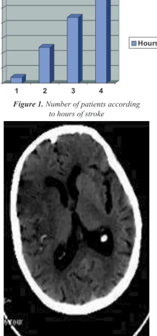
All patients were examined in short time period of 4 hours after initial symptoms (Figure 1). Patients were ex-amined by neurologist before sending to MDCT exami-nation. Patients meeting neurology standards for thromb-olytic therapy were transferred to MDCT. First we per-formed non-contrast CT (Figure 2). All patients were s-cored by ASPECTS (Alberta Stroke Program Early CT Score). Patients with score 6 or above were expected for good candidate for thrombolytic therapy (Figure 3).
Pa-Figure 2.Noncontrast computed tomography with CVI of right temporal lobe Figure 1.Number of patients according
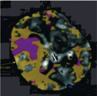
tients with score below 6 were not used in consideration for therapy. Average number of patients were 12.8 with s-tandard deviation of 3.114 and variance of 9.7. Popula-tion standard deviaPopula-tion were 2.786 with variance of 7.76. After NCCT we performed perfusion MDCT. Main area of interest was middle cerebral artery (MCA), using an artery and a vein as reference. Three parameters were m-easured — blood flow (CBF), blood volume (CBV) and mean transit time (MTT), for each patient. Also we perf-ormed white/grey separation program to determine bor-ders of white and gray matter (Figure 4).
Hemorrhagic insult was found in 9 (14.01%)
pa-tients after thrombolytic therapy. Presents of intrace- rebral hemorrhage is the most feared risk of intraveno-us intraveno-use of tissue plasminogen activator (rtPA). Accor-ding to ASPECTS, 4 were with ASPECTS 6, another 4 with ASPECTS 7 and 1 with ASPECTS 8 (Figure 5).
Four (7.81%) other patients develop new stroke of same, but mostly other side of brain. According to PECTS, 3 were with ASPECTS 6, another 1 with AS-PECTS 7 (Figure 5).
Eight (12.50%) more patients finished lethally (Figure 6. and 7). According to ASPECTS, 6 were with ASPECTS 6, another 2 with ASPECTS 7 (Figure 5).
For other 11 (17.19%) patients stroke was same size like involved core and penumbra on first examinat-ion but not bigger. Patients with ASPECTS score below 8 and patients on upper time window are more likely to Figure 3.Number of patients according to ASPECTS
Figure 5.Distribution of patients according to ASPECTS
Figure 4.White/grey mater differentiation
Figure 6.CT perfusion. CVI of right temporal lobe
be in this group: 1 patient was with ASPECTS 6, an-other 5 patients were with ASPECTS score 7, anan-other 4 with ASPECTS 8 and 4 in time window after 4 hours of stroke (Figure 8). Only one was with ASPECTS 9 and treated in first 4 hour (Figure 5).
We can positively say that in 31 (48.44%) patients penumbra was rescued. Penumbra can not be saved without therapy so we can positively say that penumb-ra was saved in 48.44% of patients with high ASPE-CTS and treated in first 4 hours (Figure 9). However, the rescued penumbra may be affected by selective neuronal loss. Rescuing penumbra is more likely in first 3 hours after the stroke, with no mater of penumb-ra size (Figure 10). ASPECTS were 10 in 12 patients, 9 in 8 patients, 8 in 7 patients, 7 in 4 patients and 6 in 1 patient (Figure 5).
DISCUSSION
Ischemic stroke occurs because of a loss of blood supply to part of the brain, initiating the ischemic cas-cade (12). Brain tissue ceases to function if deprived of oxygen for more than 60 to 90 seconds, and after ap-proximately three hours will suffer irreversible injury possibly leading to death of the tissue, i.e., infarction (13). (This is why fibrinolytics such as alteplase are given only until four hours since the onset of the s-troke.) Atherosclerosis may disrupt the blood supply by narrowing the lumen of blood vessels leading to a reduction of blood flow, by causing the formation of blood clots within the vessel, or by releasing showers of small emboli through the disintegration of atherosc-lerotic plaques. Since blood vessels in the brain are now occluded, the brain becomes low in energy, and thus it resorts into using anaerobic metabolism within the region of brain tissue affected by ischemia. Unfor-tunately, this kind of metabolism produces less a-denosine triphosphate (ATP) but releases a by-product called lactic acid. Lactic acid is an irritant which could potentially destroy cells since it is an acid and disrupts the normal acid-base balance in the brain. The isch-emia area is referred to as the “ischemic penumbra”.
In our study hemorrhagic insult after thrombolytic therapy occurred in 14.01% of patients. According to Berger et al. the 7-level mRS predictive model indicat-es that 32% of patients who experienced intracerebral hemorrhage were destined for a fatal outcome even they had not been treated with rtPA (12). According to National Institute of Neurological Disorders and Stroke, hemorrhagic insult will occur in 7 to 33% of patients depending of age, sex, recurrent stroke and ASPECTS (4). Our percent of 14.01 is tempered and b-alanced with ASPECTS.
New stroke occurred in 7.81% of patients. Accor-ding to literature, percentage of new stroke is from 4% to 32% and mostly is depending on heart condition and patient’s age (12, 13, 14). We have had low percentage of these patients because of high ASPECTS.
Lethal result was in 12.50% of patients. According to literature patients finish lethal in 10 to 28%. Accord-Figure 8.CT perfusion of massive CVI
without penumbra
Figure 9.Number of patients according to results
ing to Quareshi et al. “the problem of disentangling worsening attributable to cerebral edema, infarct expa-nsion, and other causes of ischemic stroke progression from worsening attributable to hemorrhage is difficult because the variables that independently identify patie-nts destined for thrombolysis are also variables that pre-dict symptomatic worsening and poor final outcome even if patients are not treated with rtPA” (13).
Same size of penumbra occurred in 17.19% of pa-tients. According to Nor et al., same size of penumbra occurs in 15 to 25% of patients, depending on time of initial stroke (15).
In 48.44% of patients penumbra was rescued. Ac-cording to O’Sullivan et al, penumbra can be rescued in less than half of patients and according to Fisher, percentage of rescued penumbra is 42 to 49% (16, 17).
CT angiography (CTA) is an advanced applicati-on of present-day multislice spiral CT scan machines. It allows the comprehensive evaluation of arteries any-where in the body. This is useful in the assessment of stenosis or occlusion of the carotid arteries or vertebral arteries in the neck, which can act as predisposing fac-tors for a stroke. Also, the evaluation of intracranial ar-teries is possible with a high degree of accuracy (18). CTA is now gradually replacing digital subtraction angiography (DSA) for this purpose.
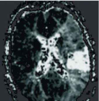
MR imaging has become a powerful clinical tool for evaluation of brain anatomy. Its application has re-cently expanded into evaluation of brain function via assessment of a number of functional or metabolic pa-rameters. One such parameter is cerebral perfusion, w-hich describes passage of blood through the brain’s vascular network. MRI diffusion as on Figure 11 (MRI diffusion after stroke) and perfusion as on Figure 12.
(MRI perfusion after stroke) imaging provide similar information with greater sensitivity and specificity. Probably the widest application of exogenous tracer methods in MR perfusion imaging has been in the ass-essment of cerebral ischemia.
CONCLUSION
The American Heart Association has provided certain guidelines and recommendations for imaging of cerebral ischemia. According to it, quantitative CTP may probably be useful to differentiate between re-versible and irrere-versible ischemic tissue in acute str-oke. On the other hand, MRI perfusion and diffusion t-echniques are probably useful in differentiating betw-een reversible and irreversible ischemic tissue in acute stroke patients. According to our results, CTP is a valu-able and important tool in stroke imaging and we perform MRI mostly after thrombolytic therapy. High percentage of rescued penumbra is most important re-sult because there is no other diagnostic tool but must be performed on selective patients and in first 4 hours.
List of abbreviations
CBF— cerebral blood flow CBV— cerebral blood volume MTT— mean transit time CVA— cerebro vascular incident TIA— transient ischemic attack
TACI— total anterior circulation infarct PACI— parcial anterior circulation infarct LACI— lacunar infarct
POCI— posterior circulation infarct Figure 11.MRI diffusion of CVI
CTP— CT perfusion
ASPECTS— Alberta Stroke Program Early CT Score
rtPA— tissue plasminogen activator ADC— apparent diffusion coefficients
DWI— diffusion weighted images TAC— time-attenuation curves MCA— middle cerebral artery ATP— adenosine triphosphate
DSA— digital supstaction angiography
Sa`etak
SPASAVANJE ISHEMIJSKE PENUMBRE — NA[A ISKUSTVA
Milosavljevi} Tamara,1Ivkovi} Aleksandar2
1Specijalna Internisti~ka bolnica Niroshi, Ni{, Srbija 2Centar za radiologiju, Klini~ki Centar Ni{, Srbija
Uvod: Preko milion mo`danih udara se dogodi u Evropi godi{nje. Mo`dani udar predstavlja jedan od n-ajva`nijih uzroka smrti kako u Evropi tako i u SAD. G-lavna uloga CT perfuzije je da odredi granicu p-enumbre i jezgra insulta. Jezgro je pretrpelo ireve-rzibilne promene dok penumbra mo`e biti spa{ena. Jezgro }e kasnijim procesom biti izlo`eno nekrozi, {to sledi i penumbri ukoliko nema tromboliti~ke terapije.
Cilj: odrediti ulogu CT mo`dane perfuzije u slu~ajevima akutnog mo`danog udara i pra}enje e-fekata tromboliti~ke terapije.
Metodi:Perfusion measurements of the brain: us-ing dynamic CT for the quantitative assessment of ce-rebral ischemia in acute stroke Pregledano je ukupno 64 pacijenta sa akutnim mo`danim udarom, koji su pri-mili tromboliti~ku terapiju nakon toga. Svi pacijenti su pregledani na 16 MSCT aparatu uz upotrebu 50 ml j-odnog kontrastnog sredstva po standardnom protokolu za CT perfuziju. MRI je ra|en nakon tromboliti~ke
terapije i upore|ivani su nalazi sa perfuzijom pre terapije.
Rezultati: Koriste}i vene i arterije kao repere vr{ena su tri merenja — protok krvi (blood flow-CBF), volumen krvi (blood volume-CBV) i srednje vreme tr-anzicije (mean transit time-MTT), i to za svakog p-acijenta. U na{oj studiji se trudimo da odredimo i grani-cu bele i sive mase radi jasnije neurolo{ke prognoze. Hemoragija je na|ena kod 9 (14.01%) pacijenata nakon tromboliti~ke terapije. ^etiri (6.25%) pacijenta su imala reinzult, na istoj ili suprotnoj strani, {to je bilo ~e{}e. Osam (12.50%) pacijenata je zavr{ilo letalno. Od ostalih 42 pacijenata mo`emo sa sigurno{}u re}i da je penum-bra spa{ena kod 31 (48.44%). Kod ostalih 11 (17.19%) insult je bio jednak jezgru i penumbri ali ne ve}i.
Zaklju~ak: CT perfuzija ima zna~ajnu ulogu u prikazivanju delova mozga podlo`nih le~enju.
Klju~ne re~i:penumbra, ishemija, CT perfuzija, insult.
REFERENCES
1. Sims NR, Muyderman H. Mitochondria, oxidative me-tabolism and cell death in stroke. Biochimica et Biophysica Acta. 2009; 1802 (1): 80–91.
2. Donnan GA, Fisher M, Macleod M, Davis SM. Stroke. Lancet. 2008; 371(2): 1612–23.
3. Mathers, CD, Boerma T, Ma Fat D. Global and regional causes of death. British medical bulletin. 2009; 92: 7–32.
4. Klotz E, König M. Perfusion measurements of the brain: using dynamic CT for the quantitative assessment of cerebral ischemia in acute stroke. Eur J Radiol. 1999 Jun; 30(3): 170–84.
5. Shuaib A, Hachinski VC. Mechanisms and management of stroke in the elderly. CMAJ . 1998; 145 (5): 433–43.
6. Stam J. Thrombosis of the cerebral veins and sinuses. The New England Journal of Medicine. 352 (17): 1791–8.
7. Guercini F, Acciarresi M, Agnelli G, Paciaroni M. Cryptogenic stroke: time to determine aetiology. Journal of Thrombosis and Haemostasis. 2008; 6 (4): 549–54.
8. Bamford J, Sandercock P, Dennis M, Burn J, Warlow C. Classification and natural history of clinically identifiable sub-types of cerebral infarction. Lancet. 1991; 337 (8756): 1521–6.
9. Bamford JM. The role of the clinical examination in the subclassification of stroke. Cerebrovascular Diseases. 2001; 10 Suppl 4: 2–4.
10. Adams HP, Bendixen BH, Kappelle LJ, et al. Classifi-cation of subtype of acute ischemic stroke. Definitions for use in a multicenter clinical trial. TOAST. Stroke. 1998; 24 (1): 35–41. 11. Goldstein LB, Simel DL. Is this patient having a stro-ke? JAMA. 2005; 293 (19): 2391–402.
12. Berger C, Fiorelli M, Steiner T, Schabitz WR, Bozzao L, Bluhmki E, Hacke W, von Kummer R. Hemorrhagic transfor-mation of ischemic brain tissue: asymptomatic or symptomatic? Stroke. 2001; 32: 1330–5.
13. Qureshi AI, Suarez JI, Yahia AM, Mohammad Y, Uzun G, Suri MF, Zaidat OO, Ayata C, Ali Z, Wityk RJ. Timing of ne-urologic deterioration in massive middle cerebral artery infarc-tion: a multicenter review. Crit Care Med. 2003; 31: 272–7.
14. Kidwell CS, Saver JL, Schubert GB, Eckstein M, Starkman S. Design and retrospective analysis of the Los Ange-les Prehospital Stroke Screen (LAPSS). Prehospital Emergency Care. 2005; 2 (4): 267–73.
Correspondence to/Autor za korespondenciju Tamara Milosavljevi},
Specijalna Internisti~ka bolnica Niroshi, Ni{ Romanijska 7/18, 18000 Ni{,
tamaradr2009ªgmail.com 060 7000 182, fax 018 520 174
and validation of a stroke recognition instrument. Lancet Neuro-logy. 2005; 4 (11): 727–34.
16. O’Sullivan, Susan. B. Stroke. In: O’Sullivan, SB, Schmitz TJ, editors. Physical Rehabilitation 5. Philadelphia: FA Davis Company; 2008. p. 719.
17. Fisher. The arterial lesions underlying lacunes. Acta Neuropathologica. 1998; 12 (1): 1–15.