Original Research
Evaluation of the Effect of Surgical Crown Lengthening on
Periodontal Parameters
Farzane Vaziri
1, Ahmad Haerian
2, Mohammad Hossein Lotfi Kamran
3,
Maryam Abrishami
41
Assistant Professor, Periodontology Department, School of Dentistry, Shahid Sadoughi
University, Yazd, Iran
2
Professor, Periodontology Department, School of Dentistry, Shahid Sadoughi University, Yazd,
Iran
3
Assistant Professor, Prosthodontics Department, School of Dentistry, Shahid Sadoughi University,
Yazd, Iran
4
Post-graduate student, Periodontology Department, School of Dentistry, Shahid Sadoughi
University, Yazd, Iran
Received 4 April 2015 and Accepted 20 June 2015
Abstract
Background: Surgical crown lengthening is needed for teeth with subgingival caries, fractured teeth, insufficient crown length, and deep subgingival margin of failed restorations. Since there is no agreement on the effects of crown lengthening surgery on gingival parameters, the purpose of this study was to evaluate periodontal parameters in patients who needed crown lengthening surgery. Methods: Twenty patients who had healthy periodontium and needed surgical crown lengthening were included in this study. After professional dental cleaning, gingival parameters including gingival index (GI), probing depth (PD), bone level (BL), and transsulcular probing (TSP) were recorded in interproximal and keratinized gingiva (KG) in mid buccal portion. The patients were evaluated one and three months after the surgery. Results: After one and three months of the surgery, the amount of PD reduced from 2.32 mm to 1.25 mm and 1.17 mm, respectively (P=0.001). The mean of BL reduction was 0.88 mm after one month (P=0.001), but there was no reduction between 1 month and 3 months. Amounts of KG at baseline andone month later were 4.2 mm and 2.9 mm, respectively (P=0.001), and remained at the same level up to three months. TSP significantly reduced (from 3.67 mm at baseline to 2.62 mm after 1 month, and to 2.27 mm after 3 months) (P=0.001, P=0.005).
Conclusion: The present study suggests that in the
presence of good oral hygiene, except BW (biological width), other parameters including PD, BL, KG, and TSP had significant changes after crown lengthening surgery in the period of 1 month and 3 months (P<0.05).
Key words: Crown lengthening surgery, periodontal treatment, healthy periodontium.
--- Vaziri F, Haerian A, Lotfi Kamran MH, Abrishami M. Evaluation of the Effect of Surgical Crown Lengthening on Periodontal Parameters. J Dent Mater Tech 2015; 4(3): 143-50.
Introduction
are recommended to prevent periodontal breakdown and to facilitate prosthetic treatment (5,6).
Removal of soft tissue and sometimes even hard tissues occurs in crown lengthening surgery. Soft tissue removal is performed when there is more than 3mm of soft tissue over the bone. In cases with less than 3mm of soft tissue or inadequate attached gingiva, bone recontouring and flap procedure are recommended. To maintain healthy periodontium, there should be a 4-mm distance from bone crest in fractured teeth and deep caries (7).
Although Bragger et al. reported that the gingival margin remained stable during the healing period and that gingival enlargement was little (8), some evaluations showed significant reduction of SOG (supra osseous gingiva) after crown lengthening in the follow-up study 2 and 6 months later (9,10). Moreover, in an animal study, different dimensions of SOG were shown in mandibular and maxillary teeth after surgery (11). Curnevale and Pontorieo reported coronal growth of gingival margin in a year after crown lengthening surgery, which was noticed more in thick gingiva. Although it was affected by individual variations, it was neither in relationship with age nor with sex (12). In a study by Deas et al, tissue rebound was significant after crown lengthening during a 6- month follow up; which was in relationship with the flap position toward the alveolar crest at suturing time (10). Perez-Smukler (2008) stated that there was clinical variation in SOG dimensions among patients with similar and different tooth types, arches, and surfaces (13).
Some studies reported stable results after the surgery (8,9,14,15,18) , while other studies reported significant tissue rebound after the surgery(10,12,16,19 )
Since the effects of crown lengthening surgery on gingival parameters were not the same in different studies, we aimed to evaluate periodontal parameters in patients who needed crown lengthening surgery.
Methods and Materials
This study was an interventional case series with the observation periods of one and three months. Individuals were referred to the Periodontology Department, School of Dentistry, Shahid Sadoughi
University, Yazd, Iran, from April 2014 to June 2014 for crown lengthening surgery. The study was approved by the Ethics Committee of the Shahid Sadoughi University (Protocol no 17/1/31915). Patients received an explanation about the purpose of the study and provided written informed consent before clinical periodontal examination.
Patients were screened for eligibility. Their teeth were approved according to the following criteria:
Need of surgical exposure for appropriate prosthetic treatment
Subgingival caries or fracture
Insufficient crown length for proper retention
Uneven gingival height
Proper prognosis and crown root ratio
Having a single root
The following criteria were approved for the patients:
No systemic disease contraindicated with gingival surgery
No drug use, no history of periodontal disease, non-smoker. The study was sampled from 20 patients (16 females and 4 males; aged 23 to 54 years; mean age 34.1 years) with 20 teeth which needed surgical exposure for proper restorative treatment.
Clinical Measurements
Two weeks before the surgery, all patients received oral hygiene instructions and also professional dental cleaning if needed.
For each patient an acrylic stent was fabricated and vertical grooves were made at the interproximal and mid-buccal aspects of the teeth for standardization of the location of the probe during measurements.
After local anesthesia (lidocaine/epinephrine 1:80.000, Daroopakhsh Co., Iran), clinical parameter including GI (gingival plaque index based on Loe and Silness, 1967; table 1), PD, BL, and TSP in the vertical grooving of the interproximal area and KG in the vertical grooving at the mid buccal portion were measured by Williams probe. BW (biologic width) was calculated by subtracting the measurement of TSP from PD (9).
GI Score Description
0 No plaque
1 A film of plaque adherence to the free gingival margin and adjacent area of the tooth. The plaque may be seen in situ only after application of disclosing solution or by using the probe on the tooth surface.
3 Abundance of soft matter within the gingival pocket and/or on the tooth and gingival margin.
Surgical Procedure
An internal bevel incision and full thickness flap procedures were performed at least one tooth mesially and distally. Bone surgery was carried out by hand and rotary instruments under saline irrigation for developing positive bone architecture. Connective tissue and periodontal ligament were removed with finishing bur. Bone tissue was removed to add biologic width (9), for proper placement of restorative margins. The flaps were positioned over the alveolar bone crest at buccal and lingual surfaces and continuous sling sutures were used for stabilizing. The surgical areas were covered with periodontal dressing (Co-Pack, GC America Inc., Alsip, IL, USA).
Antibiotics (500-mg amoxicillin capsules for 7 days) and analgesics (400-mg ibuprofen tablets) were prescribed for each patient, also 0.2% chlorhexidine gluconate mouth rinse twice a day was recommended for 7 days. The sutures were removed one week later and oral hygiene was then reinstructed. After one and three months later clinical parameter again was measured.
Statistical analysis:
Findings at baseline, one month and three months after surgery were analyzed using SPSS 18. Data were analyzed by paired sample t-test and Wilcoxon singed ranks test. P-value<0.05 was considered as significant level. Means were calculated for all the parameters at baseline and one month and three months after surgery.
Results
In this clinical study, 20 patients completed the procedure. Gingival parameters including GI, PD, BL, TSP, and KG were measured at baseline, 1 and 3 months later. During this period GI had no significant reduction (from 1.35 mm at baseline to 1.05 mm after 1 month and 3 months later) (P>0.05)( fig. 1).
Figure 1. Mean GI at baseline GI 1; GI2 1 month after surgery; GI 3 3months after surgery
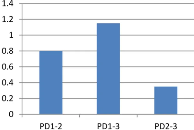
The mean PD at baseline was 2.32 mm and within 1 month and 3 months after surgery were respectively 1.52 mm and 1.17 mm. The mean PD change before the surgery and 1 month later was 0.8 mm (P=0.001), and the change in the period between 1month and 3 months after surgery was 0.35 mm, which was marginally significant (P=0.05) (fig. 2).
Figure 2. Mean PD changes at baseline and 1 month after surgery PD1_2; at baseline and 3 months after surgery PD1-3; 1 and 3 months after surgery PD2-3
The mean bone level had significant reduction one month later (from 9.09 mm to 9.97 mm) (P=0.001), but it was the same (9.97mm) between the first month and the third month after the surgery (fig. 3).
Figure 3. Mean BL changes at baseline and 1 month after surgery BL 1_2; at baseline and 3 months after surgery BL1-3; 1 and 3 months after surgery BL2-3
The mean width of KG at baseline was 4.2 mm while it reduced to 2.9 mm one month later, which was significant reduction (P=0.001); there was no difference between 1month and 3 months (fig. 4).
The mean TSP showed significant reduction after one month and this decrease continued until the third 0
0.5 1 1.5
GI1 GI2 GI3
0 0.2 0.4 0.6 0.8 1 1.2 1.4
PD1-2 PD1-3 PD2-3
-1 -0.8 -0.6 -0.4 -0.2 0
month after the surgery. (from 3.67 mm at baseline to 2.62 mm after 1 month and 2.27 mm after 3 months) (P=0.001) (P=0.005) (fig. 5).
The last parameter, the mean BW, had no significant reduction in one month (from 1.35 mm to 1.05 mm) (P=0.057), nor in the period between 1 month and three months after the surgery (from 1.05 mm to 1.05 mm) (P>0.05) (fig. 6).
Figure 4. Mean KG changes at baseline and 1 month after surgery KG1_2; at baseline and 3 months after surgery KG1-3; 1 and 3 months after surgery KG2-3
Figure 5. Mean TSP changes at baseline and 1 month after surgery TSP1_2; at baseline and 3 months after surgery TSP1-3; 1 and 3 months after surgery TSP2-3
Figure 6. Mean BW changes at baseline and 1 month after surgery BW1_2; at baseline and 3 months after surgery BW1-3; 1 and 3 months after surgery BW2-3
Discussion
Our study resulted that in the presence of stable GI and proper oral hygiene, changes of BL, TSP, PD, and KG during 1 month and 3 months after crown lengthening were significant, while BW changes were not. The results between the first and the third month after the surgery remained the same. The efficacy of crown lengthening surgery was concluded for retention and accessibility to the teeth with deep subgingival caries, teeth with fracture, teeth with insufficient crown length, and the teeth with subgingival failed restoration. With this surgery, invasion to biologic width is avoided, and so are gingival inflammation, attachment loss, and bone loss.
Tooth brushing and oral health were instructed before crown lengthening surgery.
Based on our results, GI difference before and one month after the surgery; before and three months after the surgery, between one month and three months after crown lengthening surgery was not significant. Cruz et al evaluated the effects of the surgery on plaque index (PI), bleeding on probing (BOP), PD, and final restoration outcome in the mean follow-up period of 13.57 months. Only two patients presented relative success while the rest of the participants (12 patients) presented total success. Patients with relative success presented generalized poor oral hygiene. The findings of Cruz et al research are consistent with the present study (14).
KG difference before and one month after the surgery as well as before and three months later were significant (P =0.001). KG difference one month and three months later was 0 mm; which was not significant. The effects of crown lengthening on SOG, KG, BL, and FGM have been evaluated by Ayubian in 20 patients. The author has reported significant reduction in KG and SOG and increase in BL and FGM in 2 months after the surgery. The present study led to similar results for KG and BL (9).
Studying SOG changes in 6 months after the surgery, Perez-Smukler (2007) reported that SOG reduction was significant, which confirms the results of our study (15).
PD difference before and one month after the surgery and before and three months was significant (P=0.001). PD difference one month and three months after the surgery was 0.35 mm; which was significant (P=0.005).
In the study of Deas et al, PD, BOP, PI, and AL in 43 teeth were assessed and significant tissue rebound 0
0.2 0.4 0.6 0.8 1 1.2 1.4
KG1-2 KG1-3 KG2-3
0 0.5 1 1.5
TSP1-2 TSP1-3 TSP2-3
-0.35 -0.3 -0.25 -0.2 -0.15 -0.1 -0.05 0
happened in 6 months after surgery. The amount of tissue rebound is related to flap position toward alveolar crest at the time of suturing. The closer the flap is sutured toward the alveolar crest, the more the tissue is rebounded (10). Arora et al in a study on 64 teeth which needed the surgery reported significant soft tissue rebound after 6 months. This rebound was in correlation with post suturing flap position (P<0.0001) and periodontal biotype (P<0.001). It was concluded that thick–flat biotype and suturing the flap ≤3 mm from the alveolar crest were associated with greater tissue rebound (16). The results of these studies were in discrepancy with our results. In the present study, the effects of periodontal biotype and also suturing on tissue rebound were not assessed. The same sutures were used for all patients and the flap position after suturing was not evaluated. Evaluation was done in 1 month and 3 months after the surgery and the results were stable in contrast with those of the studies done by Deas et al and Arora et al (2004) (10, 16).
Difference in BW was not significant in the evaluated period. Confirming our study results, Lanning et al (2003) reported that BW re-established its original length 6 months after surgery and a 3-mm crown height remained stable 3 and 6 months after the surgery (17).
Ganji et al reported that BW in surgical sites re-established its origin sites 3 months after the surgery and that osteoctomy and apically displaced flap were more effective than gingivectoy. In our study, we used osteoctomy and apically displaced flap, which brought about stable results (18).
In the study of Brager et al determined that gingival margin remained stable and little gingival enlargement happened during the healing period (8), which confirms the present study.
A study reported coronal growth of gingival margin in one year after crown lengthening surgery .It was observed more in the thick gingiva. The growth was affected by individual variations but neither in relationship with age nor sex (12).
Oakly et al (1999) gained different results of SOG in maxillary and mandible teeth after bone surgery in an animal study (11). However, in the present study which was on human teeth, stable results were achieved in 1 and 3 months after the surgery, though maxillary and mandible teeth were not compared.
In a study by Herrero et al on 21 teeth which needed the surgery PD, BL, PI, GI, mucogingival junction (MGJ), and gingival margin position (GMP) were evaluated before and 8 weeks after the surgery. It was reported the distance between alveolar crest and gingival margin was less than the default objective of 3 mm (mean 2.4 ± 1.4 mm). The greatest distance was at the facial aspect of teeth and the least at the distal-lingual. Herrero et al also reported that larger amounts
of bone were removed by more experienced periodontists. In the present study, analyses were done in facial surface of the teeth and the surgery was done with one practitioner. Therefore, those effects were not evaluated in the present study (19).
Limitations in our study were lack of comparison between maxillary and mandibular teeth, and also lack of evaluation of gingival thickness.
Conclusion
Present study suggests that in the presence of good oral hygiene with the exception of BW (biological width), other parameters including PD, BL, KG, and TSP have significant changes after crown lengthening surgery within a three-month period .
References
1. Bader JD, Rozier RG, Mcfall WT Jr, Ramsey DL.
Effect of crown margins on periodontal conditions
in regularly attending patients. J Prosthet Dent
1991;65:75-9.
2. Bader J, Rozier RG, Mcfall WT Jr. The effect of
crown receipt on measures of gingival status. J
Dent Res 1991;70;1386-9.
3. Newcomb GM. The relationship between the
location of sub-gingival crown margins and
gingival inflammation. J Perio-dontol 1974;45:
151-4.
4. Carnevale G, Sterrantino SF, Di febo G. Soft and
hard tissue wound healing following tooth
preparation to the alveolar crest. Int J Periodontics
Restorative Dent 1983;3:36-53.
5. Tal H, Soldinger M, Dreiangel A, Pitaru S.
Responses to periodontal injury in the dog: removal
of gingival attachment and supracrestal placement
of amalgam restorations. Int J Pe-riodontics
Restorative Dent 1988;8:44-55.
6. Rosenberg ES, Garber DA, Evian CI. Tooth
lengthening pro-cedures. Compendium ContinEduc
Gen Dent 1980;1:161-72.
7. Melnick P. Preparation of the periodontium for
restorative dentistry. In Carranza, 2012; chapter 65,
8. Brägger U, Lauchenauer D, Lang NP. Surgical
lengthening of the clinical crown. Journal of
Clinical Periodontology 1992;19:58-63.
9. Ayubian N. Evaluation of Dimensional Changes of
Supraosseous Gingiva Following Crown
Lengthening. J Periodontol Implant Dent 2010;
2(2):61-5.
10. Deas DE, Moritz AJ, McDonnell HT, Powell CA,
Mealey BL. Osseous surgery for crown
lengthening: a 6-month clinical study. J Periodontol
2004;75(9):1288-94.
11. Oakley E, Rhyu IC, Karatzas S, Gmandini-Santiago
L, Nevins M, Caton J. Formation of the biologic
width following crown lengthening in nonhuman
primates. Int J Periodontics Restora-tive Dent
1999;19:529-41.
12. Pontoriero R, Carnevale G. Surgical crown
lengthening: a 12-month clinical wound healing
study. J Periodontol 2001;72(7):841-8.
13. Jose R. Perez, Hyman Smukler, and Martha E.
Nunn. Clinical Dimensions of the Supraosseous
Gingivae in Healthy Periodontium. Journal of
Periodontology 2008;79(12):2267-72.
14. Márcio K da Cruz, Josué Martos, Luiz Fernando
Machado Silveira, Poliana M Duarte,1 and João
Batista César Neto2. Odontoplasty associated with
clinical crown lengthening in management of
extensive crown destruction. J Conserv Dent 2012;
15(1):56-60.
15. Perez JR, Smukler H, Nunn ME. Clinical
Evaluation of the supraosseous gingivae before and
after crown lengthening. J Periodontol 2007;
78:1023-30.
16. Arora R, Narula SC, Sharma RK, Tewari S.
Evaluation of supracrestal gingival tissue after
surgical crown lengthening: a 6-month clinical
study. J Periodontol 2013; 84(7):934-40.
17. Lanning SK, Waldrop TC, Gunsolley JC, Maynard
JG. Surgical crown lengthening: evaluation of the
biological width. J Periodontol 2003;74:468-74.
18. Ganji KK, Patil VA, John J. A Comparative
Evaluation for Biologic Width following Surgical
Crown Lengthening Using Gingivectomy and
Ostectomy Procedure. Int J Dent 2012;
2012:479241.
19. Herrero F1, Scott JB, Maropis PS, Yukna RA.
Clinical comparison of desired versus actual
amount of surgical crown lengthening. J
Periodontol 1995;66(7):568-71.
Corresponding Author:
Maryam Abrishami
Post graduate Student, Periodontology Department,
School of Dentistry, Shahid Sadoughi University, Yazd, Iran Phone: +985138433192
Mobile: +989153249731