Perception of changes in the gingival plane
affecting smile aesthetics
Daniela Feu*, Fabíola Bof de Andrade**, Ana Paula Camata Nascimento***,
José Augusto Mendes Miguel****, Antonio Augusto Gomes*****, Jonas Capelli Júnior******
Objective: This study investigated how 80 dental professionals and 80 lay persons, patients from private practice offices and from the School of Dentistry, Federal University of Es-pírito Santo (UFES), perceived the presence of changes in the gingival plane. Methods:
A photograph of a smiling young woman was digitally modified to produce symmetrical changes in the gingival height of the central incisors and lateral incisors, thereby causing the gingival plane to ascend progressively. Individuals were asked to choose the most pleas-ant looking picture and thereafter the interviewer questioned each individual to find out if they knew what was being changed in the sequence of pictures, i.e., whether or not they were able to identify changes in the gingival plane. Results: The results showed a significant prevalence in the selection of a harmonious gingival plane in the group of dentists and pa-tients (p<0.001 and 0.05, respectively). Furthermore, there were no significant differences between the specialties comprised in the group of dentists (p = 0.538), which was the case in the lay group (p = 0.05), showing a greater perception on the part of the group of dental office patients. Identification of changes in the gingival plane was significant in the group of dentists (p<0.001) without significant differences between group specialties. Neither was it significant in the lay group (p = 0.100). The results also highlight a significantly higher ability to identify problems in the group of dentists compared to the lay group (p<0.001).
Conclusion: It was therefore concluded that symmetrical changes greater than 2 mm can
be perceived by both dentists and lay people. Moreover, no differences were found in this perception among the dental specialties. Finally, the group of dental office patients was sig-nificantly more perceptive than UFES patients.
Abstract
Keywords: Orthodontics. Dental esthetics. Gingival plane.
* Master and PhD student in Orthodontics, State University of Rio de Janeiro (UERJ). ** Master and PhD in Public Health by the University of Pernambuco (UPE).
*** Master of Prosthodontics, Federal University of Espírito Santo (UFES) and Adjunct Professor of the discipline of prosthodontics. **** Doctor and Professor of Orthodontics, FO-UERJ and Associate Professor of Orthodontics at UERJ.
intROduCtiOn
The current situation in the field of dentistry is such that many individuals are seeking quality cosmetic improvement for their smiles. Dentists play a key role as they undertake to meet these patients’ expectations. To this end, many pros-thetic products and services have been developed over the years.5 However, it is important to note that in many situations orthodontic treatment can achieve results not attainable by cosmetic dentist-ry, especially when the problem is related to the patient’s gingival margin and heights.2
In many situations, complaint of disproportion between the gingival margins may lead patients to seek treatment, even though they may not be ca-pable of adequately pointing out the issue to the dentist.8 However, in other situations it may have been caused by corrective orthodontic treatment such as, for example, when canines are moved mesially to replace missing lateral incisors. More importantly, however, in these two situations one should be aware of the patient’s tolerance regard-ing the discrepancy they wish to address, which, in general, will guide the orthodontist in his/her therapeutic options.8
In gingival contours that are considered aes-thetic the gingival margin of the lateral incisor is located below and along a tangent drawn from the gingival margin of the central incisor to the corresponding canine region.1 The ideal gingival height of the lateral incisors is numerically 1 mm below the central incisors and canines.3,7 Unsight-ly patterns show the margins of the lateral inci-sors above the margins of the central inciinci-sors and canines—either unilaterally or bilaterally—with overerupted central incisors, and the margins be-low the lateral incisors and canines creating the appearance of a seagull.1 These unsightly contours are classified as the flat and reverse types of the gingival margin, respectively.10
In a study on the degree of aesthetic percep-tion of dentists (general practipercep-tioners and ortho-dontists) and lay persons relative to changes in the
gingival margin, it was concluded that none of the three changes, with progressive symmetrical incre-ments of 0.5 mm in the margin height of lateral incisors, totaling up to a 1.5 mm difference, could be statistically perceived by orthodontists, dentists, general practitioners or lay persons.6 Moreover, in assessing the perception of asymmetric changes in the gingival margin it was concluded that these changes are easily perceived by orthodontists, who identified unilateral changes in increments of 0.5 mm, and are also perceived by clinical dentists and lay people starting at 1.5 mm.7
With the purpose of determining the degree of perception of the aesthetic discrepancies in the gingival height of anterior teeth by dentists, and patients seeking dental treatment, this study aimed to evaluate the perception of symmetrical changes in the gingival plane—based on photo-graphs showing only smiles—by lay persons, or-thodontists, periodontists, prosthodontists and general practitioners.
MAteRiAl And MethOds
Characterization of the sampling plan
The sample of dentists consisted of four groups: » Group DI: Orthodontists.
» Group DII: Prosthodontists. » Group DIII: Periodontists.
» Group DIV: General Practitioners.
Each group comprised twenty participants to-taling eighty dentists altogether. The dentists were randomly selected from among those registered in the city of Vitória, Espírito Santo State, and the sample was stratified in order to include the same number of dentists by specialty.
The sample of lay individuals was composed of two groups:
» Group LI: Patients treated at the Integrated Clinic for Adults (UFES).
» Group LII: Patients treated in private dental offices.
FIGURE 1 - Harmonious gingival margin. FIGURE 2 - Ascending gingival margin: Cen-tral incisors reduced by 4 mm and laterals increased by 1 mm.
FIGURE 3 - Flat gingival margin: Central inci-sors reduced by 2 mm and laterals increased by 0.5 mm.
from among the patients undergoing treatment at the time of the research at the Integrated Clinic for Adults, Federal University of Espírito Santo. Patients in group II were selected from those present in dental offices visited by the researchers and who met the inclusion criteria (convenience sample).
inclusion and exclusion criteria
Dentists who were registered as specialists at the Regional Council of Dentistry-ES and whose dental offices were located in Vitória/ES were included, whereas those dentists who had either witnessed or been informed of another dentist’s evaluation were excluded from the sample.
Patients undergoing treatment at the Integrat-ed Clinic or in the dental offices visitIntegrat-ed by the researchers were included and assigned to their respective groups. Patients who had witnessed or been informed about the evaluation of another patient and those who had undergone any train-ing or experience, or who had previously worked in the dental field were excluded.
Variable
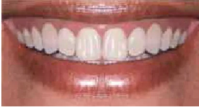
To test the aesthetic perception of these pro-fessionals and lay persons a female patient was selected, who agreed to participate in the study after signing a form of informed consent. This patient had an aesthetic gingival margin in which the central incisors and canines were at the same height as the gingival margin, and whose lateral
incisors were 1 mm below the tangent that con-nected the gingival margin of these teeth.
Digital manipulation of the patient’s smile was performed using Adobe Photoshop 7.0 by chang-ing the height of the gchang-ingival margin of the lateral and central incisors. Changes were measured rela-tive to the canines, which were therefore kept at a constant height. In the first alteration, the margin of the maxillary central incisor was reduced by 2 mm and the lateral incisor was increased by 0.5 mm in relation to the canines. In the second al-teration, the margin of the maxillary central inci-sor was reduced by 4 mm and that of the lateral incisor was increased by 1.0 mm in relation to the canines. During manipulation, the chin and nose were deleted to avert confounding factors, so that only a portion of the patient’s skin, her lips and teeth remainedapparent.4
The evaluators were given an album with pho-tos in the following order:
1. Harmonious gingival margin with the central incisors and canines at the same level, and lat-eral incisors 1 mm below the tangent between central incisors and canines (Fig 1).
2. Central incisors with margin 4 mm below the margin of the canines, and lateral incisors 1 mm above their original position, in an ascend-ing aspect (Fig 2).
FIGURE 4 - Analysis of the group of dentists. data collection
After photo manipulation, two albums were created containing the photos that were printed in size 10X15 cm on photographic paper so as to ensure that both albums would be made in the same location and with the same quality.
From then on data collection was started through blind and standardized interviews where two researchers, each responsible for half of the sample from each specialty and lay group, paid visits to show the album.
The evaluators were asked to choose the pic-ture that pleased them most and were thus given a choice of one, all or none, and 90 seconds to as-sess each picture without the possibility of see-ing again any previous pictures. After that, the interviewer questioned each evaluator in an at-tempt of fi nd out whether or not they perceived what was being changed in the sequence of pic-tures, i.e., if they were able to identify changes in the gingival plane, with the following question: “What do you think is being altered in these pic-tures?”. Each response was noted but, in order to facilitate statistical data analysis, a “yes” was assigned when changes were perceived (any al-teration in the gingival plane) and a “no” when the evaluator responded somewhat differently than expected. Up to 90 seconds were allowed for these questions.
The statistical analysis included descriptive and inferential analyses, using a 5% signifi cance level. Associations between categorical variables were tested using chi-square or Fisher’s exact tests.
Results
As regards the selection of the photographs that most pleased the evaluator, in the lay group the choice fell predominantly on the harmoni-ous gingival plane, indicating that differences between the smiles were indeed perceived (p = 0.05). Moreover, signifi cant differences were also observed in the overall results exhibited by the groups (p = 0.05), with the group of private
patients more prevalent in choosing the harmo-nious gingival smile than UFES patients (Table 1). Percentage-wise, fewer people in both groups chose the ascending gingival plane, with a sig-nifi cant difference between groups (Table 1).
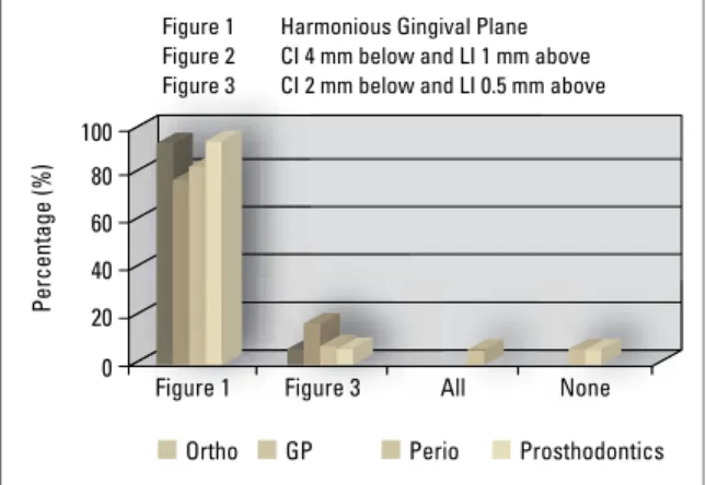
In the group of dentists there was also a sig-nifi cant prevalence in the selection of the har-monious gingival plane option (p<0.001), no selection of the ascending gingival plane op-tion while a low percentage opted for the fl at gingival plane (Fig 1). A higher percentage of subjects in the groups of orthodontists and prosthodontists selected the harmonious gingival plane option without, however, any signifi -cant differences between the groups of dentists (Table 2 and Fig 4).
The results showed a statistically signifi cant difference between the groups of dentists and lay persons (p<0.001), indicating a higher aes-thetic perception by the group of dentists (Ta-ble 3). Furthermore, selection of the harmoni-ous gingival plane option was statistically higher in the group of dentists (p = 0.01), and selection of the ascending gingival plane option was ob-served only in the groups of patients, where a statistically signifi cant difference was also found (Tables 2 and 3).
Figure 1 Harmonious Gingival Plane Figure 2 CI 4 mm below and LI 1 mm above Figure 3 CI 2 mm below and LI 0.5 mm above
100
80
60
40
20
P
e
rc
e
n
ta
g
e
(
%
)
Ortho GP Perio Prosthodontics Figure 3
Figure 1 All None
60
58%
25% 40
20
0
80
60
40
20
0
Offi ce
Patients PatientsUFES p-value
Harmonious 56.8% 31.1% 0.05
CI 4 mm below and
LI 1 mm above 2.6% 18.4% 0.05
CI 2 mm below and
LI 0.5 mm above 28.2%% 21.6% 0.05
All 12.6% 26.3% 0.05
None _ 2.6% 0.05
TABLE 1 - Dental offi ce patients vs. UFES patients. TABLE 3 - Dentists vs Patients.
TABLE 2 - Analysis of the group of dentists.
Orthodontists General practitioners Periodontists Prosthodontists p-value
Harmonious 95% 79% 84.2% 95% 0.538
CI 4 mm below and
LI 1 mm above _ _ _ _ 0.538
CI 2 mm below and
LI 0.5 mm above 5% 16% 5.3% 5% 0.538
All _ _ 5.3% _ 0.538
None _ 5.3% 5.3% _ 0.538
Dentists Patients p-value
Harmonious 88.5% 44.2% <0.001
CI 4 mm below and
LI 1 mm above _ 10.4% <0.001
CI 2 mm below and
LI 0.5 mm above 7.7% 24.7% <0.001
All 1.3% 19.5% <0.001
None 2.6% 1.3% <0.001
Identifi cation of changes in
gingival plane height
At this point in the interview the researchers wished to determine whether or not dentists and lay people were able to see the changes made in the photos. The chi-square test was performed to measure statistically signifi cant associations be-tween the groups. Statistically signifi cant results were found regarding identifi cation of the pres-ence of a non-harmonious gingival plane in the group of dentists (58.8%, p<0.001). Percentage-wise, prosthodontists were more vigilant in this identifi cation than other groups of dentists, al-though no statistical signifi cance was found in this group (p = 0.385) (Figs 5 and 6).
Values for problem identifi cation in the lay group were not signifi cant (25%, p = 0.100), but a signifi cant difference was found between the group of private practice patients and UFES patients (p = 0.010) (Table 1 and Fig 6). The difference between the group of dentists and the lay group was statisti-cally signifi cant (p<0.001) (Table 2 and Fig 5).
FIGURE 5 - Identifi cation of changes in Gingival Plane height: Dentists vs. Patients.
FIGURE 6 - Identifi cation of changes in Gingival Plane height: Evaluation of the groups of Patients and Dentists.
Dentists
Patients
Orthodontists Periodontists Offi ce patients General clinic Prosthodontists UFES patients 65%
45% 55%
70%
32.5%
disCussiOn
The orthodontist’s role in the correction and improvement of gingival aesthetic prob-lems has not been adequately explored as it should have been. An innovative approach should be disseminated across all areas of den-tistry so that all specialties are made aware of the possibility of achieving aesthetic improve-ments in gingival contour as well as biological improvements in support tissues by means of orthodontic treatment.2
With the purpose of assessing patients’ ex-pectations and the amount of discrepancy in abnormal symmetrical changes made to the gin-gival plane, a study was conducted which intro-duced progressive 0.5 mm to 1.5 mm changes in the position of lateral incisors, and concluded that these were not perceived by dentists nor lay persons. This study raised a debate on the possibility that these symmetrical changes are beyond perception at all levels.6
The present study, however, demonstrated that changes starting at a 2 mm decrease and a 0.5 mm increase, symmetrically applied to cen-tral and lateral incisors, respectively, by modify-ing the gum contour, would be significantly no-ticeable to lay people and dentists of all special-ties investigated in this study, corroborating oth-er findings in the litoth-erature1,4,9 and showing that changes in the anterior region, including at the gingival level, would be perceived by most den-tists and patients. In the group of denden-tists, as well as a significant prevalence in the selection of the harmonious gingival plane option (p<0.001), no selection of the ascending gingival plane option was made. Similarly, the significant prevalence of the harmonious smile option and low percentage rates of the options “all” and “no” in the group of lay patients showed that there is indeed the per-ception of changes in the gingival plane starting from this level of manipulation.
According to the literature symmetrical changes would only be noticeable in large
mag-nitudes.7 However, changes made in more than one tooth, which generate a conspicuously un-sightly gingival contour,1,3 become visible start-ing from 2 mm. In fact, the reverse gstart-ingival margin plane, with its ascending form, is signifi-cantly unsightly in the opinion of lay individu-als.10 On the other hand, asymmetrical changes become visible at much lower levels by dentists and laypersonsalike.7
When it comes to identifying changes in the gingival plane a significant identification by den-tists was observed, although with no statistically significant difference among the different spe-cialty groups. In addition, the group of dentists was statistically more perceptive in identifying the changes than the group of lay persons. In the patient group identification was not statisti-cally significant, but it was significantly higher in dental office patients. By the same token, from a statistical standpoint, the latter group predomi-nantly selected the harmonious gingival margin option during the first part of the interview, dem-onstrating that these patients are more aware and demanding than patients from public institutions in terms of changes in the gingival plane.
These findings showed that symmetrical changes equal to or greater than 2 mm de-serve special attention by orthodontists in their daily practice as they are identified as unsightly by the patient, especially in private practice, although the patient is not always capable of defining the problem. Moreover, the dentist who referred such patient can per-ceive and understand changes in the gingival plane, knowing that such changes can be cor-rected. Therefore, the options for orthodontic correction of these differences in the gingival margin should be presented cautiously since neglecting these issues might be construed as a treatment failure.
the gingival margin would be among the initial expectations of the patient seeking orthodon-tic treatment, or even if it would be a differen-tiating factor that could lead him/her to seek treatment. Further studies are required to shed light on these issues.
COnClusiOns
The authors concluded that for the popula-tion researched in this study:
» Symmetrical changes in the gingival plane
greater than 2 mm can be perceived by dentists and lay people.
» Dentists were significantly more percep-tive to the changes in the gingival plane than lay patients.
» No differences were found in the percep-tion of the gingival plane among the dental specialties investigated in this study. » The group of dental office patients was
significantly more perceptive than UFES patients.
1. Caudill R, Chiche G. Princípios cientíicos e artísticos aplicados à Odontologia estética. In: ______. Estabelecendo uma aparência gengival estética. 1ª ed. São Paulo: Quintessence Books; 1996. p. 13-32.
2. Chay SH, Rabie AB. Repositioning the gingival margin by extrusion. Am J Orthod Dentofacial Orthop. 2002 Jul;122(1):95-102.
3. Chiche G, Kokich V, Caudill R. Diagnosis and treatment planning of esthetic problems In: Pinault A, Chiche G, editors. Esthetics in ixed prosthodontics. Quintessence, 1994. p. 33-52.
4. Dong JK, Jin TH, Cho HW, Oh SC. The esthetics of the smile: a review of some recent studies. Int J Prosthodont. 1999 Jan-Feb;12(1):9-19.
5. Johnston CD, Burden DJ, Stevenson MR. The inluence of dental to facial midline discrepancies on dental attractiveness ratings. Eur J Orthod. 1999 Oct;21(5):517-22. 6. Kokich VO Jr, Kiyak HA, Shapiro PA. Comparing the perception
of dentists and lay people to altered dental esthetics (reprint). Advanced Esthetics Interdiciplinary Dent. 2005;1(1):20-33. 7. Kokich VO, Kokich VG, Kiyak HA. Perceptions of dental professionals and laypersons to altered dental esthetics: Asymmetric and symmetric situations. Am J Orthod Dentofacial Orthop. 2006 Aug;130(2):141-51. RefeRenCes
8. Sarver DM, Yanosky M. Principles of cosmetic dentistry in orthodontics: Part 2. Soft tissue laser technology and cosmetic gingival contouring. Am J Orthod Dentofacial Orthop. 2001;127(2):127-85.
9. Soh J, Chew MT, Chan YH. Perceptions of dental esthetics of Asian orthodontists and laypersons. Am J Orthod Dentofacial Orthop. 2006 Aug;130(2):170-6.
10. Yoon ME, Jin TH, Dong JK. A study on the smile in Korean youth. J Korean Acad Prosthodont. 1992 Aug;30(2):259-71.
Contact address
Daniela Feu
Rua Moacir Ávidos; n° 156/ apto 804
CEP: 29.055-350 – Praia do Canto, Vitória / ES, Brazil E-mail: [email protected]
Submitted: May 2007