Sarah Broderick1, Xiaoxi Wang1, Nicholas Simms3, Andrea Page-McCaw1,2*
1Department of Cell and Developmental Biology and Program in Developmental Biology, Vanderbilt Medical Center, Nashville, Tennessee, United States of America, 2Department of Cancer Biology, Vanderbilt Medical Center, Nashville, Tennessee, United States of America,3Department of Biology, Rensselaer Polytechnic Institute, Troy, New York, United States of America
Abstract
Ninjurins are conserved transmembrane proteins that are upregulated across species in response to injury and stress. Their biological functions are not understood, in part because there have been fewin vivostudies of their function. We analyzed the expression and function of one of threeDrosophilaNinjurins, NijA. We found that NijA protein is redistributed to the cell surface in larval immune tissues after septic injury and is upregulated by the Toll pathway. We generated a null mutant of NijA, which displayed no detectable phenotype. In ectopic expression studies, NijA induced cell death, as evidenced by cell loss and acridine orange staining. These dying cells did not display hallmarks of apoptotic cells including TUNEL staining and inhibition by p35, indicating that NijA induced nonapoptotic cell death. In cell culture, NijA also induced cell death, which appeared to be cell autonomous. These in vivo studies identify a new role for the Ninjurin family in inducing nonapoptotic cell death.
Citation:Broderick S, Wang X, Simms N, Page-McCaw A (2012)DrosophilaNinjurin A Induces Nonapoptotic Cell Death. PLoS ONE 7(9): e44567. doi:10.1371/ journal.pone.0044567
Editor:Partha Mukhopadhyay, National Institutes of Health, United States of America
ReceivedMay 11, 2012;AcceptedAugust 9, 2012;PublishedSeptember 28, 2012
Copyright:ß2012 Broderick et al. This is an open-access article distributed under the terms of the Creative Commons Attribution License, which permits unrestricted use, distribution, and reproduction in any medium, provided the original author and source are credited.
Funding:This work was supported by the Basil O’Connor Starter Scholar Research Award no. 5-FY07-99 and by National Institutes of Health R01 GM073883, both to APM. The funders had no role in study design, data collection and analysis, decision to publish, or preparation of the manuscript.
Competing Interests:The authors have declared that no competing interests exist.
* E-mail: [email protected]
Introduction
Ninjurins are a conserved family of transmembrane proteins first identified by upregulation in injured rat nerves [1]. There are two Ninjurin family members in mammals, Ninjurin1 and Ninjurin2 [2], and three inDrosophila, Ninjurin A, B, and C [3]. Ninjurins are small proteins of,16–27 kDa, with an N-terminal
ectodomain and two predicted transmembrane domains near the C-terminal end. In humans, mice, and Drosophila, Ninjurin transcripts are upregulated upon injury, infection, or stress suggesting that not just their structure but also their function is conserved [1,4,5,6,7,8,9] (and see supplemental data in [10,11,12]).
Although little is known about the functions of Ninjurins, many studies have implicated them as adhesion molecules, either directly through homophilic binding on the cell surface [1,13,14,15] or by regulating adhesion via their ectodomain [3]; yet these adhesion studies have been limited to cell culture models.In vivo, some data suggests that Ninjurin1 may promote hyaloid vasculature regres-sion in mouse embryos, as neutralizing antibodies against Ninjurin1 delay this regression, although the relationship between Ninjurin1 and cell deathin vivois unclear from these studies [14]. To our knowledge, no Ninjurin mutants or knock-outs have been reported in any organism.
In this study, we show that Ninjurin A (NijA) protein responds to septic injury in a developmentally regulated manner, as whole-animal levels increase in adults but not in larvae. Rather, in larvae the protein distribution is altered in immune tissues after injury, and NijA protein levels can be elevated via the Tl immune signaling pathway, suggesting that NijA may function in the immune system. We generated several deletion mutants of NijA including a molecular null allele but no phenotype was observed in
these animals. In a gain-of-function approach, however, we found thatNijAinduced cell death at a level comparable to the known apoptotic genehid, yet the NijA-induced death does not have the hallmarks of apoptosis. From cell culture studies, we conclude that NijA is likely to induce cell death in a cell-autonomous manner, rather than as a nonautonomous signaling molecule.
Results
NijA distribution is regulated in larval immune tissues
G; p = 0.0048, Fisher’s exact test). Blood cells were examinedex vivo, and NijA staining was altered in 9/9 samples of blood cells after septic wounding compared to 9 unwounded (Fig. 1H,I; p = 4.161025, Fisher’s exact test). In both fat and blood cells, there
was a marked increase in NijA localized to the cell surface, easily observed in unpermeabilized tissue since our antibody recognizes an extracellular epitope of the NijA transmembrane protein [3]. Thus NijA protein responds to septic injury in both adults and larvae.
Since fat body and blood cells are both Drosophila immune organs [17], we asked whether the immune regulator Tl was capable of regulating NijA [18]. We found that whole larvae with the constitutively activeTl10bmutation have higher levels of NijA protein, even in the absence of injury (Fig. 1C, D). Anti-NijA immunostaining of the fat body indicated that NijA levels were increased in this tissue in 9/9Tl10b
mutants compared to wild type (Fig. 1K–M; p = 4.161025, Fisher’s exact test), and this Tl-mediated upregulation appears to increase NijA levels at the cell surface. The sufficiency of Tl to upregulate NijA in larvae is consistent with the microarray findings of De Gregorioet althat spz flies, which cannot activate the Tl pathway, also cannot upregulateNijA; in contrast, larvae mutant for the Imd pathway were able to upregulateNijAlike wild type [11]. The regulation of NijA by the Tl pathway, combined with its relocalization after septic injury in the immune tissues of the blood and fat body, suggest that NijA functions in the immune system of larvae.
NijAis not required for viability
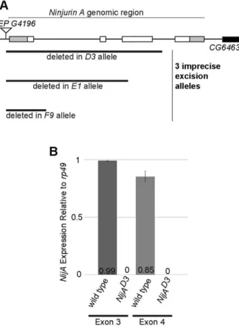
To understand the functional requirements forNijA, we made a deletion mutant by excising aP element at the genomic locus. Three imprecise excisions were generated that removed part of the NijA coding sequence: D3, E1, andF9 (Fig. 2A). The D3 allele removed the 59UTR and most of the coding region including the last internal methionine, suggesting thatD3may be a null allele. To determine whether there was internal translation of the 39
remnant of the gene in theD3allele, we performed quantitative PCR on the fourth exon, present in theD3allele, comparing its transcription level to the third exon, deleted from theD3allele and acting as a negative control. We found no transcription of either the third or fourth exon, confirming that theD3 allele is a null (Fig. 2B).NijAD3homozygous mutants were viable and fertile with Figure 1. Ninjurin A protein response to septic wounding.(A) Western blot of whole adult male lysates probed with anti-NijA. NijA increases expression two hours after infection in adults. NijAD3
null lysates demonstrate antibody specificity. Black lines indicate regions of the blot that were omitted for clarity. (B) Graph representing three replicates of the western blot pictured in (A). NijA levels increase significantly in adults after septic injury (p = 0.003). (C) Western blot of whole male larval lysates probed with anti-NijA. NijA levels do not change 2 h after septic injury in third instar larvae; in contrast larval
Toll10b
gain-of-function mutant larvae have increased levels of NijA protein. (D) Graph representing five replicates of the western blot pictured in (C). NijA levels increase significantly in constitutively activate
Toll10bmutant larvae (p,0.0001). (E–M) Anti-NijA (red) and DAPI (blue) labeling nuclei. All scale bars are 10mm. (E–G) Anti-NijA stained
non-permeabilized fat bodies of male third instar larvae show an increase in NijA at the cell surface 2 h after septic injury (compare E and F). (G)
NijAD3
larvae demonstrate the NijA antibody specificity. (H,I) Anti-NijA stained non-permeabilized hemocytes of third instar larvae ex-vivo
show an increase in NijA at the cell surface 2 h after septic injury. (K–M) Anti-NijA stained permeabilized fat bodies of male third instar larvae show increased NijA expression in gain-of-function Toll10b
mutants. Error bars in (B,D) represent standard error of the mean.
no obvious developmental abnormalities (data not shown). Thus NijAis not required for viability.
To examine the role ofNijAin the immune system, we tested viability of NijAD3 homozygous mutants after wounding or infection with gram positive or negative bacteria, but found no differences in survival or melanization (Supporting Information S1 and data not shown). The ability ofNijAD3mutants to mount an antimicrobial peptide response after septic injury was examined by measuring Drosomycin (Drs) or Drosocin (Dro), both targets of the Tl pathway [18]. Both antimicrobial peptides were elevated in the NijAD3mutant in a manner not significantly different from wild type (Supporting Information S2 A,B). To further assess a potential role forNijAin Tl signaling, we performed an epistasis test to ask whether NijAis required for the upregulation of a Tl pathway target whenTlis genetically activated by mutation in the fat body (by drivingTl10bwith thec564-GAL4driver). Examining the levels of Drs, we found that Drs elevation was similar after septic wounding of wild type or genetic activation ofTl10b
in the fat body, and these levels were not affected by theNijAD3mutation inTl10b NijA double mutants (Supporting Information S2C). To examine the cellular immune response we assayed phagocytosis and found that the capacity of hemocytes or S2 cells to
phagocytose labeled E. coli did not depend on NijA, although hemocyte phagocytosis was sensitive to a dominant locus on the NijAD3 chromosome (Supporting Information S3). Similarly, a dominant locus on theNijAD3
chromosome obscured our ability to assess a homozygous mutant phenotype in response to starvation (Supporting Information S4). Thus we were unable to identify a function forNijAusing loss-of-function approaches.
Ectopic expression ofNijAinduces nonapoptotic cell death
Because NijA is upregulated on septic injury, we focused on analyzing its function by ectopically upregulating NijA using the GAL4/UAS system for a gain-of-function approach. Under the ubiquitously expressed tubulin and actin drivers, we found that embryos died with morphological abnormalities around the time of cellularization (data not shown), which is also when zygotic transcription begins [19,20]. Lethality was also observed when we expressedNijAwithA58-GAL4in the larval epidermis [21] or in most blood cells withHe-Gal4[22] (data not shown). Attempts to control the onset of lethality by using the conditional GAL80ts inhibitor [23] or the inducibleGeneSwitch-GAL4 driver [24] both failed because even under restrictive conditions (18uor absence of RU486, respectively) leaky expression ofNijAstill caused lethality (data not shown). We expressedNijAin a tissue not required for viability, the eye, withGMR-GAL4and ey-GAL4; to our surprise, NijA expression with each driver was pupal lethal (Table 1 and data not shown). Expression ofNijAwithhml-GAL4, expressed in differentiated blood cells of the larval lymph gland and in circulation [25,26], was not lethal and allowed us to ask about the cellular consequences of NijA overexpression.
Hmlexpressing cells, visualized byHml.GFP, can be observed under the cuticle of whole third instar larvae (Fig. 3A,B); however, whenNijAis co-expressed, the cells appear to be absent (Fig. 3C). To compare the effects ofNijAto a known apoptosis inducer, we drove the expression ofhidwithhml-GAL4, and found a similar loss of GFP-labeled cells (Fig. 3D). The similarity of these results suggested thatNijAinduces cell death in differentiated blood cells. We confirmed by quantitative Western blotting that GFP was absent from animals co-expressing GFP and either NijA or hid (Fig. 3E,F). This lack of GFP indicated that larvae were missing nearly all Hml-expressing cells, both in circulation and in the lymph gland, the site of hematopoiesis in larvae.
We examined dissected lymph glands expressingNijA. Although 7/7 control (hml.GFP) lymph glands did not stain with acridine
Table 1.Organismal death resulting from GMR-driven expression of a cell-death gene is not dependent on the gene acting non-autonomously.
#GMR.UAS Expected cross
progeny
observed Mendelian# X2value
if viable
GMR-GAL4/CyO
x 3/338 (0.89%) 85.5/338 (25%) 104.8 (p,0.001)
UAS-NijA/TM3
GMR-GAL4/CyO
x 3/175 (1.7%) 58.3/175 (33%) 78.7 (p,0.001)
UAS-hid/CyO
doi:10.1371/journal.pone.0044567.t001
Figure 2.NijAD3mutants do not express mRNA from theNijA
genomic locus.(A) Schematic of theNijAlocus showing all four exons. Gray indicates untranslated regions and white indicates open reading frame. Three excision alleles (D3, E1, and F9) were generated from imprecise excisions ofEP G4196. (B) qPCR data from primers specific to exon 3 (a negative control, as it is deleted in theD3allele) or exon 4 of
NijA. TheNijAD3
mutant did not produce any detectable mRNA from exon 4 of theNijAlocus, even though exon 4 remains in the genome, indicating that theD3allele is a null. Error bars represent standard error of the mean.
orange, which can only permeate dead cells, in contrast all lymph glands expressing eitherNijAor the apoptosis inducerhid stained brightly with acridine orange (7 of each genotype, compare Fig. 3G–I9). The dying cells appeared to be restricted to differentiated cells that expresshml, as undifferentiated hemocytes expressing hemesebut not hml were still present in lymph glands expressing hml.NijA or hml.hid (Fig. 2J–L). We were able to
detect little to no GFP signal with an anti-GFP antibody in the lymph glands ofhml.NijA,GFPorhml.hid,GFPanimals (Fig. 3J9– L9), consistent with the dramatic decrease in GFP expression in whole animals by western blot analysis (Fig. 3E–F). Interestingly, NijA was not required for developmentally regulated apoptosis, however, as apoptosis in the stage 10 embryonic head region occurs normally in aNijAD3
mutant with no zygotic or maternal Figure 3. Ninjurin A over-expression in the lymph gland causes nonapoptotic cell death.(A) Wild-type w1118larva demonstrating background autofluorescence. (B)hml.GFPlarva with GFP-positive differentiated hemocytes along posterior body wall and in lymph gland. (C)
hml.NijA,GFPlarva lacked GFP-positive cells. (D)hml.hid,GFPlarva, a cell-death positive control, lacked GFP-positive cells. (E) Western blot of whole larval lysates probed with anti-GFP.hml.NijA,GFPandhml.hid,GFPlarvae were devoid of GFP. (F) Quantification of three western blots probed for anti-GFP as in (E). GFP is virtually absent fromhml.NijA,GFPandhml.hid,GFPlarvae. Error bars represent standard error of the mean. (G–I9) Live partially dissected 3rdinstar larval lymph glands (arrows) were stained with acridine orange to detect cell death. Scale bars are 200
mM. tr: trachea; id:
imaginal disc; vnc: ventral nerve cord; bl: brain lobe. (G9)hml.GFPlarval glands did not stain with acridine orange. (H9)hml.hid,GFPglands, a cell-death positive control, stained with acridine orange. (I9)hml.NijA,GFP glands stained with acridine orange, demonstrating thatNijAinduced cell death. (J–L0) Larval lymph glands were fixed, TUNEL labeled, and antibody stained. Scale bars are 50mm. (J–L) Anti-Hemese staining labeled the
lymph glands. (J9–L9) Anti-GFP staining shows no GFP-positive (hml+) hemocytes inhml.hid,GFP(K9) orhml.NijA,GFP(L9) larval glands. (J0–L0) TUNEL-labeled glands. (J0) Few TUNEL positive cells inhml.GFPnegative control glands. (K0) Many TUNEL positive cells inhml.hid,GFPpositive control glands. (L0) Few TUNEL positive-cells in thehml.NijA,GFPglands, indicating thatNijAdoes not induce apoptosis. (M) Larvae expressing the apoptotic inhibitor p35 (hml.p35,GFP)displayed GFP-positive hemocytes similar tohml.GFPin (B). (N–O) p35 did not inhibit the NijA-induced loss of the GFP-positive cells inhml.NijA,p35,GFPlarvae. (P) p35 inhibited the hid-induced loss of the GFP-positive cells inhml.hid,p35,GFPlarvae, a positive control for p35 inhibition. In (A–D, M–P), anterior is up.
NijA (Supporting Information S5). Thus NijA induces cell death when ectopically expressed.
To examine whether NijA induces cell death via apoptosis, we examined lymph glands by TUNEL staining, a nuclear label for apoptotic cells. We compared lymph glands expressingGFPand NijA to glands expressing only the GFP marker as a negative control, and to glands expressingGFPand the apoptosis-inducer hid as a positive control. We counted the number of TUNEL positive nuclei in primary lymph gland lobes: fewer than 30 nuclei was considered background, and more than 30 (often uncountable) was considered apoptotic. By these criteria, we found that 10/11 hid-expressing lymph glands were apoptotic, whereas 22/23 GFP lymph glands were TUNEL-negative and 9/13 NijA-expressing lymph glands were TUNEL-negative (Fig. 3J0–L0). The pene-trance of apoptosis inNijAlymph glands is significantly different fromhidlymph glands by Fisher’s exact test (p = 0.004). Some of the unexpected variability in ourNijAoverexpressing lymph glands may be an artifact of our cut-offs, as 3 of the 4NijA expressing lymph glands scored as ‘‘apoptotic’’ qualitatively appeared to have fewer TUNEL positive cells than the hid expressing glands;
another possibility is that there is significant cross-talk between cell death pathways (see Discussion).
As a second independent assay to assess whetherNijA induces apoptotic or nonapoptotic cell death, we co-expressedNijA with p35, an apoptotic inhibitor that blocks cell death when co-expressed with reaper or hid [27,28]. Examining GFP-labeled hemocytes in live animals, we found that p35 inhibited Hid-induced cell death as expected, but importantly p35 did not inhibit NijA-induced cell death (Fig. 3M–P). Our data indicate thatNijA induces a nonapoptotic form of cell death, as dying cells do not label with TUNEL and cell death is not inhibited by p35.
NijA appears to kill cells in a cell-autonomous manner
Because the expression of NijA in the eye withey-GAL4or GMR-GAL4was lethal to the animal, we asked whether this phenotype represented tissue nonautonomous cell death,i.e., if NijA expressed in the eye disc was effectively instructing tissues outside the eye to die. Alternatively, it was possible that even the autonomous destruction of a large tissue may release toxic factors that could cause animal lethality. To determine if we could assess tissue Figure 4. NijA induces cell death inDrosophilaS2 cell culture.(A)NijAexpression kills S2 cells. Cells were transiently transfected withpRmHa3
empty vector orpRmHa3-NijAand induced with copper for 48 h. The percentage of dead cells was determined by dividing the number of trypan blue positive cells by that of total cells counted for each sample. Data from 8 experiments are shown. Error bars indicate S.E.M, and Student’s T test was used to calculate p value. (B) Mmp1 activity is not required forNijA-induced cell death. Cells were transiently transfected and induced for 48 h.
Mmp1E255A
autonomy in this assay, we expressedhid, which is known to be an autonomous cell-death gene [27] also under GMR-GAL4 and found that the overexpression ofhid caused organismal lethality with very few escapers (Table 1 and data not shown). Either GMR-GAL4is not eye-specific or the massive developmentally-inappro-priate induction of cell death is sufficient to cause organismal death; in either case this assay cannot indicate the autonomy of NijA-induced cell death.
As another means to assess the autonomy of NijA-induced cell death, we turned to cell culture. We previously reported that when overexpressed in cultured Drosophila S2 cells, NijA inhibits cell adhesion in a nonautonomous manner within a few hours of its
induction. This NijA-mediated phenotype was dependent on the activity of the protease Mmp1, and the NijA ectodomain was sufficient to release adhesion even when Mmp1 proteolysis was inhibited [3]. We revisited these experiments and found that when S2 cells expressNijAfor longer periods, the cells died as measured by trypan blue exclusion: after 24 h, 20.2% of cell had died (not shown), and after 48 hours 33% of cells had died, a significant increase over the background death rate of about 9% in non-expressing cells (Fig. 4A). Interestingly, like in whole flies,NijAwas not required for apoptosis, as the apoptosis-inducer actinomycin D [29,30] was able to induce death at similar levels in wild-type and NijAknock-down cells (Fig. 4C,D), indicating that NijAis not an essential component of the cell death machinery. In contrast to our previous adhesion results, NijA-induced death does not require Mmp1 (Fig. 4B). Similarly, we found that the NijA ectodomain is not sufficient to trigger cell death, even though we were able to detect the tagged ectodomain in cells, cell lysates, and in the culture medium (Supporting Information S6). These Mmp1 and NijA ectodomain results suggested that NijA may induce death in a cell-autonomous manner.
To examine the autonomy of Ninjurin-induced cell death, we considered testing conditioned media, but we were concerned that dying cells may release toxic factors into the media even if the death were cell autonomous, similar to our results in whole animals. Instead, we chose to examine the relationship between NijA transfection status labeled by GFP and cell death measured by trypan blue exclusion. We co-transfected NijA and GFP expression vectors and counted the number of GFP-expressing cells remaining after 48 h induction, as transfected cells are expected to take up both vectors simultaneously (Fig. 5). When transfected with only the GFP plasmid, 39% of cells expressed GFP, and 8% of cells were dead as measured by trypan blue exclusion (Fig. 5A), consistent with the baseline rate of cell death we measured in these cultures (Fig. 4). In contrast, when cells were co-transfected withNijAandGFP, only 9% of cells expressed GFP, and 37% of the cultured cells were dead as measured by trypan blue exclusion (Fig. 5A). Thus it appeared that NijA-expressing cells were much more likely to die than their non-transfected neighbors. We continued this analysis to ask which residues were important for inducing cell death, examining four site-directed NijA mutants generated by alanine-scanning [31], in which charged residues in the ectodomain were replaced with alanines (Fig. 5A,C). We found that D140 was absolutely required, as this point mutation ablated NijA’s ability to induce death; importantly the D140A mutant protein was not able to localize correctly to the cell surface (Fig. 5D–F), suggesting that cell-surface localization is critical for inducing death. Surprisingly, two mutants seemed to increase the potency of NijA, as D124A or the double mutant K131A, K132A were more toxic to cells than wild-type NijA, resulting in no GFP-positive cells; yet because the fraction of dead cells was similar to the sum of the baseline death rate plus the transfection rate, it appears that this overactive toxic mutant still killed in a cell-autonomous manner. The double-mutant R152A,
staining, and transfection status was assessed as GFP fluorescence. Wild-typeNijAand mostNijAmutants killed cells, whereas the mock control, the
D140Amutant, and the N-terminal deletion (B) showed low levels of cell death. The sum of transfected live cells (GFP+) plus dead cells was relatively
constant across samples despite the augmented or compromised capacity to kill cells, indicating that NijA kills the cell it transfects but not others. Data from 3 replicates are shown. Error bars indicate S.E.M. (C) Schematic showing topology of NijA (form A) protein and the extracellular region recognized by our polyclonal antibody. Amino acid residue numbers are indicated. (D–G,I) Immunofluorescence localization of wild-type NijA or NijA mutant forms expressed in S2 cells and stained with anti-NijA (D–G) or anti-c-Myc (I), both extracellular epitopes. For each construct, staining was performed on permeabilized cells to show NijA protein levels, and on unpermeabilized cells to show NijA cell-surface localization. Permeabilization status was verified by anti-tubulin staining. The merge image combines images for NijA (red), tubulin (cyan), DAPI (blue) and GFP fluorescence as a transfection control (green). Bar: 10mm. (H) Diagram showing placement of the myc epitope for (I), necessary because the NijA antigenic region was
deleted in this mutant.
doi:10.1371/journal.pone.0044567.g005
Figure 6.NijAmutants behavein vivoas they do in cell culture.
(A) Third instarhml.GFP,NijA152156Alarvae were devoid of GFP-positive hemocytes, indicating that these mutants were capable of inducing cell death and that polar amino acids 152 and 156 were not required for cell death. (B) hml.NijAect,GFP larvae displayed visible GFP-positive hemocytes on the body wall, suggesting that NijAectwas not sufficient to induce cell death.
E156A killed cells similarly to wild-type NijA (Fig. 5A), and the mutant protein localized at the cell surface similarly to wild-type NijA (Fig. 5G). In a separate experiment, we asked which domains were required to induce death (Fig. 5B). The N-terminal ectodomain was required, as a deletion removing it (NijADN-term) induced only 8% death with 52% of the cells expressing GFP, similar to the GFP-alone controls. The ectodomain was also required for localization of NijA to the cell surface, as the NijADN-termprotein (carrying a myc epitope at the new N-terminus) was not detectable in unpermeabilized cells (Fig. 5I). The predicted 20-amino acid intracellular domain was partially required, as a mutant replacing most of the intracellular sequence (NijADintracell) with the myc epitope gave an intermediate level of death, with 20% dead cells and 32% GFP-expressing cells. Taken together, these data strongly suggest that NijA induces death in a cell-autonomous manner, requiring cell surface localization to kill cells.
To determine if these mechanisms applied to NijA cell death induction in vivo, we transformed flies with GAL4-inducible constructs encoding the NijA double mutant R152A, E156Aand the NijA ectodomain (UAS-NijAect). As in cell culture, the double mutant phenocopied wild-type NijA, whereas the ectodomain alone did not induce cell death comparable to wild-type NijA (Fig. 6). We conclude that when expressed at high levels,NijAkills cells by a nonapoptotic mechanism, likely in a cell autonomous manner.
Discussion
In the present study, we found that Drosophila NijA is upregulated or relocalized in tissues of the immune system upon septic injury. The NijA upregulation observed in adults may result in more protein localized to the cell surface, a similar effect to the relocalization to the cell surface observed in larvae after septic injury. NijA protein levels are upregulated via the Tl pathway, an established immunoregulatory pathway, and in activated Tl mutants, there is more NijA observed at the surface of fat body cells, supporting the idea that NijA upregulation and relocalization are functionally similar immune responses. In whole-animal genetic experiments we found that the ectopic upregulation of NijA induces cell death characterized by acridine orange staining and tissue loss. However, NijA-induced cell death is not associated with DNA fragmentation, as assessed by TUNEL labeling, and is not suppressible by p35, an apoptotic inhibitor. These data indicate that NijA induces nonapoptotic cell death. Similar death phenotypes were observed both in tissues of whole larvae and in cultured S2 cells, and the cell death appears to be autonomous. In cultured cells,NijAmutants that cannot localize to the cell surface also cannot induce cell death, suggesting thatin vivothe observed increase of NijA levels at the cell surface after septic injury may be critical to NijA function.
Originally identified as gene products upregulated on nerve injury, Ninjurins have been characterized as adhesion molecules [1,13,14,15], anti-adhesion signals [3], and mediators of cell cycle regulation [32]. This lack of consensus likely stems from the fact that these studies have been performed in cultured cells. There is scant information about Ninjurin functionin vivo, in part because animal studies have focused almost exclusively on expression analysis [1,2,3,14,15]. The only study investigating Ninjurin function in vivo was performed with neutralizing antibodies to Ninjurin1 during rat ocular development [14], and it found that Ninjurin antibodies slowed the regression of the hyaloid vascula-ture, an embryonic tissue that is removed by cell death during development.
Our in vivo gain-of-function data suggests that Ninjurins promote nonapoptotic cell death. In addition to apoptosis, the other main types of programmed cell death are autophagy and programmed necrosis (sometimes called necroptosis) [33]. Au-tophagy, a process in which the cell digests its own components, can regulate a variety of cellular processes including viral clearance and signal transduction [34,35]. Autophagy can result in cell survival in response to stress or can lead to cell death [36]. The death outcome utilizes components of the apoptotic machinery [37]. The differences between autophagic death and apoptosis are still being elucidated, and it is likely that in some contexts they can compensate for each other if one death mechanism fails [38,39]. The pathways leading to programmed necrosis also have considerable crosstalk with the pathways leading to apoptosis, and they are believed to inhibit each other [40]. Unfortunately, programmed necrosis has not been characterized inDrosophila, although it may be induced genetically [41,42]. We speculate that the interplay between these different types of cell death may explain why we observe no cell-death phenotypes in the NijA null mutants. Because cell death mechanism are known to compensate for each other, it is possible that in our NijA null mutants, cells that would have been killed by a NijA-dependent mechanism are now killed by another cell-death mechanism. Of note, genome-wide expression studies report that NijA is expressed during metamorphosis at levels,12-fold higher than during any
other time in the Drosophila life cycle ([43] as reported at flybase.org), suggesting that NijA may function during cell and tissue death in development. Interestingly, several Toll family members (Toll, 18-wheeler/tollo, Toll-6, and Toll-7) are also highly expressed during metamorphosis as well [44], and our data demonstrates that Tl can upregulate NijA.
Another possibility to explain the lack of cell death or other phenotypes in ourNijAnull mutants is genetic redundancy among the three Drosophila Ninjurin genes, NijA, NijB, and NijC. However, expression data does not support the idea of Ninjurin family redundancy. Genome-wide expression data sets, examining developmental timing and tissue-specific expression of all genes, indicate thatNijA, NijB, andNijCare not expressed in similar times or tissues in developing flies ([43,45,46] as reported at flybase.org). Nevertheless, it is possible that analysis of double and triple mutants may uncover redundant Ninjurin functions. In our preliminary examination ofNijA, NijCdouble mutants we have not found any obvious developmental abnormalities (X. Wang and A. Page-McCaw, unpubl.).
pathogen clearance from cells, but at physiologic levels NijA does not promote cell death at all.
Our genetic gain-of-function studies indicate that NijA induces cell death in a cell-autonomous manner. This is different from the nonautonomous apoptotic cell death observed in endothelial cells adjacent to Ninjurin1-positive macrophages that was interpreted as a result of altered adhesion [14], and it is different from the cell nonautonomous loss-of-adhesion signaling we observed in cultured cells [3]. It is possible that in different contexts Ninjurins can act either autonomously or nonautonomously, as their cell-membrane location would allow them to relay information from outside to inside the cell, or allow them to signal via their extracellular domain to other cells. Our study highlights a novel and potentially medially important role for the conserved Ninjurin family in inducing cell death.
Materials and Methods
Drosophilagenetics and imaging
Unless otherwise noted flies were raised at 25uunder standard conditions.Hml-GAL4, UAS-GFPandUAS-hidwere from J. Royet [25]; C564-GAL4 and Tl10b were from K.V. Anderson [50,51]; UAS-Tl10bwas from S. Cherry [52]. TheEPelementG4196was generated by Genexel (Korea) and deposited at the Bloomington DrosophilaStock Center. The site of the insertion was determined by sequencing genomic DNA to be 51 bp upstream of the transcription start site. The insertion line was outcrossed 3 times to w1118 before excising the transposon. 60 excision lines were screened in pools of five by PCR amplification of a 3561 bp genomic fragment surrounding the P insertion site; from these three imprecise excisions (shown in Figure 2) were identified by gel electrophoresis of PCR products. TheUAS-NijAline was generated by ligating the cDNA RE5744 (Berkeley Drosophila Genome Project) corresponding to NijA-RA, into thepUASTvector at the EcoR1 and BamH1 sites. The fly transformation vectors UAS-NijAect and UAS-NijAR152A,E156A were generated by ligating the inserts from the corresponding RmHa3plasmids (see below) into pUAST. Transformants were generated by Genetic Services Inc (Cambridge, MA). We examined two independent transformants of the UAS-NijA and UAS-NijAect, and in both cases we saw comparable results.
For hml.GFP analysis in whole larvae, third instars were selected from the food of a healthy vial, washed in sterile water to remove debris, and placed on a grape juice plate. GFP-labeled blood cells were scored in live larvae under a Zeiss LumarV12 fluorescence stereomicroscope prior to heat-killing for imaging. A minimum of 20 animals were scored per genotype. For imaging, each larva was placed in 20ml of PBS on a cover slip, and heated
for 5 sec at 95uC to kill the larvae. Larvae were immediately imaged by bright field and epifluorescence with a Zeiss 0.86
Neolumar objective, on magnification setting 646. Images were
cropped and edited using Adobe Photoshop.
For imaging lymph glands live-stained with acridine orange, third instar larvae were selected from the food of a healthy vial and washed in sterile water to remove debris. Each larva was placed in a 50ml drop of freshly diluted 1.661026M acridine orange in Drosophila Ringer’s solution on a Sylgard dissection plate. The dorsal cuticle was carefully torn away from posterior to the anterior to expose the internal organs. The flap of dorsal cuticle was pinned to the plate, and the fat surrounding the dorsal vessel quickly cleared away for imaging. Just prior to imaging the acridine orange solution was removed and replaced withDrosophila Ringer’s solution. Bright field and epifluorescence images were
taken on a Zeiss LumarV12 with a 1.56Neolumar objective and
processed using Adobe Photoshop.
Western blotting and antibody staining
For Western blots, lysates were made by mechanically grinding samples on ice in Laemmli buffer and heating at 95uC for 5 min. Blots were probed with guinea pig anti-NijA at 1:1500 [3], mouse anti-GAPDH at 1:2000 (IMGENEX, #IMG3073), mouse anti-Actin at 1:2000 (Abcam,#ab6276), and rabbit anti-GFP at 1:2000 (Abcam, #ab6556). Detection was performed by either HRP-mediated chemiluminescence or fluorescence imaging (Licor, Odyessy). Figure 1A and Supplementary Figure 3C were probed with HRP-labeled goat anti-guinea pig at 1:5000 (Santa Cruz) and HRP-labeled goat anti-mouse at 1:5000 (Jackson Immuno-research) secondary antibodies. Figures 1C, 2E, and 4D were probed with IRDye 680-labeled donkey anti-mouse, IRDye 800CW-labeled donkey anti-guinea pig, IRDye 680-labeled donkey anti-rabbit, or IRDye 800CW-labeled donkey anti-mouse, all diluted 1:5000 (Licor). Data was quantified using the ImageJ software. Blots were cropped and edited using Adobe Photoshop. For antibody staining, samples were dissected, fixed in 4% paraformaldehyde in phosphate buffered saline (PBS) for 20 mins, and then blocked for 30 mins in 1% bovine albumin serum (BSA) in PBS+0.2%Tween (PBST) for permeabilized samples or in 5% normal goat serum (NGS) in PBS for non-permeabilized samples. Primary antibodies were diluted in blocking reagent and incubated with sample overnight at 4uC. Primary antibodies used were guinea pig anti-NijA at 1:100 [3], mouse IgG2a anti-hemese at 1:100 (from Istvan Ando [53]), and rabbit anti-GFP at 1:50 (Abcam, #ab6556). Samples were washed and labeled with secondary antibody for 2 h in the dark at room temperature. Secondary antibodies used were Cy3-labeled goat anti-guinea pig, DyLight 649-labeled goat anti-mouse, and FITC-labeled goat anti-rabbit, all diluted 1:500 (Jackson ImmunoResearch). TUNEL labeling was performed according to the manufacturer’s directions (RocheIn situ cell death detection Kit TMR red). Tissues were mounted in Vectashield (Vector Lab) and imaged on a Zeiss Imager M2 with Apotome. Images are 2D projections of Z-sections. Projections were generated by the ImageJ software, and images were cropped and edited using Adobe Photoshop.
Hemocytes were recovered forex vivostaining as described [54]. In brief, the posterior end of a clean larva was bled onto a glass slide, and the hemolymph was recovered with a pulled glass needle. (To control the needle suction with a P20 pipettor, the dull end of the pulled needle was attached to the small end of a P20 tip by melting the plastic tip and sealing with nail polish.) The hemolymph was transferred to a Multitest slide (MP Biomedicals) in 10 ul of PBS, and the hemocytes were allowed to settle for 10 mins. Hemocytes were fixed for 7 mins in 4% paraformalde-hyde in PBS, rinsed briefly in PBS, and then blocked for 15 mins in 1% BSA in PBST. The primary antibody was applied to the samples for 2 h while the slide was in a humidified chamber. The samples were washed in several changes of PBST, and the secondary antibody was applied for 1 h in a dark humidified chamber. The samples were washed several times in PBST, and once in PBS prior to mounting in Vectasheild mounting media for viewing on a Zeiss Imager M2 with a Plan Neofluor 406oil objective.
qPCR Analysis
treated with DNase to remove contaminating DNA with the TURBO DNA-free kit (Ambion). 800 ng of total DNase-treated RNA was reverse-transcribed into cDNA pools using the iScript cDNA Synthesis Kit (BioRad) according to the manufacture’s directions in an Eppendorf AG 22331 Hamburg Thermocycler. 2ml of the cDNA pools were primed with validated primers sets for NijAExon 3 (Fwd:AACTGTTGGAGGCAACGGAG, Re-v:AAAGGAGAAACTGGGTCGTCTT. R2,0.99), NijAExon 4 (Fwd:GCGTGGGCCTTATATTGATG, Re-v:TGTTCGCCCGGCAGATAT, R2,0.99), and rp49 (R2,0.99) [56]. qPCR reactions were run using the SSO Advanced SYBR Green Super Mix (BioRAD) for SYBR green chemistry according to the manufacture’s directions in a CFX96 Real-Time C1000 Thermocycler (BioRad). Resulting Ct values were analyzed in Microsoft Excel. Ct values were fit to a standard dilution curve for correction to primer efficiency and then normalized to therp49housekeeping gene. Three replicates were performed for each condition.
Cell culture and transient transfection
DrosophilaS2 cells were obtained from the Drosophila Genomics Resources Center (Bloomington, IN) and maintained at 27uC in Schneider’s Drosophila medium (Gibco) containing 10% heat inactivated Fetal Bovine Serum (Gibco, 16140) and 100 U/ml penicillin/streptomycin. For transient transfection, 3610 5cells
were seeded per well in 750ml of complete medium in a 24-well plate. The next day, cells were transfected with 2mg plasmid DNA/well (1mg DNA/well for co-transfected GFP plasmids)
using the calcium phosphate method according to manufacturer’s protocols (Invitrogen).NijA-RAwild-type, mutant, deleted, tagged, and ectodomain constructs were each cloned into pRmHa3 for inducible expression from the metallothionine promoter; the wild-type and ectodomain constructs were described in [3]. The Mmp1E225A mutant, also in pRmHa3, was in splice isoform 1 (Mmp1-RD) and encodes an inactivating mutation at the catalytic core rendering the protein a dominant negative [3,57]. 16–24 h after transfection, cells were washed once in complete medium, and copper sulfate was added to a final concentration of 0.7 mM to induce gene expression from the metallothionine promoter in pRmHa3. Transfection with emptypRmHa3was used as the vector control. For immunostaining, S2 cells were fixed in 4% formaldehyde for 20 min, washed with PBS, permeabilized in PBS+0.1% Triton X-100 for 15 min and blocked in PBS+0.1% Triton X-100+1% NGS+1% BSA. Triton X-100 was removed from all washing and blocking solutions for nonpermeabilized staining. Primary antibodies used were guinea pig anti-NijA (1:500), rabbit anti-myc (1:500, Abcam), and mouse anti-b-tubulin (1:500) as permeablization control.
S2 cell death assay
At different time points after induction (24 h, 40 h or 48 h), cells were resuspended, mixed with 0.4% trypan blue (Gibco) and applied to a hemocytometer for counting. The percentage of dead cells was calculated by dividing the number of trypan blue positive cells by that of the total cells counted. For counting GFP positive cells, different plasmids were co-transfected with pRmHa3-GFP. 48 h after the induction, cells were resuspended and 30–50ml were applied to single wells of a 12-well multi-test slide (MP Biomedicals), allowed to settle for 2 h and then fixed and mounted in Vectashield with DAPI. Pictures were taken with a 206Plan Aprochromat objective on a Zeiss AxioImager M2 microscope. GFP positive/negative cells were counted from three randomly chosen fields. 600–2500 cells were counted for each sample. For cell death after 100 nM actinomycin D treatment, 5mg/well
dsRNA against NijA was added to 36105cells in 250ml
serum-free medium in a 24-well plate. After incubation for 1 h, 500ml complete medium was added and 48 h later, actinomycin D was added to a final concentration of 100 nM. 6 h after actinomycin D treatment, total numbers of living (trypan blue negative) cells were counted using the hemocytometer and the survival rate was determined relative to the untreated (DMSO), wild-type sample.
Supporting Information
Supporting Information S1 NijA is not required for survival to immune challenge.Survival of adult males injected with eitherM. luteus(A) orE. coli(B).NijAwas not required in adults for survival toM. luteusorE. coli. (C) Survival of third instar larvae wounded with a non-sterile fine needle.NijAwas not required in larvae to survive wounding, asNijAD3mutants were not significantly different from wild type in their ability to survive wounding (Student’s T test). Error bars represent standard error of the mean. Methods: Adult Infection: Adult males were collected from a healthy bottle 24 h after clearing, aged two days in a 25uC incubator, and injected with 69 nl of a log-phase growth culture of eitherM. luteusorE. coliusing a Nanoject apparatus (Drummond) into the lateral side of the abdomen just below the halteres. 10 animals were placed in a vial containing 10 ml of standard molasses food and allowed to recover at 25uC in a humidified incubator. An adult was scored as dead if it was not standing up, and vials were scored every 24 h. All adults survived the first 5 h. ForM. luteus three replicates were performed with 20 animals each. ForE. coli three replicates were performed of 30 animals each. Larval Wounding: Third instar larvae were collected from the food of a healthy bottle and impaled with a fine needle (Fine Science Tools) in the posterior third of the animal near the lateral side to avoid puncturing the gut or damaging the dorsal vessel. 20 larvae were allowed to recover on a grape juice plate with wet yeast in a humidified 25uC incubator. Larvae were scored as dead if they did not respond to gentle prodding with a probe and if the dorsal vessel did not beat. Three replicates were performed of 20 animals each. (PDF)
Supporting Information S3 NijA is not required for phagocytosis of E. coli. (A) Heat-killed fluorescently labeled E. coli particles were injected into third instar larvae, and after 30 min hemocytes were scored ex vivo for number of particles engulfed per cell. Cells engulfing five or more particles were considered ‘‘super’’ phagocytosing cells. BothNijAD3homozygotes and heterozygotes had significantly more super-phagocytosing cells than wild type, indicating that the effect was likely caused by a dominant locus on theNijAD3chromosome. (B) Drosophila S2 cells were incubated with fluorescently labeledE. coliand scored using flow cytometry. S2 cells treated with aNijA-RNAiconstruct were able to phagocytose at the same efficiency as wild-type cells. (C) Western blot of S2 cell lysates probed with anti-NijA demonstrat-ing a strong reduction in NijA protein inNijA-RNAi treated S2 cells. This western was repeated twice. Error bars represent standard error of the mean. Methods: In vivo phagocytosis: Wandering 3rdinstar larvae from healthy bottles were septically wounded with a fine needle (Fine Science Tools) dipped in a concentrated mix ofE. coliandM. luteus. The larvae were allowed to recover on ‘‘drinking plates’’ at 25uC in a humidified incubator for 2 h. (Drinking plates are grape juice plates with wet yeast, scored with a probe in one quarter of the plate, and the scored areas filled with distilled water.) The larvae were then injected in the lateral side using a Nanoject apparatus (Drummond) with 69 nl of 1.06106 heat-killed FITC labeled E. coli particles
(Bioparticles, Molecular Probes/Invitrogen) suspended in phos-phate buffered saline (PBS). Larvae were allowed to recover on ‘‘drinking plates’’ for 30 min at 25uC in a humidified incubator. The larval body was torn open in the posterior end with forceps and hemolymph was collected. Hemolymph from five animals was pooled in 10ml of 0.4% Trypan Blue in PBS to quench
fluorescence of the extracellular particles, and all 10ml loaded on a hemocytometer for scoring. Hemocytes were viewed on the hemocytometer with a Zeiss Imager M2 microscope with a 206
objective, and the number of fluorescent particles per cell was scored. Five or more particles per cell were considered ‘‘super’’ phagocytosers. Each sample was tested in three independent pools of five animals for each genotype. Flow Cytometry Measurements of Phagocytosis: 2.06105Drosophila S2 cells were plated in 200ml complete media in each well of a 24-well plate. Selected wells were treated with 6mg of dsRNA against NijA for 30 mins. All wells
were then supplemented with 400ml of complete media and cultured at 25uC for 48 hrs. The phagocytosis assay was conducted as previously described [1]. Briefly, to each well was added 2ml of a 1.06106particle/ml solution of heat-killed FITC labeledE. coliparticles (Bioparticles, Molecular Probes/Invitrogen) in PBS. Plates were placed on ice for 30 min then transferred to room temperature for 15 min. Cells were suspended with vigorous pipetting and mixed 1:1 with 0.4% Trypan Blue to quench extracellular fluorescence. Cells were analyzed on a FACSaria flow cytometry machine (BD Biosciences) for 10,000 events per well, and the phagocytic index (phagocytosis events multiplied by mean fluorescence of phagocytosing cells) was calculated as previously described by Kocks et. al. [2]. Six wells were run on two different days for each condition. 1. Ramet M, Manfruelli P, Pearson A, Mathey-Prevot B, Ezekowitz RA (2002) Functional genomic analysis of phagocytosis and identification of a Drosoph-ila receptor for E. coli. Nature 416: 644–648. 2. Kocks C, Cho JH, Nehme N, Ulvila J, Pearson AM, et al. (2005) Eater, a transmembrane protein mediating phagocytosis of bacterial pathogens in Drosophila. Cell 123: 335–346.
(PDF)
Supporting Information S4 NijA is not required for resistance to starvation. Although NijAD3 homozygotes are
significantly more resistant to starvation than wild type, the resistance is likely to be caused by a dominant locus on theNijAD3 chromosome because the NijAD3/+heterozygote also displays increased resistance to starvation. It appears that NijA is not required for resistance to starvation, as theNijAhomozygote does not have increased resistance compared to the heterozygote. Methods: Adult males were collected from a bottle cleared 24 h prior to collection. Ten males were placed in vials containing two Kimwipes with 1.5 ml sterile water for starvation conditions. Ten males were placed in vials containing 10 ml of standard molasses food for fed conditions. Adult males were scored as dead when they were no longer standing upright. All animals were alive on day two, and all animals were dead by day four in the starvation vials. Three independent replicates were scored concurrently. Error bars represent standard error of the mean.
(PDF)
Supporting Information S5 NijA is not required for developmentally programmed cell death in the embryo.
Stage 10 embryos were fixed and stained with anti-tubulin to show embryo morphology and anti-cleaved-caspase 3 to label apoptotic cells. Cleaved-caspase 3 staining in the anterior of the embryo appeared similar in theNijAD3mutant and the wild-type embryos. Anterior is on the left and dorsal is up. Methods: Embryos from an overnight collection of w or homozygous NijA mothers were dechorionated in 50% Clorox bleach, fixed at the interface of heptane and 4% formaldehyde (Ted Pella), and deviteillinized in methanol/heptane. Embryos were slowly rehydrated, blocked in 1% Bovine Serum Albumin (BSA) in 16PBS+0.2% Tween 20 (PBST) for 30 min at room temperature with gentle rocking, and stained overnight at 4uC with rat anti-tubulin at 1:200 (AbD Serotec, clone YL1/2) and rabbit anti-cleaved-caspase 3 at 1:50 (Cell Signaling,#9661) diluted in the blocking solution. Embryos were washed several times in PBST, and stained for 2 h at RT with FITC-labeled goat anti-rat and Cy3-labeled goat anti-rabbit, each at 1:200 in blocking solution. Embryos were washed in PBST, dehydrated with methanol, and mounted in clearing solution (2:1 Benzyl Benzoate: Benzyl Alcohol). Embryos were photographed using a Zeiss Imager M2 with Apotome. Images are a projection of a Z-series to show all of the caspase-positive cells present in the embryo. All stage 10 embryos of both genotypes were caspase-positive.
(PDF)
Supporting Information S6 The secreted ectodomain of NijAdoes not induce cell death.(A) Cells were transfected withpRmHa3-NijA(columns 1 and 2) orpRmHa3-NijA- ectodomain (columns 3 and 4) and induced for 40 h. To express C-terminal flag-tagged forms, cells were co-transfected with pRmHa3-GAL4 andUAS-NijA-flag(column 5) orUAS- NijA-ecto-flag(column 6) and induced for 48 hrs. Percentage of dead cells was determined by counting trypan blue positive cells. (B) NijA ectodomain was expressed and secreted into the medium. Western blot with anti-flag was performed on cell lysate and medium collected from cells transfected withpRmHa3-GAL4andUAS-NijA-ecto-flagor pRmHa3-GAL4 alone (control). (C) Localization of NijA-ecto-flagshown by immunofluorescence staining with anti-NijA (red) and anti-flag (green). Cell nuclei were stained in blue by DAPI.
(PDF)
Acknowledgments
(Sloan-Kettering Institute), Julien Royet (IBDML) and the Bloomington Stock Center for Drosophila stocks, Istva´n Ando´ (Biological Research Centre of the Hungarian Academy of Sciences) for antibodies, the BerkeleyDrosophila Genome Project for plasmids, and the Drosophila Genomics Resources Center for cells. We thank an anonymous reviewer for an interesting model of NijA function in the immune system, and we thank Heather Broihier and Laura Lee for comments on the manuscript.
Author Contributions
Conceived and designed the experiments: SB XW NS APM. Performed the experiments: SB XW NS. Analyzed the data: SB XW APM. Wrote the paper: SB XW APM.
References
1. Araki T, Milbrandt J (1996) Ninjurin, a novel adhesion molecule, is induced by nerve injury and promotes axonal growth. Neuron 17: 353–361.
2. Araki T, Milbrandt J (2000) Ninjurin2, a novel homophilic adhesion molecule, is expressed in mature sensory and enteric neurons and promotes neurite outgrowth. J Neurosci 20: 187–195.
3. Zhang S, Dailey GM, Kwan E, Glasheen BM, Sroga GE, et al. (2006) An MMP liberates the Ninjurin A ectodomain to signal a loss of cell adhesion. Genes Dev 20: 1899–1910.
4. Koike M, Ninomiya Y, Koike A (2008) Characterization of Ninjurin and TSC22 induction after X-irradiation of normal human skin cells. The Journal of dermatology 35: 6–17.
5. Boutros M, Agaisse H, Perrimon N (2002) Sequential activation of signaling pathways during innate immune responses in Drosophila. Dev Cell 3: 711–722. 6. Kim JW, Moon AR, Kim JH, Yoon SY, Oh GT, et al. (2001) Up-Regulation of ninjurin expression in human hepatocellular carcinoma associated with cirrhosis and chronic viral hepatitis. Mol Cells 11: 151–157.
7. Kubo T, Yamashita T, Yamaguchi A, Hosokawa K, Tohyama M (2002) Analysis of genes induced in peripheral nerve after axotomy using cDNA microarrays. J Neurochem 82: 1129–1136.
8. Di Giovanni S, De Biase A, Yakovlev A, Finn T, Beers J, et al. (2005) In vivo and in vitro characterization of novel neuronal plasticity factors identified following spinal cord injury. J Biol Chem 280: 2084–2091.
9. Buchon N, Broderick NA, Poidevin M, Pradervand S, Lemaitre B (2009) Drosophila intestinal response to bacterial infection: activation of host defense and stem cell proliferation. Cell Host Microbe 5: 200–211.
10. De Gregorio E, Spellman PT, Rubin GM, Lemaitre B (2001) Genome-wide analysis of the Drosophila immune response by using oligonucleotide microarrays. Proc Natl Acad Sci U S A 98: 12590–12595.
11. De Gregorio E, Spellman PT, Tzou P, Rubin GM, Lemaitre B (2002) The Toll and Imd pathways are the major regulators of the immune response in Drosophila. Embo J 21: 2568–2579.
12. Miyairi I, Tatireddigari VR, Mahdi OS, Rose LA, Belland RJ, et al. (2007) The p47 GTPases Iigp2 and Irgb10 regulate innate immunity and inflammation to murine Chlamydia psittaci infection. J Immunol 179: 1814–1824.
13. Araki T, Zimonjic DB, Popescu NC, Milbrandt J (1997) Mechanism of homophilic binding mediated by ninjurin, a novel widely expressed adhesion molecule. J Biol Chem 272: 21373–21380.
14. Lee HJ, Ahn BJ, Shin MW, Jeong JW, Kim JH, et al. (2009) Ninjurin1 mediates macrophage-induced programmed cell death during early ocular development. Cell death and differentiation 16: 1395–1407.
15. Ahn BJ, Lee H-J, Shin MW, Choi J-H, Jeong J-W, et al. (2009) Ninjurin1 is expressed in myeloid cells and mediates endothelium adhesion in the brains of EAE rats. Biochemical and Biophysical Research Communications 387: 321– 325.
16. Lemaitre B, Meister M, Govind S, Georgel P, Steward R, et al. (1995) Functional analysis and regulation of nuclear import of dorsal during the immune response in Drosophila. EMBO J 14: 536–545.
17. Lemaitre B, Hoffmann J (2007) The host defense of Drosophila melanogaster. Annu Rev Immunol 25: 697–743.
18. Lemaitre B, Nicolas E, Michaut L, Reichhart JM, Hoffmann JA (1996) The dorsoventral regulatory gene cassette spatzle/Toll/cactus controls the potent antifungal response in Drosophila adults. Cell 86: 973–983.
19. Edgar BA, Schubiger G (1986) Parameters controlling transcriptional activation during early Drosophila development. Cell 44: 871–877.
20. Anderson KV, Lengyel JA (1979) Rates of synthesis of major classes of RNA in Drosophila embryos. Dev Biol 70: 217–231.
21. Galko MJ, Krasnow MA (2004) Cellular and genetic analysis of wound healing in Drosophila larvae. PLoS Biol 2: E239.
22. Zettervall CJ, Anderl I, Williams MJ, Palmer R, Kurucz E, et al. (2004) A directed screen for genes involved in Drosophila blood cell activation. Proc Natl Acad Sci U S A 101: 14192–14197.
23. McGuire SE, Mao Z, Davis RL (2004) Spatiotemporal gene expression targeting with the TARGET and gene-switch systems in Drosophila. Sci STKE 2004: pl6. 24. Osterwalder T, Yoon KS, White BH, Keshishian H (2001) A conditional tissue-specific transgene expression system using inducible GAL4. Proceedings of the National Academy of Sciences 98: 12596–12601.
25. Charroux B, Royet J (2009) Elimination of plasmatocytes by targeted apoptosis reveals their role in multiple aspects of the Drosophila immune response. Proceedings of the National Academy of Sciences 106: 9797–9802. 26. Goto A, Kadowaki T, Kitagawa Y (2003) Drosophila hemolectin gene is
expressed in embryonic and larval hemocytes and its knock down causes bleeding defects. Dev Biol 264: 582–591.
27. Grether ME, Abrams JM, Agapite J, White K, Steller H (1995) The head involution defective gene of Drosophila melanogaster functions in programmed cell death. Genes Dev 9: 1694–1708.
28. White K, Tahaoglu E, Steller H (1996) Cell killing by the Drosophila gene reaper. Science 271: 805–807.
29. Primrose DA, Chaudhry S, Johnson AGD, Hrdlicka A, Schindler A, et al. (2007) Interactions of DNR1 with the apoptotic machinery of Drosophila melanoga-ster. Journal of Cell Science 120: 1189–1199.
30. Wright CW, Means JC, Penabaz T, Clem RJ (2005) The baculovirus anti-apoptotic protein Op-IAP does not inhibit Drosophila caspases or apoptosis in Drosophila S2 cells and instead sensitizes S2 cells to virus-induced apoptosis. Virology 335: 61–71.
31. Wertman KF, Drubin DG, Botstein D (1992) Systematic mutational analysis of the yeast ACT1 gene. Genetics 132: 337–350.
32. Toyama T, Sasaki Y, Horimoto M, Iyoda K, Yakushijin T, et al. (2004) Ninjurin1 increases p21 expression and induces cellular senescence in human hepatoma cells. J Hepatol 41: 637–643.
33. Kroemer G, Galluzzi L, Vandenabeele P, Abrams J, Alnemri ES, et al. (2009) Classification of cell death: recommendations of the Nomenclature Committee on Cell Death 2009. Cell Death Differ 16: 3–11.
34. Denton D, Nicolson S, Kumar S (2012) Cell death by autophagy: facts and apparent artefacts. Cell Death Differ 19: 87–95.
35. Nakamoto M, Moy RH, Xu J, Bambina S, Yasunaga A, et al. (2012) Virus recognition by toll-7 activates antiviral autophagy in Drosophila. Immunity 36: 658–667.
36. Baehrecke EH (2005) Autophagy: dual roles in life and death? Nat Rev Mol Cell Biol 6: 505–510.
37. Yu L, Alva A, Su H, Dutt P, Freundt E, et al. (2004) Regulation of an ATG7-beclin 1 program of autophagic cell death by caspase-8. Science 304: 1500– 1502.
38. Ryoo HD, Baehrecke EH (2010) Distinct death mechanisms in Drosophila development. Curr Opin Cell Biol 22: 889–895.
39. Bass BP, Tanner EA, Mateos San Martin D, Blute T, Kinser RD, et al. (2009) Cell-autonomous requirement for DNaseII in nonapoptotic cell death. Cell Death Differ 16: 1362–1371.
40. Han J, Zhong C-Q, Zhang D-W (2011) Programmed necrosis: backup to and competitor with apoptosis in the immune system. Nature Immunology 12: 1143– 1149.
41. Kanda H, Igaki T, Okano H, Miura M (2011) Conserved metabolic energy production pathways govern Eiger/TNF-induced nonapoptotic cell death. Proc Natl Acad Sci U S A 108: 18977–18982.
42. McCall K (2010) Genetic control of necrosis - another type of programmed cell death. Curr Opin Cell Biol 22: 882–888.
43. Graveley BR, Brooks AN, Carlson JW, Duff MO, Landolin JM, et al. (2011) The developmental transcriptome of Drosophila melanogaster. Nature 471: 473–479. 44. Tauszig S, Jouanguy E, Hoffmann JA, Imler JL (2000) Toll-related receptors and the control of antimicrobial peptide expression in Drosophila. Proc Natl Acad Sci U S A 97: 10520–10525.
45. Chintapalli VR, Wang J, Dow JA (2007) Using FlyAtlas to identify better Drosophila melanogaster models of human disease. Nat Genet 39: 715–720. 46. Celniker SE, Dillon LA, Gerstein MB, Gunsalus KC, Henikoff S, et al. (2009)
Unlocking the secrets of the genome. Nature 459: 927–930.
47. Bratton DL, Henson PM (2011) Neutrophil clearance: when the party is over, clean-up begins. Trends Immunol 32: 350–357.
48. Salmen S, Montes H, Soyano A, Hernandez D, Berrueta L (2007) Mechanisms of neutrophil death in human immunodeficiency virus-infected patients: role of reactive oxygen species, caspases and map kinase pathways. Clin Exp Immunol 150: 539–545.
49. Lanot R, Zachary D, Holder F, Meister M (2001) Postembryonic hematopoiesis in Drosophila. Dev Biol 230: 243–257.
50. Brennan CA, Delaney JR, Schneider DS, Anderson KV (2007) Psidin is required in Drosophila blood cells for both phagocytic degradation and immune activation of the fat body. Curr Biol 17: 67–72.
51. Schneider DS, Hudson KL, Lin TY, Anderson KV (1991) Dominant and recessive mutations define functional domains of Toll, a transmembrane protein required for dorsal-ventral polarity in the Drosophila embryo. Genes Dev 5: 797–807.
54. Sorrentino RP, Melk JP, Govind S (2004) Genetic analysis of contributions of dorsal group and JAK-Stat92E pathway genes to larval hemocyte concentration and the egg encapsulation response in Drosophila. Genetics 166: 1343–1356. 55. Taylor K, Kimbrell DA (2007) Host immune response and differential survival
of the sexes in Drosophila. Fly (Austin) 1: 197–204.
56. Leulier F, Lhocine N, Lemaitre B, Meier P (2006) The Drosophila inhibitor of apoptosis protein DIAP2 functions in innate immunity and is essential to resist gram-negative bacterial infection. Mol Cell Biol 26: 7821–7831.