Matured Hop Bittering Components Induce
Thermogenesis in Brown Adipose Tissue
via
Sympathetic Nerve Activity
Yumie Morimoto-Kobayashi1*, Kazuaki Ohara1, Chika Takahashi1, Sayoko Kitao1, Guanying Wang1, Yoshimasa Taniguchi1¤, Mikio Katayama1, Katsuya Nagai2
1Research Laboratories for Health Science & Food Technologies, KIRIN Company, Ltd., Yokohama, Kanagawa, Japan,2ANBAS Corporation, Osaka, Japan
¤ Current address: Central Laboratories for Key Technologies, KIRIN Company, Ltd., Yokohama, Kanagawa, Japan
Abstract
Obesity is the principal symptom of metabolic syndrome, which refers to a group of risk fac-tors that increase the likelihood of atherosclerosis. In recent decades there has been a sharp rise in the incidence of obesity throughout the developed world. Iso-α-acids, the bitter compounds derived from hops in beer, have been shown to prevent diet-induced obesity by increasing lipid oxidation in the liver and inhibition of lipid absorption from the intestine. Whereas the sharp bitterness induced by effective dose of iso-α-acids precludes their acceptance as a nutrient, matured hop bittering components (MHB) appear to be more agreeable. Therefore, we tested MHB for an effect on ameliorating diet-induced body fat accumulation in rodents. MHB ingestion had a beneficial effect but, compared to iso-α-acids and despite containing structurally similar compounds, actedviadifferent mechanisms to reduce body fat accumulation. MHB supplementation significantly reduced body weight gain, epididymal white adipose tissue weight, and plasma non-esterified free fatty acid lev-els in diet-induced obese mice. We also found that uncoupling protein 1 (UCP1) expression in brown adipose tissue (BAT) was significantly increased in MHB-fed mice at both the mRNA and protein levels. In addition, MHB administration in rats induced theβ-adrenergic signaling cascade, which is related to cAMP accumulation in BAT, suggesting that MHB could modulate sympathetic nerve activity innervating BAT (BAT-SNA). Indeed, single oral administration of MHB elevated BAT-SNA in rats, and this elevation was dissipated by sub-diaphragmatic vagotomy. Single oral administration of MHB maintained BAT temperature at a significantly higher level than in control rats. Taken together, these findings indicate that MHB ameliorates diet-induced body fat accumulation, at least partly, by enhancing thermo-genesis in BATviaBAT-SNA activation. Our data suggests that MHB is a useful tool for developing functional foods or beverages to counteract the accumulation of body fat.
OPEN ACCESS
Citation:Morimoto-Kobayashi Y, Ohara K, Takahashi C, Kitao S, Wang G, Taniguchi Y, et al. (2015) Matured Hop Bittering Components Induce Thermogenesis in Brown Adipose Tissuevia Sympathetic Nerve Activity. PLoS ONE 10(6): e0131042. doi:10.1371/journal.pone.0131042
Editor:Qinghua Sun, The Ohio State University, UNITED STATES
Received:September 25, 2014
Accepted:May 29, 2015
Published:June 22, 2015
Copyright:© 2015 Morimoto-Kobayashi et al. This is an open access article distributed under the terms of theCreative Commons Attribution License, which permits unrestricted use, distribution, and reproduction in any medium, provided the original author and source are credited.
Data Availability Statement:All relevant data are within the paper and its Supporting Information files.
Introduction
Metabolic syndrome, which is closely linked to atherosclerosis, is now recognized as a major worldwide public health problem [1]. Obesity is associated with insulin resistance, hyperlipid-emia and hypertension, and is a major symptom of metabolic syndrome [2]. Because excess energy intake is a key cause of obesity, appropriate dietary modification and increased energy expenditure are obvious therapeutic approaches [3].
BAT is a major organ for cold- and diet-induced adaptive thermogenesis [4,5]. Uncoupling proteins (UCPs) can uncouple respiration from ATP synthesis by short-circuiting the inward flow of protons across the inner mitochondrial membrane. Although UCP1, UCP2 and UCP3 are all expressed in BAT, UCP1 is thought to be the key regulator of adaptive thermogenesis in this tissue [6]. Thus UCP1 contributes to maintaining body weight by helping to control energy expenditure. BAT has long been recognized to be abundant in small rodents but absent or negligible in adult humans. However, recent studies, using fluorodeoxyglucose (FDG)-PET in combination with computed tomography (CT) [7], revealed that adult humans have metabolically active BAT. Since the publication of these findings, considerable effort has been devoted to understanding the regula-tion of BAT activity with the aim of managing obesity and related metabolic disorders.
Hops, the immature inflorescences of the female hop plant (Humulus lupulusL.), have been widely used for beer production to add flavor and bitterness. Iso-α-acids, major bitter compo-nents in beer, are converted fromα-acids in hops by isomerization during the brewing process. Several studies have established the many health benefits of ingesting iso-α-acids [8–10]. In terms of prevention of obesity, isomerized hop extract, which consists primarily of iso-α-acids, was shown to prevent diet-induced obesity by the modulation of lipid oxidation in the livervia
PPARαactivation and inhibition of intestinal lipid absorption [11]. However, it is difficult to add an effective dose of iso-α-acids to foods because of their bitterness. It is now well estab-lished that theα- andβ-acid content of hops rapidly decreases during storage, while other bit-tering components accumulate [12]. Although these components were thought to be derived fromα- andβ-acids, there is little published information concerning their identity. Recently, however, we revealed that these bittering components primarily consist ofα-acid oxides, which possess a commonβ-tricarbonyl moiety in their structures similar toα-,β- and iso-α-acids [13–15]. A recent study indicated that the oxidation products ofα-acids may result in a more agreeable bitterness than iso-α-acids [16], suggesting that these bittering components may be more useful for food applications.
The physiological effects of bittering components in oxidized hops have not been reported. Here, we prepared bittering components from oxidized hops, namely matured hop bittering components (MHB), and then evaluated the effects of MHB on body fat accumulation. Fur-thermore, the underlying mechanism was investigated.
Materials and Methods
Materials and chemicals
Hop pellets and isomerized hop extract (as standards for iso-α-acids) were purchased from HopSteiner (Mainburg, Germany). Tricyclooxyisohumulones A and B were prepared as described [14]. As standards ofα- andβ-acids, ICE2 was purchased from American Society of Brewing Chemists (St. Paul, MN).
Preparation and HPLC analysis of MHB
MHB was prepared as described previously from hop pellets [15]. MHB was analyzed and quantified by HPLC using a previously reported method [14,15].
High resolution mass spectra of the components of MHB were measured using a Thermo Scientific LTQ Orbitrap mass spectrometer (Thermo Fisher Scientific, San Jose, CA).
Animals
Male C57BL/6J mice and male Wistar rats were purchased from Charles River Japan (Kana-gawa, Japan) or Kiwa Laboratory Animals Co. Ltd. (Wakayama, Japan). High fat diet (HFD)-feeding study was with obesity-prone C57BL/6J mice, other single oral administration studies were with Wistar rats due to technical difficulties using mice. Animals were maintained under a constant 12-h light/dark cycle (light from 8:00 a.m. to 8:00 p.m.). Mice were acclimatized by feeding AIN93G (Research Diets, Inc., New Brunswick, NJ), and rats were acclimatized by feeding CE-2 (Clea Japan, Tokyo, Japan) or type MF (Oriental Yeast Co., Tokyo, Japan), for at least five days. In the acclimatization period, the animals were allowed free access to water and food, and were then used for each experiment. The animal care and handling procedures of the study for recording BAT-SNA were approved by the Institutional Animal Care and Use Com-mittee of the ANBAS Corporation in accordance with the Guidelines for Animal Experiments Issued by the Science Council of Japan on June 1, 2006 (Permit Number: 317 and 345). Other studies were conducted according to the Guidelines for Ethical Animal Care, Handling, and Termination from Kirin Company, which are in line with International and Japanese Guide-lines for Animal Care and Welfare, and were approved by the Institutional Animal Care and Use Committee of the Kirin Company (Permit Number: YO13-00026, YO13-00058 and YO14-00085). All surgery was performed under anesthesia, and all efforts were made to mini-mize suffering.
Administration of a MHB-supplemented HFD to mice
Six-week-old male C57BL/6J mice were divided into two groups (n = 12 mice/group) that were matched for body weight. Mice were allowed free access to water, and fed a‘western’-type HFD containing 21% fat by weight; i.e., 42% of calorie intake as fat (TD88137, Harlan Teklad, Madison, WI) [17–20], with MHB for 9 wk. Mice fed a HFD containing MHB were adminis-tered increasing amounts of MHB up to 0.2% in stages i.e., 0.025, 0.05 and 0.1% for 1 wk, respectively, and then 0.2% for the remaining period. For the control group, mice were fed a HFD with the vehicle of MHB for 9 wk. Since our preliminary study in mice indicated that MHB supplementation in the diet tended to slightly reduce food intake, feeding amount of control group was slightly adjusted to prevent the divergence of food intake between the two groups during the experimental period. The calorie content of the HFD with or without 0.2% MHB supplementation were both 19.7 kJ/g. Energy values for each diet were calculated from the macronutrient composition using values of 17, 17 and 38 kJ/g for carbohydrate, protein and lipid, respectively. Mice were individually housed in cages. Food intake was measured every 1–3 days during the course of the study using Roden CAFE food dispensers (Oriental Yeast Co., Tokyo, Japan) to minimize dispersion of the diet. Body weight was measured twice/ wk during the course of the study. On the final day of the experiment, blood was collected by orbital sinus puncture under diethyl ether anesthesia, and mice were sacrificed by exsanguina-tion from the orbital sinus and cervical dislocaexsanguina-tion under diethyl ether anesthesia. Tissues were dissected from each mouse and immediately frozen in liquid nitrogen.
Plasma analysis in mice fed a HFD
NEFA C Test Wako, and Glucose C Test Wako (Wako Pure Chemicals, Osaka, Japan) accord-ing to the manufacturer’s instructions.
Measurement of fecal lipid content in mice fed a HFD
Feces from the cages in which mice were individually housed were collected twice during the eighth wk of the experiment. After freeze-drying, the total lipid content of the feces was extracted in chloroform:methanol (2:1, v/v), as described previously [21]. The amount of extracted lipid fraction was measured gravimetrically, followed by subtraction for the amount of MHB in that fraction. The amount of MHB was measured by the method described above.
Quantitative real-time PCR in tissues of mice fed a HFD
Total RNA was extracted from mouse tissues with Isogen (Nippon Gene, Toyama, Japan), and purified using RNeasy (Qiagen, Hilden, Germany) according to the manufacturer’s instruc-tions. cDNA was synthesized from total RNA by reverse transcription using ThermoScript RT-PCR System (Invitrogen, Carlsbad, CA). Quantitative real-time PCR was performed with a LightCycler 480 instrument (Roche Diagnostics, Tokyo, Japan) using SYBR Premix Ex Taq (Takara Bio, Shiga, Japan). Levels of mRNA were normalized to that ofglyceraldehyde -3-phos-phate dehydrogenase(GAPDH) mRNA. Primer sequences forUCP1,peroxisome proliferator-activated receptor gamma coactivator-1α(PGC-1α),carnitine palmitoyltransferase 1β(CPT1β),
acyl-CoA oxidase(ACO),PR domain containing 16(PRDM16),peroxisome proliferator-acti-vated receptorγ(PPARγ),liver X receptorα(LXRα),sterol regulatory element-binding protein-1c(SREBP-1c),acetyl-CoA carboxylase 1(ACC1),fatty acid synthase(FAS),medium-chain acyl-CoA dehydrogenase(MCAD),UCP3andGAPDHare provided inS1 Table.
Immuno-blot analysis of UCP1 in BAT of mice fed a HFD
The mitochondrial fraction was prepared using Mitochondria Isolation Kit (Biochain, Newark, CA) according to the manufacturer’s instructions. The mitochondrial protein content was measured with a DC protein assay kit (Bio-Rad, Hercules, CA). Bovine serum albumin was used for generating a standard curve. The mitochondrial fraction (2.5μg) isolated from BAT in
each mouse were separated by SDS-PAGE. Anti-UCP1 rabbit polyclonal antibody (1:2000, Cal-biochem, Darmstadt, Germany) and anti-Ubiquinol-cytochrome C reductase core protein 1 (UQCRC1) rabbit polyclonal antibody (1:2000, Abcam Plc., Cambridge, UK) were used as pri-mary antibodies and Anti-rabbit IgG, HRP-linked whole antibody (1:20000, GE Healthcare Life Science, Amersham, UK) as a secondary antibody. Antibody reactivity was detected by ECL Prime Western Blotting Detection Reagent (GE Healthcare Life Science). The density of the UCP1 protein band was quantified by densitometric and image analysis, and normalized to that of the UQCRC1 band as internal standard by use of LAS-4000 (Fujifilm, Tokyo, Japan). Data were presented as the value relative to the control group.
Recording of BAT-SNA in rats given a single oral administration of MHB
collected, amplified, filtered and monitored on an oscilloscope. Nerve activity was analyzed by conversion of the raw data to standard pulses by using a window discriminator. In subgroups of rats, subdiaphragmatic vagotomy was performed before the recording of BAT-SNA. Sham surgery was performed in the same way but without cutting the vagus nerve. MHB was dis-solved in 3.6 mM potassium carbonate, and administrated into the gastric cavity of the rats using the oral cannula. For the control group, rats were administered vehicle in which the pH was adjusted to the same value as that of the MHB solution (pH 7) using 1 N HCl.
Measurement of the cAMP level in BAT of rats given a single oral
administration of MHB
After fasting for 3 h, 9-wk-old male Wistar rats were pretreated with theβ-adrenergic antago-nist propranolol (Sigma-Aldrich, ST. Louis, MO) (10 mg/kg, i.p.) or saline to investigate involvement of theβ-adrenergic receptor. Thirty minutes after the pretreatment, rats were orally administered MHB solution (10 mg/kg)viaa stomach tube (n = 10 rats/group). As a control group, rats were administered vehicle with the pH adjusted to the same value as that of the MHB solution (pH 7) using 1 N HCl. Rats were killed 3 h after administration of MHB by exsanguination from the abdominal aorta under diethyl ether anesthesia, and interscapular BAT was then removed. For the preparation of the lysates of BAT and the measurement of cAMP level in the lysates, we used DetectX High Sensitivity Direct cAMP Chemiluminescent Immunoassay Kit (Arbor Assays, Ann Arbor, MI) following the manufacturer’s instructions. In each case, the cAMP level is presented as the ratio of cAMP to protein.
Measurement of BAT temperature (T
BAT) in rats given a single oral
administration of MHB
Eight days before the measurement of TBAT, a temperature transmitter (TA11TA-F10, Data
Sciences International (DSI), St. Paul, MN) was inserted between an interscapular BAT pad and the trapezius muscle, and the incision was sutured with silk thread under inhalation anes-thesia of isoflurane (Escain, Mylan Japan, Tokyo, Japan). The output signal was processed using a receiver (RPC-1, DSI), a data exchange matrix, and an ambient pressure reference monitor (APR-1, DSI). The data obtained by this system were analyzed by Dataquest ART 4.0 Acquisition system (DSI). On the experimental day, 9-wk-old male Wistar rats were anesthe-tized with urethane (1.5 g/kg, i.p.) after fasting for 3 h (n = 4 rats/group). For oral administra-tion of MHB soluadministra-tion (10 mg/kg), an oral cannula was inserted into the gastric cavity. Rats were placed on fixed temperature heating pads (34°C) during the experiments. After the administration of MHB, TBATwas measured for a 3 h period. As a control group, rats were
administered vehicle with the pH adjusted to the same value as that of the MHB solution (pH 7) using 1 N HCl.
Statistical analysis
All values are means ± SEM. Statistical differences were analyzed by the appropriate statistical methods specified in the figure legends below.Pvalues<0.05 were considered statistically signif-icant. Statistical analysis was performed by using KaleidaGraph (Synergy Software, Reading, PA).
Results
HPLC analysis of MHB
(S1B and S1C Fig). MHB was recently found to be primarily composed of oxidativeα-acid derivatives [13–15]. Actually, from direct comparison of HPLC retention times and high reso-lution mass spectra with authentic standards, MHB was confirmed to contain tricyclooxyiso-humulones A and B, as oxides ofα-acids (S1D and S1EandS2Figs).
MHB ameliorated HFD-induced body fat accumulation in mice
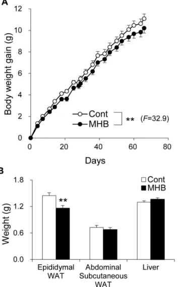
The body weight of mice fed HFD with and without MHB supplementation was monitored. As shown inFig 1A, weight gain was significantly suppressed in the group fed a diet containing MHB compared to mice fed HFD without supplement (P<0.01). There was no significant dif-ference in food intake between the two groups (Table 1andS3A Fig). Moreover, there was no significant correlation between total food intake and body weight gain (S3B Fig, Pearson corre-lation coefficientr= 0.21,p= 0.32). Epididymal white adipose tissue (WAT) weight of
MHB-Fig 1. MHB ameliorated HFD-induced body fat accumulation in mice.(A) Body weight gain of HFD-fed mice with or without MHB supplementation. Data are presented as means±SEM. n = 12 mice/group. **P<0.01 (by analysis of variance (ANOVA) with repeated measures). (B) Epididymal WAT, abdominal subcutaneous WAT and liver weight. Data are presented as means±SEM. n = 12 mice/group.**P<0.01 (by unpaired Student’st-test).
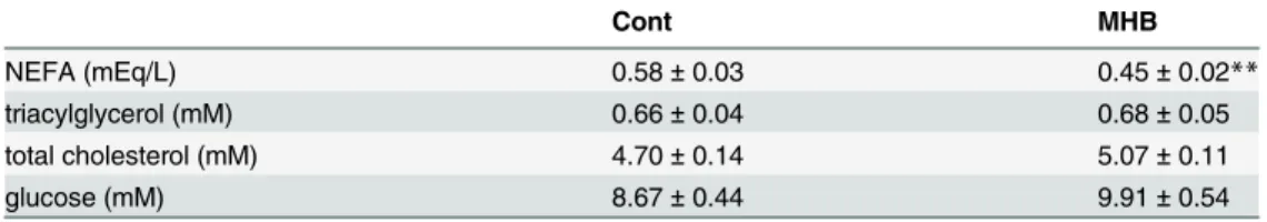
fed mice was also significantly decreased to 80.4% that of the control mice (Fig 1B,P<0.01). There were no significant differences in plasma TG, total cholesterol and glucose levels between the two groups. However, the plasma NEFA level in mice fed HFD containing MHB was sup-pressed by 21.7% (P<0.01) compared to that of the control group (Table 2). There was no sig-nificant difference in fecal excretion of lipids between the two groups (Table 1), suggesting that MHB does not affect the absorption rate of dietary fat under our experimental conditions.
MHB induced an increase in the mRNA levels of genes related to
thermogenesis and fatty acid oxidation in BAT of mice
To elucidate the underlying mechanisms of the effects of MHB, quantitative RT-PCR was car-ried out using tissues from mice after 9 wk feeding of MHB. Supplementation with MHB sig-nificantly increased the mRNA levels ofPGC-1α,UCP1,CPT1βandACOgenes by 1.6-fold, 1.2-fold, 1.3-fold, 1.2-fold in BAT, respectively (Fig 2A). The mRNA levels ofPRDM16and
PPARγwere also significantly increased by feeding MHB. There was also a concomitant increase in the UCP1 protein level in BAT mitochondria isolated from MHB fed mice (1.3-fold,P<0.01) (Fig 2B). Although the mRNA levels ofFASin the liver andACC1in the gastrocnemius muscle were significantly increased in the MHB-fed mice, no inclusive changes of gene expression were observed in the gastrocnemius muscle and liver between the groups (S4 Fig). These results indicate that MHB induces an increase in the mRNA levels of genes related to thermogenesis and fatty acid oxidation in BAT.
Single oral administration of MHB led to elevated BAT-SNA in rats
Because BAT is mainly innervated by sympathetic nerves [5], we orally administered MHB at a dose of 10 mg/kg and investigated changes in BAT-SNA in rats. MHB significantly elevated BAT-SNA compared to the control group (Fig 3A,P<0.01). There was no significant statisti-cal difference between the initial values of the two groups (Mann-WhitneyUtest). Subdiaph-ragmatic vagotomy completely abolished the observed effect of MHB (Fig 3B). There was no significant statistical difference between the initial values of the sham and vagotomized rats
Table 1. Total food intake of mice fed on HFD with or without MHB supplementation and the effect of MHB on fecal lipid content.
Cont MHB
fecal lipids excretion (mg/7 days) 38.63 ± 0.91 39.61 ± 2.23
total food intake (g/66 days) 172.40 ± 0.84 169.32 ± 1.52
Data are presented as means±SEM. n = 12 mice/group. No significant differences were observed by unpaired Student’st-test.
doi:10.1371/journal.pone.0131042.t001
Table 2. Effect of MHB on plasma components in mice fed on HFD with or without MHB supplementation.
Cont MHB
NEFA (mEq/L) 0.58±0.03 0.45±0.02**
triacylglycerol (mM) 0.66±0.04 0.68±0.05
total cholesterol (mM) 4.70±0.14 5.07±0.11
glucose (mM) 8.67±0.44 9.91±0.54
Data are presented as means±SEM. n = 12 mice/group.
**P<0.01 (by unpaired Student’st-test). NEFA, non-esterified fatty acid.
(Mann-WhitneyUtest). This finding suggests that vagal afferent nerves are involved in MHB induced elevation of BAT-SNA.
Fig 2. Measurement of mRNA expression levels and Immuno-blot analysis in the interscapular BAT of mice fed HFD supplemented with MHB.(A) mRNA expression levels in BAT. mRNA expression levels were normalized to the expression level ofGAPDHas a reference. (B) Immuno-blot analysis of UCP1 in BAT mitochondria. Representative immuno-blots are shown on the histogram. Representative signals shown are from the same immuno-blot membrane. UCP1 protein level was normalized to the level of UQCRC1 protein as a reference. Data are presented as means±SEM. n = 12 mice/group. *P<0.05,**P<0.01 (by unpaired Student’st-test).
doi:10.1371/journal.pone.0131042.g002
Fig 3. Single oral administration of MHB elevated BAT-SNA in rats.(A) Time course of BAT-SNA changes taken every 5 min and mean BAT-SNA over 0- to 90- min period. Urethane-anesthetized rats were administered 10 mg/kg of MHB. Data were calculated as a percentage of baseline, and as means±SEM. n = 3 rats/group.**P<0.01 (by ANOVA with repeated measures). (B) Effects of 2 mg/kg MHB on mean BAT-SNA over 0- to 90- min period in sham-operated or vagotomized rats. Data are presented as means±SEM. n = 3 rats/group.**P<0.01 (by ANOVA with repeated measures).
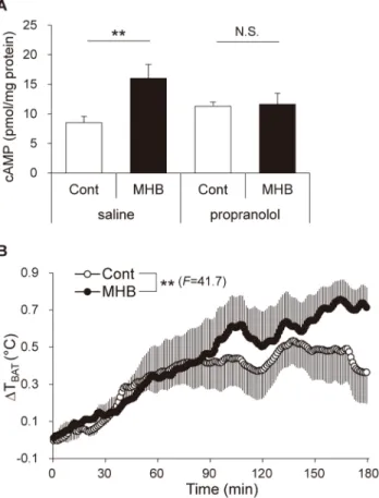
Single oral administration of MHB increased the cAMP level and the
temperature of BAT in rats
In general, norepinephrine, which is secreted from the sympathetic nerve endings, induces cAMP accumulationviatheβ3-adrenergic signaling cascade in BAT [4,24]. To confirm the involvement of this mechanism, we measured the cAMP level in BAT 3 h after MHB adminis-tration at a dose of 10 mg/kg with or without pretreatment ofβ-adrenergic antagonist propran-olol in rats. MHB induced an increase in the cAMP level (1.9-fold,P<0.01), while
pretreatment with propranolol completely suppressed cAMP induction by MHB (Fig 4A). To confirm whether elevation of BAT-SNA by MHB promotes thermogenesis in BAT, we mea-sured the temperature of BAT (TBAT) in rats using a temperature transmitter. Single oral
administration of MHB (10 mg/kg) significantly elevated TBATcompared to that in the control
group as shown inFig 4B(P<0.01). There was no significant statistical difference between the initial values of the two groups (Mann-WhitneyUtest).
Fig 4. Single oral administration of MHB increased the level of cAMP in BAT and elevated its temperature in rats.(A) cAMP level in the lysates of BAT obtained from rats administered MHB pretreated with propranolol or saline. Thirty minutes after the pretreatment with 10 mg/kg propranolol or saline, rats were administered 10 mg/kg MHB and killed after 3 h. Data are presented as means±SEM. n = 10 rats/group. **P<0.01, N.S., not significant (by unpaired Student’st-test). (B) Time course of BAT temperature changes (ΔTBAT) taken every 1 min relative to the baseline. The baseline was determined as the mean TBATover a 5-min period before the administration of MHB. Urethane-anesthetized rats were administered 10 mg/kg of MHB. Data are presented as means±SEM. n = 4 rats/group.**P<0.01 (by ANOVA with repeated measures).
Discussion
Some of the beneficial effects associated with ingesting specific components of hops have been reported previously. These include the cancer chemopreventive activity of xanthohumol [25,26], estrogenic activity of 8-prenylnaringenin [25,27], cyclooxygenase-2 inhibition of humulone (anα-acid) [28], and prevention of diet-induced obesity of iso-α-acids [11]. Here, we show that ingestion of MHB, derived from hops, can induce thermogenesis in BAT and ameliorate diet-induced body fat accumulation. Taken together, these results provide scientific support for the empirical utilization of hops over many years in herbal medicines [29].
MHB did not induce gene expressions ofACOandMCAD, which are involved in β-oxida-tion and known to be induced by PPARαin the liver [30], or induce a suppressive effect on lipid absorption, while they are thought to be associated with the mechanism by which iso-α-acids prevent diet-induced obesity [11]. Intriguingly, these results indicate that the body fat accumulation-ameliorating mechanism of MHB could be different from that of iso-α-acids, despite the structural similarity of MHB components with iso-α-acids [14,15]. Thus, in order to reveal the differences between the mechanism of iso-α-acids and MHB, a more comprehen-sive investigation will be needed to identify all the active ingredients in MHB. Further struc-tural information of multiple compounds found in MHB will reveal additional valuable data concerning the mechanism by which MHB reduces body fat accumulation.
Here, we found MHB supplementation induced a higher level ofPGC-1αexpression in BAT. PGC-1αis known to be a crucial regulator of gene expression related to thermogenesis and fatty acid oxidation such asUCP1,ACOandCPT1βgenes [31,32]. Indeed, the expression of these genes in BAT was increased by MHB supplementation in the diet. By contrast, inges-tion of MHB had no inclusive effect on the expression levels of genes involved in the beneficial effect of MHB on body fat accumulation in the gastrocnemius muscle and liver. Our results suggest that the effect of MHB is elicited, at least in part, by targeting BAT.
BAT thermogenesis is thought to be mediated by UCP1. Thus, control of UCP1 expression is considered to be an important factor for thermoregulation, energy expenditure regulation and maintenance of body weight [4,33]. Accordingly, activation of thermogenesis by increased expression of UCP1 in BAT could explain, at least in part, the body fat accumulation-amelio-rating effect of MHB. The main energy source of thermogenesis is free fatty acids, which are first released from brown adipocytes and then from circulating fatty acids and lipoproteins [4,34]. MHB reduced the plasma NEFA level in mice fed HFD, suggesting that MHB may facil-itate free fatty acid consumption in BAT by activation of thermogenesis.
In this study, single oral administration of MHB activated BAT-SNA in addition to elevat-ing the level of cAMP and maintainelevat-ing the temperature of BAT at a higher level than that of the control group in rats. Moreover, vagotomized rats completely lost the MHB induced activa-tion of BAT-SNA, and the block on theβ-adrenergic signaling cascade eliminated cAMP eleva-tion brought on by MHB. These data suggest that induceleva-tion of thermogenesis in BAT by MHB is mediated by theβ-adrenergic signaling cascadeviaBAT-SNA activation, which follows the vagal afferent nerve activation. The mechanism of the vagal afferent nerve activation by MHB is unclear at the present time. One possibility is the involvement of taste receptors expressed in endocrine cells within the gut mucosa [35]. In the gut, taste receptors release hormones or neu-rotransmitters in response to basic tastants, which can communicate with the brain directly,
In addition, it has been reported that chronic sympathetic stimulation, such as repeated administration ofβ3-adrenergic receptor agonists and repeated cold exposure, results in increased amounts of UCP1 protein and increased BAT [36]. MHB supplementation for 9 wk increased the level of UCP1 in BAT both at the mRNA and protein levels, suggesting that these effects may have been mediated by the chronic sympathetic stimulationviaBAT-SNA. The cAMP signaling pathway has also been reported to lead to increasedPPARγgene expression in brown adipocytes [37]. MHB supplementation for 9 wk in mice significantly increasedPPARγ
gene expression level in BAT, suggesting that it is possible that MHB induces an increase in the
PPARγgene expression levelviacAMP signaling pathway. It has been reported that the cAMP signaling and PPARγsignaling pathways synergistically increase theUCP1gene expression level [37]. Thus, it is possible that the PPARγsignaling pathway is also related to the induction ofUCP1gene expression by MHB. It has been reported that acute activation of theβ 3-adrener-gic signaling cascade induces thermogenesis by enhancing UCP1 activityviaactivation of lipol-ysis, while chronic activation of this cascade induces thermogenesis by increasing the UCP1 expression level as well as the biogenesis of mitochondria themselves [36]. Our findings, together with those of previous studies, suggest that continuous activation of BAT-SNA by MHB may induce thermogenesis by both an acute effect, which enhances UCP1 activity, and a chronic effect, which increases the UCP1 expression level. This enhanced thermogenesis induced by MHB may contribute to body fat reduction.
In our study, TBATchanges were observed 90 min after a single oral administration of
MHB, whereas continuous BAT-SNA elevation was detected after only 10 min of MHB admin-istration. The reason for this apparent discrepancy is unclear at the present time. One possibil-ity is that these temporal differences may reflect the time-lag of sequential physiological responses between BAT-SNA activation and TBATelevation. Alternatively, single oral
adminis-tration of MHB may elevate TBATby distinct mechanisms other than those involving
BAT-SNA. For example, MHB may contain direct activators of BAT thermogenesis, such as a β3-adrenergic agonist [38] and a TGR5 agonist [39], or affect the endocrine/paracrine activa-tors, such as heart-derived natriuretic peptides (NP) [40] and fibroblast growth factor 21 (FGF21) [41]. More detailed investigations will be needed to fully understand the effect of MHB on BAT activation. Besides, it has been reported that UCP1-positive brown fat-like adi-pocytes, named‘beige’adipocytes, in WAT are induced under various physiological and phar-macological conditions by activating adipocyte receptors such asβ3-adrenergic receptors and PPARγ[42]. The emergence of beige adipocytes in WAT is associated with protection against obesity and related disorders in rodent models [42]. In future studies, it should be explored whether MHB has an effect on inducing these beige adipocytes in WAT, not only on inducing thermogenesis in BAT.
In conclusion, our results reveal that single oral administration of MHB activates BAT-SNA, which induces thermogenesis in BAT of rats. In addition, administration of MHB-supple-mented HFD to mice increases the expression level of UCP1 in BAT and ameliorates diet-induced body fat accumulation. Therefore, MHB could be a useful tool in providing functional foods and beverages to reduce diet-induced body fat accumulation and related metabolic disor-ders. However, additional studies will be needed to better understand the mechanism by which MHB ameliorates body fat accumulation.
Supporting Information
tricyclooxyisohumulone A at 270 nm. (E) HPLC chromatogram of standard of tricyclooxyiso-humulone B at 270 nm. Chemical structures are shown inS2 Fig.
(TIF)
S2 Fig. Structure ofα- andβ-acids and their derivatives.α-acids: cohumulone (1a),n -humu-lone (1b), adhumulone (1c);β-acids: colupulone (2a),n-lupulone (2b), adlupulone (2c);trans -iso-α-acids:trans-isocohumulone (3a),trans-iso-n-humulone (3b),trans-isoadhumulone (3c);
cis-iso-α-acids:cis-isocohumulone (4a),cis-iso-n-humulone (4b),cis-isoadhumulone (4c); tri-cyclooxyisohumulone A (5); tricyclooxyisohumulone B (6).
(TIF)
S3 Fig. Food intake and correlation between total food intake and body weight gain in mice fed HFD supplemented with MHB.(A) Food intake of HFD-fed mice with or without MHB supplementation. Data are expressed per body weight, and as means ± SEM. n = 12 mice/ group. No significant difference was observed by ANOVA with repeated measures. (B) Corre-lation between total food intake and body weight gain in mice fed on HFD with (●) and with-out () MHB supplementation. n = 12 mice/group. There was no significant correlation
between total food intake and body weight gain (Pearson correlation coefficientr= 0.21,
p= 0.32). (TIF)
S4 Fig. Measurement of mRNA expression levels in the liver and gastrocnemius muscle of mice fed HFD supplemented with MHB.(A) mRNA expression in the liver. (B) mRNA expression in the gastrocnemius muscle. The mRNA level was normalized to that ofGAPDH. Data are presented as the relative level to the control group, and as means ± SEM. n = 12 mice/ group.P<0.05 (by unpaired Student’st-test).
(TIF)
S1 Table. Primer sequences used for real-time PCR. (DOC)
Acknowledgments
We thank Dr. Hiroaki Yajima of Kirin Company for valuable discussion, Ms. Yasuko Matsu-kura and Ms. Harumi Taniguchi of Kirin Company for excellent technical assistance in the preparation of MHB, and our group members for their technical support and valuable discussion.
Author Contributions
Conceived and designed the experiments: YMK KO MK KN. Performed the experiments: YMK CT SK GW YT KN. Analyzed the data: YMK CT SK GW YT KN. Contributed reagents/ materials/analysis tools: YT. Wrote the paper: YMK KO MK KN.
References
1. Mathieu P, Pibarot P, Despres JP (2006) Metabolic syndrome: the danger signal in atherosclerosis. Vasc Health Risk Manag 2: 285–302. PMID:17326334
2. Kahn BB, Flier JS (2000) Obesity and insulin resistance. J Clin Invest 106: 473–481. PMID:10953022 3. Spiegelman BM, Flier JS (2001) Obesity and the regulation of energy balance. Cell 104: 531–543.
PMID:11239410
5. Lowell BB, Spiegelman BM (2000) Towards a molecular understanding of adaptive thermogenesis. Nature 404: 652–660. PMID:10766252
6. Azzu V, Brand MD (2010) The on-off switches of the mitochondrial uncoupling proteins. Trends Bio-chem Sci 35: 298–307. doi:10.1016/j.tibs.2009.11.001PMID:20006514
7. Saito M, Okamatsu-Ogura Y, Matsushita M, Watanabe K, Yoneshiro T, et al. (2009) High incidence of metabolically active brown adipose tissue in healthy adult humans: effects of cold exposure and adipos-ity. Diabetes 58: 1526–1531. doi:10.2337/db09-0530PMID:19401428
8. Yajima H, Ikeshima E, Shiraki M, Kanaya T, Fujiwara D, et al. (2004) Isohumulones, bitter acids derived from hops, activate both peroxisome proliferator-activated receptorαandγand reduce insulin resis-tance. J Biol Chem 279: 33456–33462. PMID:15178687
9. Nozawa H, Nakao W, Zhao F, Kondo K (2005) Dietary supplement of isohumulones inhibits the forma-tion of aberrant crypt foci with a concomitant decrease in prostaglandin E2 level in rat colon. Mol Nutr Food Res 49: 772–778. PMID:15968705
10. Miura Y, Hosono M, Oyamada C, Odai H, Oikawa S, et al. (2005) Dietary isohumulones, the bitter com-ponents of beer, raise plasma HDL-cholesterol levels and reduce liver cholesterol and triacylglycerol contents similar to PPARαactivations in C57BL/6 mice. Br J Nutr 93: 559–567. PMID:15946420 11. Yajima H, Noguchi T, Ikeshima E, Shiraki M, Kanaya T, et al. (2005) Prevention of diet-induced obesity
by dietary isomerized hop extract containing isohumulones, in rodents. Int J Obes (Lond) 29: 991–997. 12. Ono M, Kakudo Y, Yamamoto R, Nagami K, Kumada J (1987) Simultaneous analysis of hop bittering
components by high-performance liquid chromatography. II. Evaluation of hop deterioration. J of Am Soc Brew Chem 45: 61–69.
13. Taniguchi Y, Matsukura Y, Ozaki H, Nishimura K, Shindo K (2013) Identification and quantification of the oxidation products derived fromα-acids andβ-acids during storage of hops (Humulus lupulusL.). J Agric Food Chem 61: 3121–3130. doi:10.1021/jf3047187PMID:23469991
14. Taniguchi Y, Taniguchi H, Matsukura Y, Kawachi Y, Shindo K (2014) Structural elucidation of humulone autoxidation products and analysis of their occurrence in stored hops. J Nat Prod 77: 1252–1261. doi: 10.1021/np4008427PMID:24875004
15. Taniguchi Y, Matsukura Y, Taniguchi H, Koizumi H, Katayama M (2015) Development of preparative and analytical methods of the hop bitter acid oxide fraction and chemical properties of its components. Biosci Biotechnol Biochem (in press).
16. Almaguer C, Gastl M, Arendt EK, Becker T (2012) Contributions of hop hard resins to beer quality. Bre-wingSci—Monatsschr Brauwiss 65: 118–129.
17. Smith SJ, Cases S, Jensen DR, Chen HC, Sande E, et al. (2000) Obesity resistance and multiple mechanisms of triglyceride synthesis in mice lacking Dgat. Nat Genet 25: 87–90. PMID:10802663 18. Li B, Nolte LA, Ju JS, Han DH, Coleman T, et al. (2000) Skeletal muscle respiratory uncoupling pre-vents diet-induced obesity and insulin resistance in mice. Nat Med 6: 1115–1120. PMID:11017142 19. Hemmeryckx B, Himmelreich U, Hoylaerts MF, Lijnen HR (2011) Impact of clock gene Bmal1 deficiency
on nutritionally induced obesity in mice. Obesity (Silver Spring) 19: 659–661.
20. Bullen JW Jr., Bluher S, Kelesidis T, Mantzoros CS (2007) Regulation of adiponectin and its receptors in response to development of diet-induced obesity in mice. Am J Physiol Endocrinol Metab 292: E1079–1086. PMID:17164441
21. Folch J, Lees M, Sloane Stanley GH (1957) A simple method for the isolation and purification of total lip-ides from animal tissues. J Biol Chem 226: 497–509. PMID:13428781
22. Shen J, Niijima A, Tanida M, Horii Y, Maeda K, et al. (2005) Olfactory stimulation with scent of lavender oil affects autonomic nerves, lipolysis and appetite in rats. Neurosci Lett 383: 188–193. PMID: 15878236
23. Shen J, Nakamura H, Fujisaki Y, Tanida M, Horii Y, et al. (2009) Effect of 4G-α-glucopyranosyl hesperi-din on brown fat adipose tissue- and cutaneous-sympathetic nerve activity and peripheral body temper-ature. Neurosci Lett 461: 30–35. doi:10.1016/j.neulet.2009.05.067PMID:19497350
24. Bachman ES, Dhillon H, Zhang CY, Cinti S, Bianco AC, et al. (2002)βAR signaling required for diet-induced thermogenesis and obesity resistance. Science 297: 843–845. PMID:12161655
25. Stevens JF, Page JE (2004) Xanthohumol and related prenylflavonoids from hops and beer: to your good health! Phytochemistry 65: 1317–1330. PMID:15231405
26. Kim SY, Lee IS, Moon A (2013) 2-Hydroxychalcone and xanthohumol inhibit invasion of triple negative breast cancer cells. Chem Biol Interact 203: 565–572. doi:10.1016/j.cbi.2013.03.012PMID:23562496 27. Milligan S, Kalita J, Pocock V, Heyerick A, De Cooman L, et al. (2002) Oestrogenic activity of the hop
28. Shimamura M, Hazato T, Ashino H, Yamamoto Y, Iwasaki E, et al. (2001) Inhibition of angiogenesis by humulone, a bitter acid from beer hop. Biochem Biophys Res Commun 289: 220–224. PMID: 11708802
29. Zanoli P, Zavatti M (2008) Pharmacognostic and pharmacological profile of Humulus lupulus L. J Eth-nopharmacol 116: 383–396. doi:10.1016/j.jep.2008.01.011PMID:18308492
30. Schoonjans K, Staels B, Auwerx J (1996) Role of the peroxisome proliferator-activated receptor (PPAR) in mediating the effects of fibrates and fatty acids on gene expression. J Lipid Res 37: 907–
925. PMID:8725145
31. Puigserver P (2005) Tissue-specific regulation of metabolic pathways through the transcriptional coac-tivator PGC1-α. Int J Obes (Lond) 29 Suppl 1: S5–9.
32. Kajimura S, Seale P, Spiegelman BM (2010) Transcriptional control of brown fat development. Cell Metab 11: 257–262. doi:10.1016/j.cmet.2010.03.005PMID:20374957
33. Boss O, Farmer SR (2012) Recruitment of brown adipose tissue as a therapy for obesity-associated diseases. Front Endocrinol (Lausanne) 3: 14.
34. Saito M (2014) Human brown adipose tissue: regulation and anti-obesity potential [Review]. Endocr J 61: 409–416. PMID:24401694
35. Janssen S, Depoortere I (2013) Nutrient sensing in the gut: new roads to therapeutics? Trends Endocri-nol Metab 24: 92–100. doi:10.1016/j.tem.2012.11.006PMID:23266105
36. Saito M (2013) Brown adipose tissue as a regulator of energy expenditure and body fat in humans. Dia-betes Metab J 37: 22–29. doi:10.4093/dmj.2013.37.1.22PMID:23441053
37. Chen HY, Liu Q, Salter AM, Lomax MA (2013) Synergism between cAMP and PPARγSignalling in the Initiation of UCP1 Gene Expression in HIB1B Brown Adipocytes. PPAR Res 2013: 476049. doi:10. 1155/2013/476049PMID:23554809
38. Inokuma K, Okamatsu-Ogura Y, Omachi A, Matsushita Y, Kimura K, et al. (2006) Indispensable role of mitochondrial UCP1 for antiobesity effect ofβ3-adrenergic stimulation. Am J Physiol Endocrinol Metab 290: E1014–1021. PMID:16368788
39. Ono E, Inoue J, Hashidume T, Shimizu M, Sato R (2011) Anti-obesity and anti-hyperglycemic effects of the dietary citrus limonoid nomilin in mice fed a high-fat diet. Biochem Biophys Res Commun 410: 677–681. doi:10.1016/j.bbrc.2011.06.055PMID:21693102
40. Bordicchia M, Liu D, Amri EZ, Ailhaud G, Dessi-Fulgheri P, et al. (2012) Cardiac natriuretic peptides act via p38 MAPK to induce the brown fat thermogenic program in mouse and human adipocytes. J Clin Invest 122: 1022–1036. doi:10.1172/JCI59701PMID:22307324
41. Fisher FM, Kleiner S, Douris N, Fox EC, Mepani RJ, et al. (2012) FGF21 regulates PGC-1αand brow-ning of white adipose tissues in adaptive thermogenesis. Genes Dev 26: 271–281. doi:10.1101/gad. 177857.111PMID:22302939