Recebido em 30.07.2002. / Received in July, 30thof 2002.
Aprovado pelo Conselho Consultivo e aceito para publicação em 11.02.2003. / Approved by the Consultive Council and accepted for publication in February, 11thof 2003. * Trabalho realizado no Centro Dermatológico de Guarulhos. / Work done at "Centro Dermatológico de Guarulhos".
1Ex-professor instrutor de Dermatologia da Faculdade de Medicina da Santa Casa de São Paulo. / Ex- Professor of Dermatology, School of Medicine, Santa Casa, São Paulo. 2Acadêmicas de medicina da Universidade Metropolitana de Santos. / Medical Students at the Metropolitan University of Santos.
©2004by Anais Brasileiros de Dermatologia
Coxins interfalangeanos sobre paquidermodactilia
*Interphalangeal pads on pachydermodactyly
*José Marcos Pereira
1Fernanda Corrêa Netto Pereira
2Vivian Corrêa Netto Pereira
2Resumo: Coxins interfalangeanos são nodulações ceratósicas, de limites precisos, com
aproximada-mente um centímetro de diâmetro, geralaproximada-mente sobre as articulações interfalangenas das mãos. A paqui-dermodactilia é uma tumefação uniforme da pele que ocorre nas falanges proximais das mãos. É relata-do o caso de um jovem com associação de paquidermodactilia e coxins interfalangeanos, fato não encontrado na literatura médica. Embora haja descrição que considera serem essas manifestações diver-sas da mesma doença, acredita-se que sejam entidades distintas. O ato compulsivo de atritar a pele parece ser o denominador comum mais aceito para justificar as duas doenças. Os autores postulam que o coxim interfalangeano seria uma resposta epidérmica, enquanto a paquidermodactilia, uma resposta dérmica a um mesmo fator traumático sobre a pele. O paciente foi tratado com infiltração intralesional de triancinolona, com melhora clínica expressiva das duas manifestações.
Palavras-chave: dedos; fibrose.
Summary:Knuckle pads are keratotic nodulations within precise limits and approximately one centimeter in diameter, usually on the interphalangeal joints of the hands. Pachydermodactyly is a uniform swelling of the skin occurring in the proximal phalanges of the hands. A case involving a young man suffering from several knuckle pad lesions concomitant with pachydermodactyly was studied. This association has not been previously described in the literature. Although it has been reported that both conditions are different manifestations of the same disease, they are believed to be distinct disorders. The compulsive act of rubbing the skin seems to be a common denominator mostly accepted as the cause of both diseases. The authors affirm that knuckle pads may be acquired as an epidermal response, while pachydermodactyly, is a dermal response to the same traumatic factor to the skin. The patient was treated with intralesional infiltration of triamci-nolone resulting in a remarkable clinical improvement in both manifestations.
Keywords: fingers; fibrosis.
Caso Clínico / Case Report
INTRODUÇÃO
Coxim interfalangeano (CI), também muito con-hecido como knuckle pads, e paquidermodactilia (PD) são duas dermatoses pouco estudadas. Segundo Verbov,1
cri-ador do termo PDem 1975, as duas entidades representari-am manifestações clínicas diferentes da mesma doença.
CI são nodulações hiperceratósicas, de superfície áspera, discretamente acastanhadas ou acinzentadas, com cerca de um centímetro de diâmetro, em geral arredondadas, endurecidas, de limites precisos e contornos regulares. São assintomáticas e de evolução crônica. As localizações mais freqüentes são as articulações inter-falangeanas proximais e metacarpointer-falangeanas. São menos
INTRODUCTION
Interphalangeal pad (IP), also frequently known as knuckle pads and pachydermodactyly (PD) are two der-matoses that have been the subject of few studies. According to Verbov,1
who created the term PDin 1975, the two entities could represent different clinical manifesta-tions of the same disease.
freqüentes em qualquer outra articulação dos dedos das mãos e raramente ocorrem nos artelhos.
A histopatologia é bastante característica, verifican-do-se hiperceratose, acantose e prolongamento das cristas papilares.15Também já foram observados acantose
psoriasi-forme,22 proliferação dos capilares e fibroblastos,9
infla-mação perivascular e aumento das bandas de colágeno na derme.22
A primeira publicação foi realizada em 1893 por Garrod,2 que descreveu três pacientes com nodulações
sobre o dorso dos dedos. Como um dos pacientes tinha con-tratura de Dupuytren e outro tinha um avô também com a contratura, o autor associou as nodulações à contratura de Dupuytren. Em 1904, o mesmo autor3fez estudo detalhado
sobre a doença, tendo usado o termo padsem 12 casos, dos
quais seis tinham contratura de Dupuytren. A expressão clássica knuckle padsfoi criada por Jones,4em 1923. O CI
é conhecido desde o Renascimento, pois é mostrado nitida-mente nas obras de Michelangelo, Vasari, Bronzino e Durer.5
A doença já recebeu inúmeras designações, entre elas helodermia, tylositas articuli, fibroma subcutâneo, keratosis supracapitularis, coussinets des phalanges, pul-villus digiti, Garrod' pads, discreta queratoderma, tumor
benigno do córion, fibrocondroma, hipoplasia congênita e
cojinetes articulares laterales.
Desde a publicação de Garrot,2cerca de 335 casos
foram descritos.2-29 Predomina no sexo masculino (56%),
com idade variável entre cinco e 69 anos. São também descritos casos familiares de CI.8,23
Apenas o trabalho de Mikkelsen14mostrou a
incidên-cia do CIna população. O autor, na cidade de Haugesund, na Noruega, em 1977, examinou 1.871 pessoas normais (752 homens e 1.119 mulheres), entre 20 e 89 anos de idade, e encontrou 164 com CI, sendo 68 homens (9%) e 96 mulheres (8,6%), ou seja, aproximadamente 9% da popu-lação, sendo a doença mais freqüente acima dos 40 anos.
O CIfoi associado a várias alterações, entre elas dis-função do metabolismo da vitamina A,13 fibrodisplasias,
ceratoses palmoplantares e ictiose,9 surdez e leuconíquia
(casos familiares),11,25pseudoxantoma elástico10e
acrocera-toelastoidose;28a associação mais freqüente, porém, é com
a contratura de Dupuytren. Caroli e cols.21examinaram 352
pacientes com contratura de Dupuytren (318 homens e 34 mulheres) e encontraram CIem 54 deles (15,3%), sendo 51 homens (95,3%) e três mulheres (5,5%). Em outro trabalho, Mikkelsen14 examinou 869 pacientes com contraturas de
Dupuytren (623 homens e 246 mulheres), encontrado 385 com CI, sendo 303 homens (48,7%) e 82 mulheres (33,3%), mostrando incidência muito alta do CIem pacientes com contratura de Dupuytren.
A etiopatogenia do CI ainda é desconhecida. Segundo Ronchese30a CIfaz parte da chamada fibromatose
periférica de Touraine, que inclui a contratura de Dupuytren, induratio penis plastic (doença de Peyronie),
metacarpophalangeal articulations. They are less frequent in the other articulations of the fingers and rarely occur in the toes.
The histopathology is very characteristic with hyperkeratosis, acanthosis and prolongation of the papil-lary crests.15
Psoriasiform acanthosis,22
proliferation of the capillaries and fibroblasts,9
perivascular inflammation and increase in the bands of collagen in the dermis have also been observed.22
The first publication was made in 1893 by Garrod,2
who described three patients with dorsal nodulations on the fingers. Since one of the patients presented Dupuytren's contracture and another had a grandfather also with con-tracture, the author associated the nodulations to Dupuytren's contracture. In 1904, the same author3
made a detailed study on the disease, and used the term pads in 12 cases, of which six had Dupuytren's contracture. The clas-sic denomination of knuckle pads was created by Jones,4
in 1923. IPwas known during the Renaissance, because it is clearly depicted in the works of Michelangelo, Vasari, Bronzino and Durer.5
The disease has already received countless designa-tions, including heloderma, tylositas articuli, subcutaneous fibroma, keratosis supracapitularis, coussinets des
pha-langes, pulvillus digiti, Garrod's pads, discreet keratoder-ma, benign tumor of the chorion, fibrochondrokeratoder-ma, congen-ital hypoplasia and cojinetes articulares laterales.
Since the publication of Garrot,2
approximately 335 cases have been described.2-29
There is a male sex bias (56%), with ages ranging from five to 69 years. Familial cases of IPhave also been described.8,23
Only the work by Mikkelsen14
has described the inci-dence of IP in the population. In 1977, the author, examined 1,871 normal people (752 men and 1,119 women), aged between 20 and 89 years, in the city of Haugesund (Norway) and found 164 presented IP, of which 68 were men (9.0%) and 96 (8.6%) women, or in other words, approximately 9% of the population and the disease was most frequent among those over 40 years.
IPis associated to several alterations, including dys-function in the metabolism of vitamin A,13
fibrodysplasia, palmoplantar keratoses and ichthyosis,9
deafness and leukonychia (familial cases),11,25
pseudoxanthoma elas-ticum10
and acrokeratoelastoidosis;28
the most frequent association, however, is with the Dupuytren's contracture. Caroli and cols.21
examined 352 patients with Dupuytren's contracture (318 men and 34 women) and found IP in 54 (15.3%), of which 51 were men (95.3%) and three women (5.5%). In another work, Mikkelsen14
examined 869 patients with Dupuytren's contracture (623 men and 246 women), found 385 with IP, of which 303 were men (48.7%) and 82 women (33.3%), showing a very high incidence of IP in patients with Dupuytren's contracture.
The etiopathogenesis of IPis still unknown. According to Ronchese30
peripher-al fibromatosis, which includes Dupuytren's contracture, induratio penis plastic (Peyronie's disease), cutaneous fibro-mas and scleroderma. Caroli and cols.21
consider that Dupuytren's contracture and CIare expressions of the same fibromatous alteration, since fibroblasts, myofibroblasts, macrophages and extracellular matrix are findings common to both diseases.
Lagier and cols.12
classify IPinto two forms: false or pseudo-IP, resulting from attrition and represented by acan-thosis and hyperkeratosis; and true IP, characterized by a proliferation of fibroblasts in the dermis. Sehgal and cols.15
divided the disease into two types: Primary IP (asympto-matic) and secondary (sympto(asympto-matic), which could be asso-ciated to Dupuytren's contracture, palmoplantar keratosis, ichthyosis or professional trauma. Trauma certainly seems to have a great influence on the onset of IP; consequently, it is frequently associated to work.31
Richards and cols.18
examined 41 workers in a poultry slaughterhouse and found 23 presented IP. While in the offices of the same company, they examined 41 employees and no cases of IPwere found. The disease is also observed frequently in pugilists27
and violinists.19
In children there is a prevalence of the idio-pathic form, while in adults the IPoriginates from trauma.29
Patients with IPusually consult the dermatologist for aesthetic reasons, however there is no specific treatment for the disease. Some cases in childhood disappear spontaneous-ly by adult age,17
though this in general does not occur with the idiopathic form. Regarding IPof traumatic origin, remis-sion of the leremis-sion can occur following removal of the attri-tion.22
Several types of therapy have been attempted, including 50% phenol and cryotherapy,7
surgical excision20
and occlu-sion with silicone gel;24
however, recurrence is frequent. Pachydermodactyly (PD) is a thickening of the skin of the fingers, mainly close to the proximal phalanges. It is most common in adolescents.
The first description of this disease was made in 1973, by Bazex,32
in a 23-year-old man and denominated it
pachidermie digitale des primières phalanges. In 1975, Verbov1
presented the case of a man aged 19 years and called the picture pachydermodactyly. Although the patient of Verbov did not present IPlesions, due to the histopatho-logy characterized by hyperkeratosis, acanthosis and increase in fibrous tissue in the dermis, the author postula-ted that PDcould be a variant of IP.
PDhas also been denominated pachyderma, discreet keratoderma, hyperkeratose-acanthosis and fibromatosis.
Since the first publication on PD, approximately 50 cases have been described in the literature1,32-62 of which 77.5% involved males and the remaining 22.5% were female. The majority of patients were between 14 to 29 years of age, although cases have been reported among patients as young as 5 and as old as 63 years. The disease duration was up to 15 years.
In the majority of cases PDinvolves the second, third and fourth fingers of both hands. A case has been described of
fibromas cutâneos e esclerodermia. Caroli e cols.21
conside-ram que a contratura de Dupuytren e CIsão expressões de uma mesma alteração fibromatosa, pois fibroblastos, miofi-broblastos, macrófagos e matriz extracelular representam achados comuns a ambas as doenças.
Lagier e cols.12 classificam o CI em duas formas:
falso ou pseudo-CI, resultado de atrito e representado por acantose e hiperceratose; e o CIverdadeiro, caracterizado por proliferação de fibroblastos na derme. Sehgal e cols.15
dividem a doença em dois tipos: CIprimário (assintomáti-co) e secundário (sintomáti(assintomáti-co), o qual seria associado à contratura de Dupuytren, ceratose palmoplantar, ictiose ou trauma profissional. Realmente, o trauma parece ter grande influência sobre o aparecimento do CI; assim sendo, é fre-qüentemente associado à ocupação.31 Richards e cols.18
examinaram 41 trabalhadores em um abatedouro de galin-has e encontraram 23 com CI. Já no escritório da empresa, examinaram 41 funcionários e não encontram CI. A doença também é observada com freqüência em lutadores de box27
e violinistas.19Na criança predomina o CIidiopático, e no
adulto, o CIde origem traumática.29
Pacientes com CI consultam o dermatologista por razões estéticas, porém não existe um tratamento específico para a doença. Alguns casos, em crianças, desapareceram espontaneamente na idade adulta,17o que em geral não ocorre
com a forma idiopática. Em se tratando de CI de origem traumática, uma vez removendo o atrito, pode haver remissão da lesão.22Várias tentativas terapêuticas foram feitas, entre
elas fenol 50% e crioterapia,7remoção cirúrgica20e oclusão
com gel de silicone;24porém, a recidiva é freqüente.
A paquidermodactilia (PD) é um espessamento da pele dos dedos das mãos, principalmente junto às falanges proximais. É mais comum em jovens adolescentes.
A primeira descrição dessa doença foi feita em 1973, por Bazex,32que a encontrou em um homem de 23 anos de
idade e a chamou de pachidermie digitale des primières phalanges. Em 1975, Verbov1 apresentou o caso de um
homem com 19 anos e chamou o quadro de paquider-modactilia. Embora o paciente de Verbov não tivesse lesões de CI, devido à histopatologia caracterizada por hipercer-atose, acantose e derme com aumento de tecido fibroso, o autor postulou que PDseria uma variante do CI.
A PDtambém já foi denominada paquidermia, dis-creta ceratodermia, acanto-hiperceratose e fibromatose.
Desde a primeira publicação acerca da PD, cerca de 50 casos foram descritos na literatura,1,32-62, sendo 77,5% dos
casos em homens e 22,5% em mulheres. A maioria dos pacientes estavam com idades entre 14 a 29 anos, embora tenham sido descritos casos com 5 anos e 63 anos de idade. A duração da doença foi de meses até 15 anos.
Na maioria dos casos o PDacomete o segundo, ter-ceiro e quarto dedos de ambas as mãos. Foi descrito o caso de uma mulher com a doença em apenas um dedo.42Pode
haver liquenificação da pele sobre a lesão,39 eritema e
dolorosa.32,37,57 O processo pode estender-se pelas
articu-lações metacarpofalangeanas, configurando a PD transgres-siva.40Há relatos de casos familiares com lesões em mãe e
filha48e em irmãs.57
A histopatologia clássica mostra hiperceratose, acantose, intenso espessamento da derme por aumento de fibroblastos e depósito de colágeno. A coloração pelo alcian blue evidenciou depósito de mucina.37,39,40,42,47,57 Podem ser
também observados aumento da espessura da membrana basal,33 aumento das glândulas sudoríparas écrinas, várias
fibrosadas, e intenso depósito de mucopolissacarídeos,34
pobre demarcação entre derme papilar e reticular,35
marca-da eosinofilia36e infiltrado linfocitário.37
Estudo histoquímico das fibras de colágeno mostrou serem colágeno I, III e V. Pela microscopia eletrônica as fibras de colágeno são mais finas do que as dos tecidos normais.35,59
A etiopatogenia da PD é desconhecida. Alguns autores consideram-na decorrente de ato compulsivo, no qual os pacientes atritam os dedos de uma mão contra os da outra.44,50
A PD já foi associada à síndrome de Dupuytren,35
síndrome do túnel do carpo,35ginecomastia,37atrofia
macu-losa varioliforme,55 artrite crônica juvenil, disfunção da
tireóide, dermatite seborréica e glossite,34 esclerose
tuberosa,46,57síndrome de Ehlers-Danlos.57Porém, a
associ-ação mais freqüente é com trauma repetitivo.41,44,50,61,62
Baldazzi e cols.57classificaram a PD em cinco
for-mas principais: 1 - forma clássica (associada ao trauma); 2 - forma localizada ou monopaquidermodaquitilia; 3 - forma transgressiva (dorso das mãos); 4 - familiar; e 5 - sin-tomática, associada à esclerose tuberosa.
O diagnóstico diferencial deve ser feito com36
paqui-dermoperiostose, acropaquidermodactilia na psoríase, osteíte cistóite múltipla, tumor fibroso, acromegalia, artrite juvenil crônica, sinovite, nódulos reumatóides, tofo gotoso, xantomatoses, fibrossarcomas, fibromatose juvenil, nódu-los de Heberden e líquen mixedematoso.
Algumas tentativas terapêuticas já foram realizadas para a PD, entre elas remoção cirúrgica,34,61 infiltração
intralesional com triancinolona36,37,56e diminuição do atrito
por controle do ato compulsivo.44,50,62
No Brasil não há registro sobre a ocorrência de PD. O CIfoi descrito em uma menina por Rutowitsch & Lima.62
O objetivo deste trabalho é mostrar a associação do
CIcom a PD, fato ainda não descrito na literatura, e o exce-lente resultado com a terapêutica instituída para ambas as doenças.
CASO CLÍNICO
MVNS, 17 anos, do sexo masculino, branco, natu-ral de São Paulo, SP, com queixa de inchaço nos dedos das mãos e pequenos nódulos sobre a pele há oito anos. No exame dermatológico, o paciente apresentava aumen-to do volume dos dedos, principalmente na região da
a women with the disease in a single finger.42
there can be lichenification of the skin in the lesion,39
erythema and desqua-mation38,40
and, in some cases, the lesion can be painful.32,37,57
The process can extend to the metacarpophalangeal articulations, configuring PDtransgrediens.40
There are reports of familial cases with lesions in the mother and daughter48
and in sisters.57
The classic histopathology shows hyperkeratosis, acanthosis, intense thickening of the dermis due to an increase in the fibroblasts and deposits of collagen. Staining by Alcian blue reveals a mucin deposit.37,39,40,42,47,57
An increase can also be observed in the thickness of the basal mem-brane,33
augmented eccrine sudoriparous glands, various of which are fibrose, and intense deposit of mucopolysaccha-rides,34
poor demarcation between the papillary and reticular dermis,35
marked eosinophilia36
and lymphocytic infiltrate.37
Histochemical study of the collagen fibers showed them to be collagen I, III and V. Under electron microscopy the collagen fibers are finer than those of normal tissues.35,59
The etiopathogenesis of PDis unknown. Some authors consider it is due to compulsive neuroses, in which patients rub the fingers of one hand against those of the other.44,50
PD has already been described associated to Dupuytren's disease,35
carpal tunnel syndrome,35
ginecomas-tia,37
varioliform macular atrophy,55
juvenile chronic arthritis, thyroid dysfunction, seborrheic dermatitis and glossitis,34
tuberous sclerosis,46,57
Ehlers-Danlos syndrome.57
However, the most frequent association is with repetitive trauma.41,44,50,61,62
Baldazzi and cols.57
classified PD into five main forms: 1 - classic form (associated to trauma); 2 - localized form or monopachydermodactyly; 3 - transgressing forms (dorsal region of the hands); 4 - familial; and 5 - sympto-matic, associated to tuberous sclerosis.
The differential diagnosis should be made between:36
pachydermoperiostosis; acropachydermodactyly in psoria-sis, multiple osteitis cystica, fibrous tumor, acromegaly, chronic juvenile arthritis, synovitis, rheumatoid nodules, gouty tophus, xanthomatosis, fibrosarcomas, juvenile fibro-matosis, Heberden's nodes and lichen myxedematosus.
Various therapies have been attempted for PD, including surgical removal,34,61
intralesional infiltration with triamcinolone36,37,56
and reducing the attrition by con-trolling the compulsive neurosis.44,50,62
In Brazil, no case of PD has been registered. Rutowitsch & Lima62
described CIin a girl.
The objective of this work was to report the associa-tion of CIwith PD, a which has hitherto not been described in the literature, and the excellent result with the therapeu-tics instituted for both diseases.
CASE REPORT
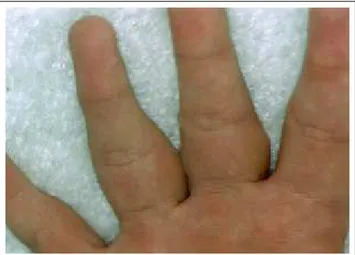
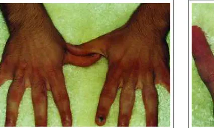
falange proximal, acometendo do segundo ao quinto dedos de ambas as mãos (Figuras 1 e 2). A alteração era assintomática, porém impossibilitava-o de flexurar total-mente os dedos, o que lhe atrapalhava em algumas ativi-dades do cotidiano. Sobre as articulações metacarpo-falangeanas e face lateral da tumefação observavam-se algumas lesões nodulares, com limites precisos, ceratósi-cas (Figura 3). O próprio paciente admitia que suas lesões eram causadas pelo atrito, pois constantemente cruzava e friccionava os dedos de uma mão contra os da outra. Embora as lesões hiperceratósicas também fossem assin-tomáticas, o paciente coçava-as com freqüência. Além disso, acusou o controle do videogame como outro fator de atrito. Foi feito exame anatomopatológico de fragmen-to em fuso, pegando ao mesmo tempo a lesão nodular e a região edemaciada (Figura 4). A epiderme apresentava acantose e hiperceratose (correspondendo à nodulação). Na derme observavam-se aumento numérico de fibroblas-tos, proliferação e espessamento das fibras colágenas envolvendo as glândulas sudoríparas écrinas. As fibras de colágeno estavam
dissoci-adas pela presença de muci-na. Não havia processo infla-matório. O raio X das mãos não mostrou comprometi-mento ósseo ou periósteo. O diagnóstico clínico e
his-Figura 3: Coxins inter-falangeanos sobre paquider-modactilia. Notar nódulos de coxins interfalangeanos sobre as regiões espessadas
Figure 3: Interphalangeal pads on pachydermodactyly. Note the nodules of the interphalangeal pads on the thickened regions
phalange, involving the second to fifth fingers of both hands (Figures 1 and 2). The alteration was asymptomatic, how-ever it totally impeded flexing of the fingers, which hindered certain daily activities. Several keratose and nodular lesions were observed, with precise limits, on the metacar-pophalangeal articulations and lateral face of the tumefac-tion (Figure 3). The patient himself affirmed that the lesions were caused by attrition, because he constantly crossed and rubbed the fingers of one hand against the other. Although the hyperkeratose lesions were also asymptomatic, the patient scratched them frequently. Furthermore, he report-ed that the video game control was another factor of attri-tion. Anatomicopathological exam was done on a spindle shaped specimen, including at the same time the nodular lesion and edematous area (Figure 4). The epidermis pre-sented acanthosis and hyperkeratosis (corresponding to the nodulation). In the dermis there was a numeric increase in the fibroblasts, proliferation and thickening of the collagen fibers were also observed, involving the eccrine sudori-parous glands. The collagen fibers were dissociated by the presence of mucin. There was no inflammatory process. X-ray of the hands did not reveal any involvement of the bones or periostea. The clinical and histological diagnosis was interphalangeal pad on
pachy-Figura 1: Paquidermodactilia - espessamento cutâneo das regiões
falangeanas proximais, do segundo ao quinto dedos / Figure 1:
Pachydermodactyly; cutaneous thickening in the proximal pha-langeal regions, from the second to fifth fingers
Figura 2: Paquidermodactilia - detalhe da região palmar
dos dedos / Figure 2: Pachydermodactyly; detail of the
tológico foi de coxim inter-falangeano sobre paquider-modactilia. O paciente foi orientado a não mais atritar os dedos e a realizar duas ses-sões quinzenais de infiltração intralesional de triancinolona na PD e sob o CI, com
desa-parecimento total das lesões de CI, normalização pratica-mente total da pele na região do PD (Figuras 5 e 6) e recu-peração ampla do movimento dos dedos. Após um mês de observação, não havia indícios de recidiva.
DISCUSSÃO
Os autores classificam o paciente como forma clás-sica de PD, segundo Baldazzi e cols.,57e CIsecundário,
con-forme Sehgal e cols.15 Tanto o paciente como seus
fami-liares não apresentavam indícios de contratura de Dupuytren. Ficou evidente que as manifestações clínicas do paciente eram de origem traumática.
Embora o paciente de Verbov1tivesse PDsem lesões
características de CI, pelos achados histopatológicos ele considerou a PDum tipo de manifestação clínica do CI. Em toda a literatura consultada, essa é a única descrição que considera ambas as alterações fazendo parte de uma mesma doença.
Até o momento foram descritos cerca de 335 casos de CIe 50 de PD, mostrando que são doenças clinicamente
dermodactyly. The patient was counseled to avoid rubbing his fingers the fingers and per-form biweekly sessions of intralesional infiltration with triamcinolone in the PD and under the IP. There was a total disappearance of the CI
lesions and a practically total normalization of the skin in the area of PD (Figures 5 and 6) together with an ample recovery in the finger movement. After one month of follow-up, there were no indications of recurrence.
DISCUSSION
The authors classify the patient as having the classic form of PD, according to the definition of Baldazzi and cols.,57
and secondary CI, according to Sehgal and cols.15
Neither the patient nor his relatives presented indications of Dupuytren's contracture. It was evident that the patient's clinical manifestations were of traumatic origin.
Although the patient of Verbov1
had PDwithout the characteristic lesions of IP, due to the histopathological findings he considered PDto be a type of clinical manifes-tation of IP. In all the literature consulted, this is the only description that considers both alterations as being part of the same disease.
To date, approximately 335 cases of IPand 50 of PD
have been described, demonstrating that they are clinically
Figura 4: Histologia do coxim interfalangeano sobre paquidermodactilia (hematoxilina e eosina
-100X)
Figure 4: Histology of the interphalangeal pads on pachydermodactyly (hematoxylin & eosin - 100X)
Figura 5: Coxins interfalangeanos sobre paquidermodactilia.
Região dorsal após tratamento / Figure 5: Interphalangeal pads
on pachydermodactyly; dorsal region after treatment
Figura 6: Coxins interfalangeanos sobre paquidermodactilia.
Região palmar após tratamento / Figure 6: Interphalangeal pads
distinct diseases, IPpresenting histological alterations that are eminently epidermal, and PDdermal alterations. There was no description regarding a simultaneous occurrence of both diseases. Except for the patient described by Yanguas and cols.,45
that presented PDand apparently had a single
IP lesion, as observed in the photograph, however the authors did not make any mention to this fact in the text.
It seems there can be no doubt that both diseases are triggered, or at least aggravated, by some compulsive rub-bing action of the skin. The authors consider that both dis-eases are two different responses to the same etiological agent.
Patients can have different cutaneous responses to attrition of the skin. For instance, a patient that frequently scratches a lesion can present a predominantly epidermal reaction, forming a neurodermatitis. While another with pruritus and consequent itch, can have a dermal response, forming a keloid scar.
The case described here, besides PD, presented sev-eral lesions of metacarpophalangeal IP, some of which were on the PDitself, or in other words, on the lateral face of the fingers, showing that following rubbing his skin, the patient had concomitant epidermal and dermal responses, forming IPand PD, respectively. Returning to the previous example, it is as if the patient had developed a neuroder-matitis on a keloid scar, a fact that has already been observed several times by the authors.
The patient was effectively clinically cured by under-standing the problem and avoiding rubbing his hands together with intralesional infiltration with triamcinolone. There was a total recovery in the movement of the fingers. We consider that the two entities are much more fre-quent than reported in the literature. A closer observation of all the ways that the skin can suffer attrition, whether due to compulsive neuroses or repetitive movements in the work-place, and a study of each individual's form of reaction should clarify the etiopathogenesis of both diseases and, consequent-ly, lead to the development of an effective treatment. q
diferentes, o CI apresentando alterações histológicas emi-nentemente epidérmicas, e a PD, dérmicas. Não existe rela-to da ocorrência simultânea de ambas as doenças. Apenas o paciente descrito por Yanguas e cols.,45 apresentando PD,
aparentemente tinha uma única lesão de CI, observada por fotografia, porém, textualmente, os autores não fizeram qualquer menção ao fato.
Parece não haver dúvida de que ambas as doenças são desencadeadas, ou pelo menos agravadas, por algum ato compulsivo de atritar a pele. Os autores acreditam que ambas as doenças sejam duas respostas diferentes a um mesmo agente etiológico.
Atritando a pele, pacientes têm respostas cutâneas diferentes. Por exemplo, um paciente que coça muito uma lesão pode ter uma reação predominantemente epidérmica, formando uma neurodermite. Outro, frente a prurido e con-seqüente coçadura, pode ter uma resposta dérmica, forman-do uma cicatriz queloideana.
O caso aqui descrito, além da PD, apresentava várias lesões de CI metacarpofalangeanas e algumas sobre a própria PD, ou seja, na face lateral dos dedos, mostrando que o paciente, atritando a pele, teve ao mesmo tempo respostas epidérmica e dérmica, formando CIe PD, respec-tivamente. Voltando ao exemplo anterior, é como se o paciente tivesse desenvolvido uma neurodermite sobre uma cicatriz queloideana, fato já observado algumas vezes pelos autores.
O paciente teve cura clínica efetiva com a conscien-tização do problema, pois deixou de atritar as mãos, e as infiltrações intralesionais de triancinolona. A recuperação da movimentação dos dedos foi total.
Acreditamos que as duas entidades são muito mais freqüentes do que a literatura relata. Uma melhor obser-vação de todos os atos que levam a algum atrito da pele, seja compulsivo ou repetitivo profissional, e um estudo da forma reacional de cada indivíduo deverão esclarecer a etiopatogenia de ambas as doenças e, consequentemente,
proporcionar um tratamento efetivo. q
AGRADECIMENTO
Ao professor doutor Nilceo S Michalany, pela realização dos exames anato-mopatológicos.
ACKNOWLEDGEMENT
2000; 6:450-452.
30. Ronchese F. Knuckle pads and similar looking disorders. G Ital Dermatol Venereol 1966; 107:1227-1236.
31. Hueston JT, Wilson WF. Knuckle pads. Aust NZ J Surg 1973;42:274-277.
32. Bazex A, Dupré A, Teillard J. Pachydermie digitale des pre-mieres phalanges par hyperplasie dermique et aplasie hypoder-mique. Bull Soc Fr Dermatol Syphiligr 1973;80:455-458. 33. Garrel J, Sonneck JM, Neveux Y, Millet P, Doss N, Lanternier G. Pachydermie digitale isolée. Ann Dermatol Venereol 1982; 109:769-770.
34. Fleeter TB, Myrie C, Adams JP. Pachydermodactyly: a case report and discussion of the pathologic entity. J Hand Surgery 1984;9:764-766.
35. Reichert CM, Costa J, Barsky SH, Claysmith AP, Liotta LA, Enzinger FM, Triche TJ. Pachydermodactyly. Clin Orthopaed Rel Res 1985;194:253-257.
36. Hudson PM. Pachydermodactyly. Br J Dermatol 1989; 121:111.
37. Curley RK, Hudson PM, Marsden RA. Pachydermodactyly: a rare form of digital fibromatosis - report of four cases. Clin Exp Dermatol 1991;16:121-123.
38. Martin JC, Rennie JAN, Kerr KM. Pachydermodactyly: con-fused with JCA. Ann Rheum Dis 1992; 51:1101-1102.
39. Draluck JC, Kopf AW, Hodak E. Pachydermodactyly: first report in a woman. J Am Acad Dermatol 1992;27:303-305. 40. Sola A, Doval JV, Sola J, Quintanilla E. Pachydermodactyly transgrediens. Int J Dermatol 1992; 31:796-797.
41. Iraci S, Bianchini L, Innocenzi D, Tomassoli M, Nini G. Pachydermodactyly: a case of an unusual type of reactive digital fibromatosis. Arch Dermatol 1993;129:247-248.
42. Bardazzi F, Fanti PA, Padova MP, Varotti C. Localized pachy-dermodactyly in a woman. Acta Derm Venereol (Stockh) 1994;74:152-153.
43. Meunier L, Pailler C, Barneon G, Meynadier J. Pachydermodactyly or acquired digital fibromatosis. Br J Dermatol 1994; 131:744-746.
44. Lautenschlager S, Itin PH, Rufli T. Pachydermodactyly: reflecting obsessive-compulsive behavior? Arch Dermatol 1994; 130:387.
45. Yanguas I, Goday JJ, Soloeta R. Pachydermodactyly: report of two cases. Acta Derm Venereol (Stockh) 1994;74:217-218. 46. Lo WL, Wong CK. Localized pachydermodactyly in tuberous sclerosis. Clin Exp Dermatol 1993; 18:146-147.
47. Rai A. An unusual case of peri-articular soft tissue finger swelling in na adolescent male: pachydermodactyly or pachyder-moperiostose? Br J Rheum 1994;33:677-679.
48. Russo F, Pichardo RP, Camacho F. Familial pachyder-modactyly. Acta Derm Venereol 1994; 74:386-387.
49. Brousse C, Rybojad M, Piette AM, Gepner P, Chapman A. Pachydermodactyly: report of a case. Rev Med Interne 1994;15:412-414.
50. Hagedon M, Graf HG, Grosshans E. Pachydermodactyly: sequela of obsessive nurosis. Hautarzt 1994; 45:88-90.
51. Dupin N, Gautier MS, Rabary G, Beltzer-Garelly E, Binet O. Pachydermodactyly. Ann Dermatol Venereol 1994;121:632-634. 52. Perez B, Gomez MI, Sanchez E, Munoz E, Ledo A. Pachydermodactyly: a case report. J Dermatol 1995;22:43-45. 53. Kopera K, Soyer HP, Kerl H. An update on pachydermodacty-ly and a report of three addicional cases. Brit J Dermatol 1995;133:433-437.
54. Costa MM, Romeu JC, Costa T. Pachydermodactyly a rare
REFERÊNCIAS / REFERENCES
1. Verbov J. Pachydermodactyty: a variant of the true knuckle pad. Arch Dermatol 1975;111:524.
2. Garrod AE. On na unusual form of nodules upon joints of the fingers. St Bartholomew's Hospital Report 1883; 29:157-161. 3. Garrod AE. Concerning pads upon the finger joints and their clinical relationships. Brit Med J 1904; II: 8.
4. Jones HW. Two cases of Knuckle pads. Br Med J 1923; I:759. 5. Ramos e Silva J. Coussinets des phalanges (pulvillus digit). Ann Dermatol 1956; 83:22-33.
6. Carol WLL, Prakken JR, Zwijndregt HA. Tylositas articuli. Acta Dermatolvenereologica. 1940; 21:87-97.
7. Wise F. Knuckle pads. Arch Dermatol Syphilol. 1944;49:144-145. 8. Veltman G. Etiology of Knuckle pads. Dermatologica 1954; 108:20-33.
9. Allison Junior JR, Allison JR. Knuckle pads. Arch Derm 1966; 93:311-316.
10. Stankler L. Pseudoxanthoma elasticum with a knuckle pad on the thumb. Acta derm-venereol 1967; 47:263-266.
11. Bart RS, Pumphrey RE. Knuckle Pads, leukonichia and deaf-ness. N Engl J Med 1967; 276:202-207.
12. Lagier R, Meinecke R. Pathology of Knuckle pads. Virchows Arch A Path Anat Histol 1975;365:185-191.
13. Morginson WJ. Discrete keratodermas over the knuckle and finger articulations. Arch Dermatol 1955; 71:349-353.
14. Mikkelsen OA. Knuckle pads in Dupuystren's disease. The Hand 1977; 9:301-305.
15. Sehgal VN, Singh M, Saxena MHK, Nayar M. Primary knuck-le pads. Clin Exp Dermatol 1979;4:337-339.
16. Kouskoukis CE. Stump the experts. J Dermatol Surg Oncol 1985; 11:209 e 349.
17. Paller AS, Hebert AA. Knuckle pads in children. AJDC 1986; 140:915-917.
18. Richards TB, Gamble JF, Castellan RM, Mathias CGT. Knuckle pads in live-chicken hangers. Contact Dermatitis 1987; 17:13-16.
19. Bird HA. Development of Garrod's pads in the fingers of a professional violinist. Ann Rheum Diseases 1987;46:169-170. 20. Marino JM,Vilar ML, Pozo T, Quinones PA. Cojinetes articu-lares laterales: una localización atípica de knuckle pads. Actas Dermo Sifilog 1990;81:473-474.
21. Caroli A, Zanasi S, Marcuzzi A, Guerra D, Cristiani G, Ronchetti IP. Epidemiological and structural findings supporting the fibromatous origen of dorsal knuckle pads. J Hand Surg (Brit) 1991; 16:258-262.
22. Mackey SL, Cobb MW. Knuckle pads. Cutis 1994; 54:159-160. 23. Ramer JC,Vasily DB, Ladda RL. Familial leuconychia, knuck-le pads, haring loss na palmoplantar hyperceratosis. J Med Genet 1994; 31:68-71
24. Guberman D, Lichtenstein A, Vardy DA. Knuckle pads. A for-gotten skin condition: report of a case and review of the literature. Cutis 1996; 57:241-242.
25. Kose O, Baloglu H. Knuckle pads, leukonychia and deafness. Int J Dermatol 1996;35:728-729.
26. Irwin LR, Naylor IL, Holms W. The contractility of knuckle pads. J Hand Surg(Brit) 1997; 22:110-112.
27. Kanerva L. Knuckle pads from boxing. Eur J Dermatol 1998; 8:359-361.
ti delle falangi: dermatosi autoprovocanti? G Ital Dermatol Venereol 1993;128:393-397.
61. Itin PH, Lautenschlager S. Pachydermodactyly: a psychocuta-neous disorders. Dermatology 1995;190:1-3.
62. Rutowitsch M, Lima LAF. Coxim interfalangeano. An Bras Dermatol 1971; 46:361-368.
cause of finger joint swelling. J Rheumatol 1995;22:2374-2375. 55. Callot V, Wechsler J, Hovnanian A, Revuz J. Pachidermodactyly and atrophia maculosa varioliforme cutis. Dermatology 1995;190:56-58.
56. Kim TH, Cho YH, Park HB. Two cases of pachydermodacty-ly. J Dermatol 1996; 23:419-424.
57. Bardazzi F, Neri I, Fanti PA, Patrizi A. Pachydermodactyly in two woung girls. Pediatr Dermatol 1996;13;288-291.
58. Cartier H, Guillet MH, Schollhammer M, Guillet G. Pachydermodactylie de l'adolescent: expression d'un mal-être? Arch Pédiatr 1996;3:1091-1094.
59. Kang BD, Hong SH, Kim IH, Kim WK, Oh CH. Two cases of pachydermodactyly. Int J Dermatol 1997; 36:768-772.
60. Bardazzi F, Neri I, Raone B, Patrizi A. Pachydermodactyly: seven new cases. Ann Dermatol Venereol 1998; 125:247-250. 61. Aoki K, Iida H, Umeda T, Katayama I, Nishioka K. A case of pachydermodactyly. Jpn J Plast Reconstr Surg 1994; 37:109-113. 62. Aloi F, Solaroli C, Tomasini C. Pachidermodattilia e
cursinet-ENDEREÇO PARA CORRESPONDÊNCIA: / MAILINGADDRESS: Dr. José Marcos Pereira