Croat. Chem. Acta88 (2) (2015) 133–137. http://dx.doi.org/10.5562/cca2548
Original Scientific Article
Antibacterial Activity of
Pinus pinaster
Bark Extract and its Components
Against Multidrug-resistant Clinical Isolates of
Acinetobacter baumannii
Mirna Ćurković-Perica,a,* Jasna Hrenović,a Nuša Kugler,b Ivana Goić-Barišić,c and Mirta Tkaleca
aUniversity of Zagreb, Faculty of Science, Department of Biology, Division of Microbiology, Zagreb, Croatia bZel-en razvojni center energetike, d.o.o, Sevnica, Slovenia cUniversity Hospital Centre Split and University of Split School of Medicine, Department of Clinical Microbiology,
Split, Croatia
RECEIVED OCTOBER 10, 2014; REVISED MARCH 19, 2015; ACCEPTED APRIL 14, 2015
Abstract. The aim of this research was to test the antibacterial activity of Pinus pinaster aqueous bark ex-tract (PABE) and its basic components against multidrug-resistant isolates of Acinetobacter baumannii belonging to European clone I and II, isolated previously from the clinical outbreaks. The minimum bactericidal concentration of PABE against both clones of A. baumannii was 200 mg ml–1, while lower
concentrations showed high antibacterial activity. After 24 h of treatment with 100, 50 or 10 mg ml–1 of
extract, the reduction in the number of A. baumannii isolates belonging to European clone I and II was 85.8 ± 2.5 %, 78.5 ± 1.1 %, 66.3 ± 2.5 % and 90.2 ± 1.7 %, 78.6 ± 1.2 %, 69.8 ± 0.7 %, respectively. Sev-eral basic components: caffeic acid, catechin, epicatechin, gallic acid and vanillin, detected in the extract by high performance liquid chromatography, contributed to the antibacterial activity of the extract against both clones of A. baumannii. However, the antibacterial activity of extract was higher than that of each tested basic component suggesting that proanthocyanidins, which were present in quite a large amount in the extract, might have also contributed to the activity of the extract. Antibacterial activity of PABE against A. baumannii reveals that complex and inexpensive natural product might be useful in combat against natu-rally competent bacteria that easily acquire resistance against antibiotics.
Keywords: antimicrobials, aqueous extract, bacteria, multidrug-resistant pathogens, natural compounds
INTRODUCTION
Bark extracts of P. pinaster (PBEs) have a long history of
ethnomedicinal use.1 As a mixture of a large variety of
substances, PBEs are reported to exhibit a wide range of biological activities, including antioxidative, antiinflama-tory, antitumor, antiatherogenic, antiviral, antimicrobial,
etc.2 In papers published thus far, different extraction
procedures, using different solvents were applied, affect-ing the composition of extracts and biological activity.
Species of the genus Acinetobacter, like A.
bau-mannii are naturally competent3 and easily acquire DNA
from the environment/other species. Therefore, it is not
surprising that Acinetobacter spp. displays mechanisms
of resistance to all existing antibiotic classes as well as a prodigious capacity to acquire new determinants of
re-sistance.4 A. baumannii is a nonfermentative,
Gram-negative, nonmotile, oxidase-negative coccobacillus which
is found in many health care environments5 and is a very
effective human colonizer. This species emerged over the last decade as a leading cause of hospital-acquired
infec-tions; there are numerous reports of multidrug-resistant
(MDR) A. baumannii from hospitals around the world6
including Croatia.7,8 Problems caused by A. baumannii in
the hospital setting are emphasized by high degree of resistance to drying and disinfectants, leading to long-term persistence and the occurrence of outbreaks in the
hospital environment.6 Global outbreaks of infections
caused by A. baumannii are also frequent.4
Current concerns regarding inefficiency of antibi-otics or deleterious effects of synthetic chemicals used for medical purposes have encouraged a worldwide research on natural products potentially useful in devel-opment of alternative treatments for common and emerging pathogens. Research on natural products rep-resents convenient strategy to find extracts and bioac-tive compounds, based on which new, effecbioac-tive and less
expensive treatments can be applied.9 The aim of this
study was to determine the antibacterial and bactericidal activity of P. pinaster aqueous bark extract (PABE) and
its components against MDR A. baumannii isolated
EXPERIMENTAL
Plant Material and Preparation of Extracts
In Croatia Pinus pinaster is mostly planted in coastal
region. For this research bark was collected from 15 trees growing on the island of Rab. Bark samples from 15 trees were randomly pooled to form 3 samples (barks collected from 5 trees in each sample), air dried at 50–55 °C for eight days to constant weight and grinded to pieces (fraction < 1 mm) using Colortronic mill. Grinded bark (100 g) was extracted with 500 ml of water at 100 °C for 30 minutes. The aqueous extract was collected and grinded bark was once again extract-ed with 500 ml of fresh water at 100 °C for 30 minutes. The first and second extract were pooled and dry-powdered extract was prepared from aqueous extract by spray-drying at 200 °C. From 100 g of grinded bark approximately 10 g of powdered extract was obtained. For treatment of bacteria dry-powdered extract was solved in sterile distilled water.
HPLC Analysis of Pinus pinaster Aqueous Bark Extract (PABE)
The following substances were detected and quantified: caffeic acid (cas: 331-39-5), catechin (cas: 225937-10-0), epicatechin (cas: 490-46-0), ferulic acid (cas: 537-98-4), gallic acid (cas: 149-91-7), taxifolin (cas: 24198-97-8) and vanillin (cas: 121-33-5). Authentic standards were purchased from Fluka (Germany) and Sigma-Aldrich (Germany). High performance liquid chromatography analysis (HPLC) system (Shimadzu, SIL-20AC XR) equipped with a quaternary pump, multi-wave UV/Vis detector, autosampler, fraction collector and Kinetex 2.6u C18 100A column; (100 × 4,60 mm, Phenomenex) was used for all analyses. Absorbance was measured from 200 nm to 400 nm. The injection volume was 20 μl and
the flow rate 1.0 ml min–1 from 0–29 minute and 38–45
minute, and 1.5 ml min–1 from 29–38 minute, at 30 °C.
Elution profile consisted of solvent A (vol. ratio:
deion-ised H2O : H3PO4 = 999 : 1) and solvent B (vol. ratio:
methanol : H3PO4 = 999 : 1). The solvent composition
(A/B) changed according to the following gradient: 97/3 at 0 min, 97/3 at 2 min, 85/15 at 6 min, 84/16/0 at 15 min, 82/18 at 29 min, 47/53 at 30 min, 10/90 at 31 min, 10/90 at 38 min and finally 97/3 at 39 min. Con-centrations of tested compounds were determined, based on the chromatographic data of the standards. The cali-bration curves (peak area vs. concentration) for individual compounds were obtained for a wide concentration range. Analyses of each extract were done in triplicates. The
concentration of each substance was expressed in mg g–1
of dry extract. Retention times were: 3.74 min for gallic acid; 10.722 min for catechin; 13.272 min for caffeic
acid; 14.385 min for vanillin; 17.457 min for epicatechin; 20.213 min for p-cumaric acid; 25.125 min for ferulic acid; 26.074 min for taxifolin.
Determination of Proanthocyanidins (PA) Content The proanthocyanidins were determined by
hydrochlo-ric acid–butanol method.10 Ten milligram of pine bark
extract was dissolved in 10 ml of water. To 0.20 ml of
this solution 3 ml of a 95 % solution of n-butanol/HCl
(95/5; v/v) was added, followed by addition of 0.1 ml of
a 1.4 % (w/v) Fe(SO4) 7 H2O in 2 M HCl. The tubes
were incubated for 40 min at 95 °C. After incubation, the samples were cooled and analyzed by measuring absorb-ance at 550 nm. Cyanidin chloride (Sigma-Aldrich, Germany) was used as a standard. The final PA in pine bark extracts was expressed on a dry basis (mg/gDW) and data were shown as a mean value of three biological and three technical replicates ± SE.
Bacterial Strains
Antibacterial activity of PABE against two different
isolates of A. baumannii with affiliation to European
clone I (EU I) and II (EU II) was tested and compared. Clinical isolates of A. baumannii were collected during two different outbreaks, first from 2002–2007 (EU I) and second from 2009–2010 (EU II) in Clinical Hospital
Center Split, Croatia,7,8 a referral hospital for a wide
area of southern Croatia. Isolation of A. baumannii was performed on blood agar plates (Bio Rad). Initial identi-fication was made using the ATB 32GN and Vitek 2 sys-tems (bioMerieux, Marcy-l’Etoile, France) according to Clinical and Laboratory Standards Institute
recommenda-tions, CLSI.11 Susceptibility to β-lactams (ceftazidime,
cef-triaxone, cefepime, imipenem, meropenem), β-lactam/ β-lactamase inhibitor combinations (ampicillin/sulbac-tam, piperacillin/tazobactam), aminoglycosides (amika-cin, gentamicin) and fluoroquinolones (ciprofloxacin) was determined by disc-diffusion tests, and susceptibil-ity to colistin by E-tests (AB Biodisk, Solna, Sweden). MICs were determined by broth microdilution
accord-ing to CLSI.11 Isolate from EU clone II was resistant to
all antimicrobials (imipenem MIC 64 μg ml–1,
mero-penem 128 μg ml–1, amikacin 64 μg ml–1, gentamicin 64
μg ml–1, ceftazidime 256 μg ml–1, cefepime 128 μg ml–1,
ciprofloxacin 32 μg ml–1, piperacillin/tazobactam 128
μg ml–1 and ceftriaxone 256 μg ml–1) except colistin
(MIC: 0.5 mg ml–1) and ampicillin/sulbactam (MIC: 1
mg ml–1). Isolate from EU clone I had lower minimum
inhibitory concentrations (MIC) for carbapenems
(imi-penem 6 mg ml–1, meropenem 16 mg ml–1) than isolate
belonging to EU clone II (imipenem and meropenem
≥ 64 mg ml–1).12 The mechanisms of carbapenem
Antibacterial Activity Tests
The antibacterial activities of PABE against isolates of A. baumannii belonging to the EU clone I and II were tested according to the microdilution method, following the
CLSI recommendations.11 The bacteria were pre-grown
on nutrient agar (Biolife, Italy) for 16 h at 36.0 ± 0.1°C to obtain the cultures in log phase of growth. The bacterial biomass was then suspended in the nutrient broth (Bi-olife, Italy). For the experiments with powdered extract, 200, 100, 50, 10 or 1 mg of extract was added into 0.5 ml of sterile deionised water and vigorously shaken (40 Hz/10 min, Kartell TK3S). A 0.5 ml of bacterial suspen-sion was added in the tubes, which resulted in following concentrations of powdered extract: 200, 100, 50, 10 or 1
mg ml–1. The tubes which served as the positive control
contained 0.5 ml of bacterial suspension and 0.5 ml of sterile deionised water. Both experiments were set in triplicate. The tubes were incubated in a dark during 24 h at 36.0 ± 0.1 °C with shaking at 120 rpm to assure the complete mixing. To confirm the antibacterial activity of basic components of extract, the experiments with pure caffeic acid, catechin, epicatechin, ferulic acid, gallic acid, taxifolin and vanillin solutions in 2 % ethanol were set up in triplicate. The tested concentrations of basic components were equivalent to those contained in 200
mg ml–1 of powdered extract. The positive controls were
set up without and with 2 % of ethanol, which showed no difference in the number of bacteria (data not shown).
The number of viable cells was determined at the beginning of experiment and after 24 h of treatment. Tubes were vigorously shaken and 0.1 ml of sample was plated (spread plate method) directly onto the nutrient
agar, while another sample was serially diluted (10–1 to
10–8) and inoculated onto the nutrient agar plates in
triplicate. The inoculated plates were incubated at 36.0 ± 0.1 °C for 24 h. After the incubation period, the bacte-rial colonies were counted and the number of viable
cells was reported as Colony forming units (CFU) ml–1.
The numbers of CFU were logarithmically transformed. The antibacterial activity of the extract, each compound and mixture of compounds (caffeic acid, catechin, epi-catechin, ferulic acid, gallic acid, taxifolin and vanillin) was expressed as the reduction of log CFU and as the percent reduction of log CFU as compared to the corre-sponding control. The minimum bactericidal concentra-tion (MBC) values were determined after 24 h of
exper-iments according to the CLSI directions.11 The
mini-mum inhibitory concentration (MIC) was not deter-mined due to the turbidity and colour of extracts. The comparisons between the numbers of log CFU were done using the ANOVA and subsequently the post-hoc Duncan test was performed for the calculations concern-ing pair-wise comparisons. Statistical decisions were made at a significance level of p < 0.05.
RESULTS AND DISCUSSION
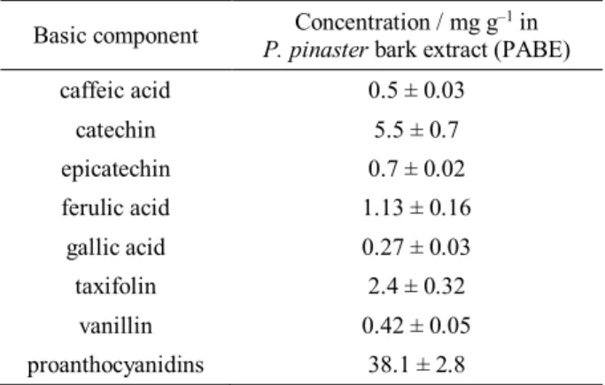
Concentrations of basic substances (determined by HPLC) as well as proanthocyanidins were expressed as
mg g–1 of spray-dried powdered PABE (Table 1).
Statis-tically significant differences in the concentrations of basic compounds and proanthocyanidins between ex-tracts from 3 pooled samples (barks collected from 5 trees in each sample) were not determined, and there-fore the extracts were pooled for further experiments/ treatments.
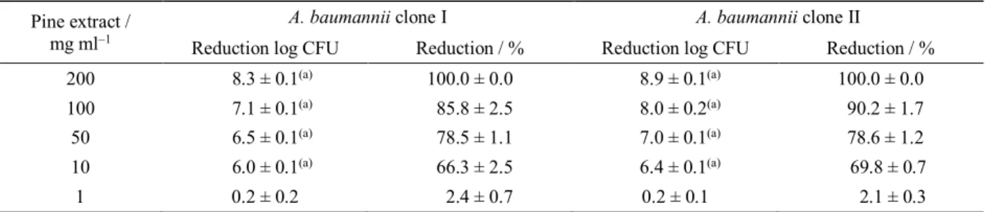
Antibacterial activities of PABE against isolates of A. baumannii belonging to EU clone I and II are shown in Table 2. The extract showed antibacterial activity
with MBC of 200 mg ml–1 against both clones.
To elucidate which basic components contribute to the bactericidal activity of powdered extract at
concentra-tion of 200 mg ml–1, experiments with pure solutions of
components were performed (Table 3). For clone I caffeic acid, catechin, epicatechin, ferulic acid, gallic acid and vanillin resulted in statistically significant reduction of log CFU as compared to corresponding control, but the reduction of log CFU was generally low (0.4–0.9 log CFU reduction). The antibacterial activity of basic com-ponents against clone II (Table 3) was similar to those against clone I, except that the ferulic acid had no anti-bacterial activity against clone II. When basic substances
were tested together against two clones of A. baumannii,
the reductions of log CFU were 36.9 and 23.5 % for clone I and II, respectively. These results suggest that besides tested substances, some other substances such as proanthocyanidins, which were present in quite a large amount in the extract (Table 1), might have contributed to the antibacterial activity of extract, too.
Table 1. Concentrations of basic substances and proanthocya-nidins in Pinus pinaster aqueous bark extract (PABE) ex-pressed as mg g–1 of spray-dried powdered PABE. Data shown
are mean values of three biological and three technical repli-cates ± SE.
Basic component P. pinasterConcentration / mg g bark extract (PABE) –1 in caffeic acid 0.5 ± 0.03
catechin 5.5 ± 0.7
epicatechin 0.7 ± 0.02
ferulic acid 1.13 ± 0.16 gallic acid 0.27 ± 0.03
taxifolin 2.4 ± 0.32
vanillin 0.42 ± 0.05
Several research groups published the results on antimicrobial activity of PBEs. As opposed to simple aqueous extract (PABE) which was used in this
re-search, in those researches mostly Pycnogenol® -
com-mercial water/ethanol extract was tested and it was
shown that 250 μg ml–1 of Pycnogenol® counteracted
the growth of several tested positive and
Gram-negative bacteria,13 inhibited growth of Helicobacter
pylori in suspension (12.5 μg ml–1), while in ten times
higher concentration reduced the adherence of the
bac-terium to gastric cells.14 However its efficiency against
A. baumannii was not tested. Grimm et al.15,16 showed
that substances which are basic components of PBE are quickly absorbed after oral ingestion and their distribu-tion in the body tissues is fast. Fourteen hours after volunteers were given 300 mg or 200 mg of PBE, fifteen compounds from PBE were detected in the
their plasma.15,16 Maximum concentrations of four
iden-tified compounds in the plasma; catechin, caffeic acid,
ferulic acid and taxifolin were 107 ng ml–1, 17 ng ml–1,
15 ng ml–1 and 33 ng ml–1, respectively. Of those four
substances, caffeic acid and catechin revealed
antibacte-rial activity against A. baumannii clones in our research, but epicatechin, gallic acid and vanillin also contributed
to the activity of PABE against both clones of A.
bau-manii, while ferulic acid slightly contributed to the activity of PABE against EU clone I. The above men-tioned substances are known to possess antimicrobial activity against different pathogens. Vanillin is proved
to express antifungal activity17 and antibacterial activity
against Cronobacter species;18,19 catechin was effective
against pathogenic bacteria, Escherichia coli O157:H7,
Pseudomonas aeruginosa and the food spoilage fungus
Penicillium chrysogenum20,21 and, it also showed
syner-gistic antibacterial effect with antibiotics.22
Further-more, the combination of catechin and epicatechin gal-late potentiated the activity of beta-lactam antibiotics
against methicillin-resistant Staphylococcus aureus.23
Furthermore, epicatechin showed synergistic effect with the aflavin against clinical isolates of A. baumannii and
Stenotrophomonas maltophilia.24 Caffeic acid is also
known to exhibit antimicrobial activity, however its centration in PABE was 0.05 % making it even less con-centrated in solution which was applied to A. baumannii,
Table 3. Reduction in the number of A. baumannii isolates belonging to European clone I and II after 24 h of treatment with basic components of Pinus pinaster bark extract (in concentration contained in 200 mg ml–1 ofpowdered extract) as compared to the
corresponding control. c0 A. baumannii clone I(107 CFU ml–1) = 7.35 ± 0.92; c0 A. baumannii clone II(107 CFU ml–1) = 4.90 ±
0.14. Data shown are mean values of three biological and three technical replicates ± SE.
Component Concentration / µg ml–1 Reduction log CFU A. baumannii clone I Reduction / % Reduction log CFU A. baumannii clone II Reduction / %
Caffeic acid 100 0.5 ± 0.0(a) 5.6 ± 0.5 0.3 ± 0.1(a) 3.9 ± 1.0
Catechin 110 0.9 ± 0.0(a) 9.7 ± 0.1 0.1 ± 0.0(a) 1.2 ± 0.1
Epicatechin 140 0.4 ± 0.0(a) 4.5 ± 0.1 0.3 ± 0.0(a) 3.3 ± 0.0
Ferulic acid 226 0.6 ± 0.0(a) 6.2 ± 0.4 0.0 ± 0.0 –0.1 ± 0.1
Gallic acid 54 0.4 ± 0.0(a) 4.3 ± 0.3 0.8 ± 0.0(a) 8.7 ± 0.2
Taxifolin 400 0.1 ± 0.0 0.9 ± 0.2 0.0 ± 0.0 0.0 ± 0.1
Vanillin 84 0.5 ± 0.0(a) 6.2 ± 0.1 0.3 ± 0.0(a) 3.4 ± 0.1
All components 3.4 ± 0.0(a) 36.9 ± 0.2 2.2 ± 0.0(a) 23.5 ± 0.0
Table 2. Reduction in the number of A. baumannii isolates belonging to European clone I and II after 24 h of treatment with Pinus pinaster aqueous bark extract as compared to the corresponding control. c0 A. baumannii clone I(107 CFU ml–1) = 2.93 ±
0.57; c0 A. baumannii clone II(107 CFU ml–1) = 1.54 ± 0.26. Bactericidal activity was obtained at 200 mg ml–1, and antibacterial
activity at 10 mg ml–1. Data shown are mean values of three biological and three technical replicates ± SE.
Pine extract / mg ml–1
A. baumannii clone I A. baumannii clone II
Reduction log CFU Reduction / % Reduction log CFU Reduction / %
200 8.3 ± 0.1(a) 100.0 ± 0.0 8.9 ± 0.1(a) 100.0 ± 0.0
100 7.1 ± 0.1(a) 85.8 ± 2.5 8.0 ± 0.2(a) 90.2 ± 1.7
50 6.5 ± 0.1(a) 78.5 ± 1.1 7.0 ± 0.1(a) 78.6 ± 1.2
10 6.0 ± 0.1(a) 66.3 ± 2.5 6.4 ± 0.1(a) 69.8 ± 0.7
1 0.2 ± 0.2 2.4 ± 0.7 0.2 ± 0.1 2.1 ± 0.3
and it was reported previously that concentrations high-er than 0.4 % whigh-ere needed to inhibit the growth of some
microorganisms, including L. monocytogenes, E. coli,
and S. aureus.25 Gallic acid which also contributed to
antibacterial activity of PABE is a basic constituent of many other plant extracts. Tea extracts which contain gallic acid and catechins proved to be effective against
H. pylori without affecting beneficial bacteria.26 These
data suggest that PABE or its components like gallic acid and catechin could not only be efficient against
MDR A. baumannii, but they could also be used in
combination with probiotic bacteria. In our research the
tested in vitro concentrations of basic PABE
compo-nents that showed antibacterial activity against A. bau-mannii were at least three orders of the magnitude high-er than the concentration of substances found in plasma of patients after oral ingestion of the same quantity of PBE.15,16 Therefore, the concern exists that the
concen-tration of the bioactive substances in plasma of patients would not be high enough to treat bacteraemia, but since A. baumannii causes also skin and wound
infec-tions,27,28,29 application of PABE in sufficient
concentra-tion in such cases is certainly possible. Except investi-gated basic substances, proanthocyanidins that were determined in PABE could have also contributed to the antibacterial activity. In grape seeds, proanthocyanidins were determined as active antibacterial agents toward 10 different pathogenic Gram-positive and
Gram-nega-tive bacterial strains.30 Plant-derived proanthocyanidins
may inhibit the growth of pathogenic bacteria by
bind-ing strongly to proteins at bacterial cell surfaces.31
In this research we revealed that PABE at the
con-centration 200 mg ml–1 showed bactericidal activity
against two MDR clinical isolates of A. baumannii
be-longing to EU clone I and II and antibacterial activity at lower concentrations, in vitro. This finding suggests that PABE and its components could potentially find an application in treatment of MDR A. baumannii-infected patients.
Acknowledgements. This research was supported by the Croa-tian Ministry of Science, Education and Sports.
REFERENCES
1. A. Maimoona, I. Naeem, Z. Saddiqe, and K. Jameel,
J. Ethnopharmacol.133 (2011) 261–277.
2. P. Rohdewald P, Int. J. Clin. Pharmacol. Ther.40 (2002) 158–168. 3. M. S. Ramirez, M. Don, A. K. Merkier, A. J. S. Bistue, A. Zorreguieta, D. Centron, and M. E. Tolmasky, J. Clin. Microbiol.
48 (2010) 1488–1490.
4. F. Perez, A. Hujer, K. Hujer, B. K. Decker, P. N. Rather, and R. A. Bonomo, Antimicrob. Agents Ch.51 (2007) 3471–3484. 5. L. Malone and D. H Kwon, Int. J. Antimicrob. Ag.41 (2013) 70–74. 6. I. Roca, P. Espinal, X. Vila-Farres, and J. Vila, Front. Microbiol.
3 (2012) 1–30.
7. I. Goić-Barišić, B. Bedenić, M. Tonkić, A. Novak, S. Katić, S. Kalenić, V. Punda-Polić, and K. J Towner, J. Clin. Microbiol.47
(2009) 3348–3349.
8. I. Goić-Barišić, K. J. Towner, A. Kovačić, K. Sisko-Kraljević, M. Tonkić, A. Novak, and V. Punda-Polić, J. Hosp. Infect.77
(2011) 368–369.
9. H. J. Doughari, P.A. Ndakidemi, I. S. Human, and S. Benade,
Microbes Environ.26 (2011) 101–111.
10. L. J. Porter, L. N. Stich, and B. G. Chan, Phytochemistry 25
(1986) 223–230.
11. CLSI - Clinical and Laboratory Standards Institute (2007) Per-formance standards for antimicrobial susceptibility testing, 17th
informational supplement, M100-S17. CLSI, Wayne, PA. 12. I. Goić-Barišić and V. Kaliterna, Med. Glas. 8 (2011) 312–313. 13. M. A. C. Torras, C. A. Faura, F. Schönlau, and P. Rohdewald,
Phytother. Res.19 (2005) 647–648.
14. P. Rohdewald and W. Beil, Phytother. Res.22 (2008) 685–688. 15. T. Grimm, Z. Chovanova, J. Muchova, K. Sumegova, A.
Lip-takova, Z. Durackova, and P. Hogger, J. Inflam.3 (2006) 1. 16. T. Grimm, R. Skrabala, Z. Chovanova, J. Muchova, K.
Sume-gova, A. Liptakova, Z. Durackova, and P. Hogger, BMC Clin. Pharmacol.3 (2006) 4.
17. D. J. Fitzgerald, M. Stratford, M. J. Gasson, and A. Narbad, J.
Agricult. Food Chem.53 (2005) 1769–1775.
18. D. J. Fitzgerald, M. Stratford, M. J. Gasson, J. Ueckert, A. Bos, and A. Narbad, J. Appl. Microbiol.97 (2004) 104–113. 19. G. P. Yemis, F. Pagotto, S. Bach, and P. Delaquis, J. Food
Pro-tect.74 (2011) 2062–2069.
20. S. Muthuswamy, and H. P. V. Rupasinghe, J. Food Agricult.
En-viron.5 (2007) 81–85.
21. OM Vandeputte, M. Kiendrebeogo, S. Rajaonson, B. Diallo, A. Mol, M. El Jaziri, and M. Baucher, Appl. Environ. Microbiol.76
(2010) 243–253.
22. Y. S. Lee, C. H. Han, S. H. Kang, S. J. Lee, S. W. Kim, O. R. Shin, Y. C. Sim, S. J. Lee, and Y. H. Cho, Int. J. Urol.12 (2005) 383–389.
23. R. X. Qin, K. K. Xiao, B. Li, W. W. Jiang, W. Peng, J. Zheng, and H. Zhou, Int. J. Mol. Sci.14 (2013) 1802–1821.
24. J. W. Betts, S. M. Kelly, and S. J. Haswell, Int. J. Antimicrob. Ag.38 (2011) 421–425.
25. M. P. Almajano, R. Carbo, M. E. Delgado, and M. H. Gordon,
J. Food Sci.72 (2007) C258–263.
26. C. Ankolekar, D. Johnson, M. D. Pinto, K. Johnson, R. Labbe, and K. Shetty, J. Med. Food14 (2011) 1321–1329.
27. C. Bachmeyer, N. Landgraf, F. Cordier, P. Lemaitre, and L. Blum, Clin. Exp. Dermatol.30 (2005)256–258.
28. B. Corradino, F. Toia, S. di Lorenzo, A. Cordova, and F. Moschella, Int. J. Lower Extremity Wounds9 (2010) 152–154. 29. B. La Scola, and D. Raoult, Emerg. Infect. Dis. 10 (2004)
1671–1673.
30. R. Mayer, G. Stecher, R. Wuerzner, R. C. Silva, T. Sultana, L. Trojer, I. Feuerstein, C. Krieg, G. Abel, M. Popp, O. Bobleter, and G. K. Bonn, J. Agric. Food Chem.56 (2008) 6959–6966. 31. Z. Xu, P. Du, P. Meiser, and J. Claus, Nat. Prod. Commun.7