Study MRSA Colonization
Pawel Tulinski1, Ad C. Fluit2, Jos P. M. van Putten1, Alain de Bruin3, Sarah Glorieux4, Jaap A. Wagenaar1,5, Birgitta Duim1*
1Department of Infectious Diseases and Immunology, Faculty of Veterinary Medicine, Utrecht University, Utrecht, The Netherlands, 2Department of Medical Microbiology, University Medical Center Utrecht, Utrecht, The Netherlands,3Department of Pathobiology, Faculty of Veterinary Medicine, Utrecht University, Utrecht, The Netherlands,4Laboratory of Virology, Faculty of Veterinary Medicine, Ghent University, Merelbeke, Belgium,5Central Veterinary Institute of Wageningen UR, Lelystad, The Netherlands
Abstract
Staphylococcus aureus is an opportunistic pathogen able to colonize the upper respiratory tract and skin surfaces in
mammals. Methicillin-resistantS. aureusST398 is prevalent in pigs in Europe and North America. However, the mechanism of successful pig colonization by MRSA ST398 is poorly understood. To study MRSA colonization in pigs, anex vivomodel consisting of porcine nasal mucosa explants cultured at an air-liquid interface was evaluated. In cultured mucosa explants from the surfaces of the ventral turbinates and septum of the pig nose no changes in cell morphology and viability were observed up to 72 h. MRSA colonization on the explants was evaluated followed for three MRSA ST398 isolates for 180 minutes. The explants were incubated with 36108CFU/ml in PBS for 2 h to allow bacteria to adhere to the explants surface. Next the explants were washed and in the first 30 minutes post adhering time, a decline in the number of CFU was observed for all MRSA. Subsequently, the isolates showed either: bacterial growth, no growth, or a further reduction in bacterial numbers. The MRSA were either localized as clusters between the cilia or as single bacteria on the cilia surface. No morphological changes in the epithelium layer were observed during the incubation with MRSA. We conclude that porcine nasal mucosa explants are a valuableex vivomodel to unravel the interaction of MRSA with nasal tissue.
Citation:Tulinski P, Fluit AC, van Putten JPM, de Bruin A, Glorieux S, et al. (2013) An Ex VivoPorcine Nasal Mucosa Explants Model to Study MRSA Colonization. PLoS ONE 8(1): e53783. doi:10.1371/journal.pone.0053783
Editor:Tara C. Smith, University of Iowa, United States of America
ReceivedJune 13, 2012;AcceptedDecember 4, 2012;PublishedJanuary 11, 2013
Copyright:ß2013 Tulinski et al. This is an open-access article distributed under the terms of the Creative Commons Attribution License, which permits unrestricted use, distribution, and reproduction in any medium, provided the original author and source are credited.
Funding:The work was supported by European Union Framework 7 Programme HEALTH project CONCORD (CONtrol of COmmunity-acquired MRSA: Rationale and Development of counteractions), grant number 222718. The funders had no role in study design, data collection and analysis, decision to publish, or preparation of the manuscript.
Competing Interests:The authors have declared that no competing interests exist.
* E-mail: [email protected]
Introduction
Staphylococcus aureusis an opportunistic pathogen colonizing the upper respiratory tract and skin surfaces of humans as well other mammalian species. The nose is considered to be the primary ecological niche of S. aureus colonization in humans [1]. Nasal carriage of S. aureus has been identified as a risk factor for the development of various infections in humans [1].
In 2004 a new distinct clone of methicillin-resistant S. aureus
(MRSA) ST398 has been found in pigs in the Netherlands [2]. Since then, MRSA ST398 has been detected in pigs, veal calves and poultry around the world [3,4]. The transmission of MRSA ST398 from livestock to humans has been reported in many countries [5,6] and contact with livestock is recognized as a risk factor for human colonization [4,7]. Additionally, ST398 isolates may cause infections in humans [8]. However, the mechanisms underlying successful colonization of pigs are poorly understood. Determination of the essential bacterial colonization factors is crucial to develop new treatment strategies to prevent colonization and consequently reduce MRSA ST398 interspecies transmission. Animal models are useful to study MRSA colonization. Murine [9] and rat models [10] have been developed specifically for studyingS. aureuscolonization in humans. However, the study of Gonzalez-Zorn showed that the murine nasal cavity is not a
natural habitat ofS. aureusand that this model may not be optimal to study S. aureus colonization [11]. Recently, in vivo pig colonization models have been applied [12–14]. Inoculation of pigs however, yielded variable results possibly due to unstable colonization [13,14] or too low numbers of bacteria to detect with the sampling and/or isolation method used [13].In vivo S. aureus
colonization may be further difficult to control due to the presence of undefined local microbial and environmental factors. A suitable alternative system to gain better understanding of nasal coloniza-tion may be the use of porcine nasal mucosa explants in which bacterial and host factors can be evaluated under controlled conditions. At present there is noex vivomodel to study pig nasal colonization although some models based on the nasal primary tissue culture are used in virological studies [15–17]. In the present study we for the first time established porcine nasal mucosa explants as a novel tool to study MRSA ST398 colonization in pigs using bacterial observation of CFU changes in time or mainte-nance on the explants as indicators of colonization.
Materials and Methods
Netherlands). Pigs were euthanized and exsanguinated following experimental/teaching surgery at the UMCU (Utrecht, the Netherlands). The pig head, as medical waste, was removed from the carcass, and immediately used for isolation of mucosa tissue.
Isolation of the nasal mucosa explants was performed according to the protocol of Glorieux at al. 2007 with some modifications [17]. Briefly, after removal of the head of the sow, the nose was sawed off the skull at a level just distal of the eyes. The mucosa membrane was carefully stripped from the surfaces of the ventral turbinates and septum using surgical blades (Swann-Morton No. 24), and placed in Dulbecco’s phosphate buffered saline with calcium and magnesium (DPBS) (Gibco, the Netherlands) supplemented with 1 mg/ml streptomycin (Invitrogen, the Neth-erlands), 1000 U/ml penicillin (Invitrogen), 1 mg/ml kanamycin (Invitrogen) and 5mg/ml fungizone (Invitrogen).
The stripped mucosa of each tissue was divided into equal explant pieces of 1 cm2using 12 mm biopsy punches (AcuDerm Inc, USA). The epithelium was placed upwards on fine-meshed gauze for culture at an air-liquid interface. The explants were cultured in serum-free medium (50% RPMI GlutaMAXTM/50% DMEM GlutaMAXTM (Gibco, the Netherlands)) supplemented with 100mg/ml gentamicin (Invitrogen), 0.1 mg/ml streptomycin
and 100 U/ml penicillin (Invitrogen). The medium was added to half the height of the explant thickness to create an air liquid interface [17]. A schematic presentation of the model is shown in Figure S1. Culture was at 37uC in a 5% CO2atmosphere. Ciliary beating was checked on a daily basis (each 24 h) using light microscopy as described before [17].
Morphometric Analysis
The nasal mucosa explants were evaluated by light microscopy and by SEM after 0, 24, 48 and 72 h of cultivation. For light microscopy the explants were fixed in a phosphate-buffered 3.7% formaldehyde solution for 24 h. After fixation, the samples were embedded in paraffin. Sections (4mm thick) were cut,
deparaffi-nised in xylene, rehydrated in descending grades of alcohol, stained, and dehydrated in ascending grades of alcohol and xylene. Haematoxylin–eosin staining was used to estimate the epithelial thickness. Using Soft Imaging System Leica LAS AF Lite software (Leica Microsystems, Germany) the effect ofex vivoculture of nasal mucosa explants on the epithelial morphometry was evaluated by measuring the epithelial thickness at five randomly selected places in five random fields.
Viability was analyzed using the ApopTagH PeroxidaseIn Situ
Apoptosis Detection Kit (Chemicon, Germany) which determines DNA fragmentation. Detection is based on the terminal deoxynucleotidyl transferase mediated dUTP Nick End Labelling (TUNEL assay). Detection was performed according to the manufacturer’s instructions. TUNEL-positive cells were counted from in five randomly chosen fields of 100 cells in epithelium as well as in underlying connective tissue.
A Microscope (Olympus Microscope BX60) was Used to Analyze All Samples at 2006Magnification
For SEM analysis explants were fixed in HEPES-buffer with 2.5% glutaraldehyde for 24 h. Afterwards, the samples were post-fixed in 1% osmium tetroxide for 2 h at room temperature. Next, the fixed samples were dehydrated in an increasing gradient of alcohol and transferred to a critical point drier. The dried samples were followed by Pt/Pd sputter coating. Cells were viewed in a field emission scanning electron microscope at 5 kV (Philips XL30S SEM FEG, Germany) at 25006magnification.
Bacterial Strains
Three different MRSA ST398 isolates were used to assess the ability to maintain on the nasal mucosa explants: MRSA S0462 (spa-type: t011, SCCmecIV) was isolated from a carrier pig, MRSA S0385-1 (spa-type: t011, SCCmec V) was derived from an endocarditis patient [18], whereas S0385-2 was a laboratory variant with a phage integrated in the beta-toxin gene (hlb), lacking the lyses of red blood cells. Additionally, human derived MRSA strain Mu50 (spa-type: t002, SCCmecII) was used. All strains were obtained from the UMCU collection (Utrecht, the Netherlands).
Bacterial Colonization
The explants were inoculated with MRSA isolates as described previously [19]. Briefly,S. aureusstrains were grown overnight in BHI at 37uC. A 2% aliquot was inoculated into fresh 10 ml broth and grown at 37uC under shaking (200 rpm) to mid-exponential phase (approximately 4 h). Bacteria were harvested by centrifu-gation at 3,7506g for 5 min, washed 3 times in DPBS, and suspended to an OD600of 0.6 (approximately 36108CFU/ml) in DPBS. Explants were washed with the cultivation medium without antibiotics and kept without antibiotics for at least 18 h prior inoculation. Explants were taken from the gauze and placed into a 24-well plate with the epithelial surface upwards. Next, explants were inoculated with 1 ml of bacterial suspension and allowed to adhere to the explants for 2 h at 37uC and 5% CO2. After incubation explants were washed three times with 1 ml of PBS and placed onto a 6-cell culture insert with 0.4mm pores (Falcon,
Becton Dickinson, the Netherlands), to prevent bacterial migration and growth in the lower chamber. To the lower chamber 3 ml of fresh cultivation medium without antibiotics was added, to the top chamber the medium was added until just a thin film of medium covered the explants (approximately 500ml). Next, explants were
cultivated for up to 3 h at 37uC and 5% CO2. At different post adhering time points (0, 30, 60, 90, and 180 min) of the assay the explants were washed three times in 1 ml of PBS by pipetting. Bacteria were isolated from the explants by scraping the epithelium surface using cell scrapers (Falcon, Becton Dickinson, the Netherlands), and resuspended in 1 ml of PBS with 0.1% Triton X-100. Bacterial suspensions were plated in serial dilution in PBS on Blood Agar Plates (Oxoid, UK). The plates were incubated overnight at 37uC and CFU were enumerated after 24 h. The colonization assay for each strain was repeated independently five times. Bacterial localization on the explants was determined by immunohistochemistry using mouse
anti-Staphylococcus aureus protein A monoclonal antibody (Sigma– Aldrich, USA). At time 0 and 180 min after inoculation, explants were fixed in a phosphate-buffered 3.7% formaldehyde solution for 24 h. After fixation, the samples were embedded in paraffin. Sections (4mm thick) were cut, deparaffinised in xylene and
Results
Viability and Morphology of the Explants
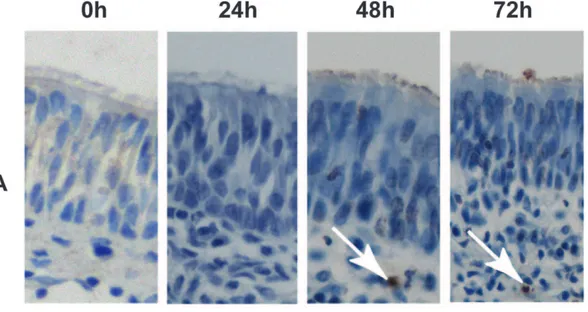
The influence of cultivation on the viability and morphology of porcine nasal mucosa explants was first investigated. Viability was estimated by evaluating ciliary beating on the epithelial cells using a light microscope and by quantification of the number of apoptotic cells using the terminal deoxynucleotidyl transferase mediated dUTP Nick End Labelling (TUNEL) assay.
Cultivation of the mucosa explants for up to 72 h did not show any biological difference in the number of apoptotic cells (TUNEL-positive cells) in the epithelium (less than 1% of apoptotic cells) and basal body (less than 5% of apoptotic cells) of the explants (Figure 1A, Table 1). Morphometric analyses indicated that the explants also did not show changes in epithelial thickness after 72 h of exvivocultivation (Figures 1B and 2).
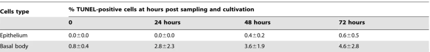
Scanning electron microscopy revealed the presence of both ciliated and non-ciliated cells at the surface of the explant.
Representative scanning electron micrographs (SEM) of explants at 0 and 72 h ofex vivoculture illustrate ciliary cells and non-ciliary cells (Figure 3). Duringex vivoculture, no morphological changes of the epithelium layer were observed. The cilia of the epithelial cells continued to beat up to 72 h after the start of cultivation.
Persistence of MRSA on the Porcine Mucosa Explants Next we investigated as to whether porcine mucosa explants could be exploited to study MRSA ST398 colonization. The ability of S. aureusto colonize the porcine mucosa explants was defined as persistence or outgrowth of MRSA on the explants. Three MRSA ST398 isolates were tested. One strain was isolated from a carrier pig (S0462), one human isolate originated from an endocarditis patient (S0385-1) and S0385-2 was a laboratory variant showing a different hemolysis pattern.
Initial inoculation of the explants (1 cm2) was performed with 36108 colony forming units (CFU)/ml. After 2 h of incubation and washing of the explants, approximately 86106CFU/cm2
Figure 1. Evaluation of porcine mucosa explants after cultivation by means of light microscopy.Sections of 4mm thickness were stained
by immunohistochemistry to evaluate the apoptosis of cells. TUNEL-positive cells in the epithelium are indicated with white arrows (panel A). Panel B shows the thickness of the epithelium after staining with haematoxylin-eosin (indicated with a white arrow).
(5%) adhered to the explants. The presence of the isolates on the mucosa explants was followed for an additional 180 min. During the first 30 min, S0462, S0385-1 and S0385-2 showed an initial decline in the number of CFU to approximately 36106, 26105, and 1.56105CFU/cm2respectively (Figure 4). Then, the number of recovered bacteria from isolate S0385-1 remained almost stable until the end of the experiment at approximately 46105CFU/ cm2. Bacterial adhesion for isolate S0462 remained stable until 90 min of the experiment. Then a significant increase to approximately 46107CFU/cm2was observed. Bacterial recovery for MRSA S0385-2 showed a gradual decline during the experiment up to approximately 26102CFU/cm2 at the end of the incubation period. As a control, growth of the strains in culture medium alone without antibiotics at 37uC in 5% CO2 without shaking for 3 h did not show any inhibition (data not shown). These results were reproducible in five independent experiments. To verify whether the porcine nasal mucosa explantsex vivomodel can be used to study colonization by other MRSA strains, MRSA Mu50 of human origin was used. This strain showed a similar colonization pattern as S0385-1 on the explants. The growth curve is shown in Supplementary figure S2.
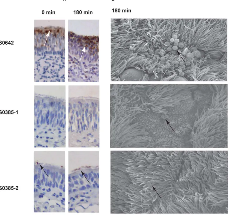
To visualize MRSA ST398 on the explants during the colonization assay, immunohistochemistry using anti-Staphylococcus aureus protein A monoclonal antibody was used (Figure 5). We were able to determine the localization of strain S0462. Isolate S0462 adhered to the surface under cilia and between epithelial cells of the top part of mucosa explants. After 180 min of incubation, the isolate S0462 formed clusters of colonies on the
surface of the explants, and migration of the bacterial cells to the bottom part of the epithelium layer was observed. The immuno-chemistry of the explants inoculated with strains S0385-1 and S0385-2 yielded either no or a weak signal, respectively. Nevertheless, these strains showed comparable numbers of bacteria as S0462 at post adhering time 0 min. Isolate S0385-2 adhered and remained on to the surface of the cilia itself. After 180 min of incubation with bacteria cells, isolate S0462 showed clustering of bacteria on the surface of the explants and further migration of the bacteria in the epithelium layer. Isolate S0385-2 remained on the surface of the cilia. To visualize the interaction of the bacteria with the epithelium in more detail, SEM was performed on the explants at 180 min of the incubation. Bacteria of the strain S0462 were present as clusters on the epithelial surface between the cilia (Figure 5, panel B). Strain S0385-2 remained on the cilial surface and did not appear in clusters. SEM on bacteria of the S0385-1 showed bacteria located on the epithelial surface. Interaction of MRSA ST398 isolates with the tissue did not result in visible changes in morphology of the inoculated epithelium.
Discussion
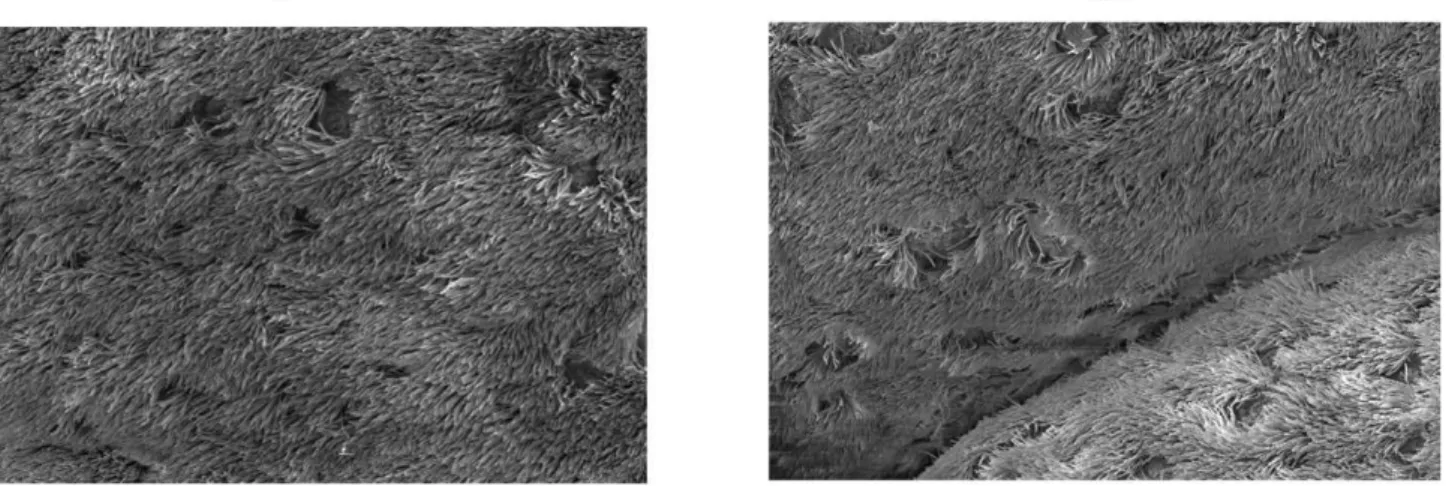
We evaluated porcine nasal mucosa explants as a model system to study S. aureus colonization in pigs. The model was adapted from a study on interaction of viruses with the respiratory tract [17].In vitroadhesion ofS. aureusto monolayer cell cultures have been used, especially to study bacterial interaction with human cells [19]. The limitation of this system is the lack of diversity in Table 1.Percentage of apoptotic cells as a parameter for the effect of cultivation.
Cells type % TUNEL-positive cells at hours post sampling and cultivation
0 24 hours 48 hours 72 hours
Epithelium 0.060.0 0.060.0 0.460.2 0.660.5
Basal body 0.860.4 2.862.3 3.661.9 4.662.8
doi:10.1371/journal.pone.0053783.t001
Figure 2. Average epithelial thickness of the porcine mucosa explants at different time points.Data are presented as mean6standard deviation (error bars) of five independent experiment.
cell types and often the lack of the presence of mucus [20]. The nasal environment contains different types of epithelial cells [21]. The ex vivoexplants model system applied here was designed to overcome these limitations and to better display the many characteristics and cell types of the porcine nasal mucosa cells
in vivo.
The porcine nasal mucosa explants were cultivated at an air-liquid interface which creates a physiological environment corresponding to natural conditions. As described earlier [17], serum-free medium was used to cultivate the explants. It has been reported that use of fetal calf serum results in enlarged epithelial cells, loss of cell–cell contacts and a loose epithelium. With the conditions employed in our hands, we successfully maintained porcine nasal respiratory explants for at least 72 h without any signs of gross changes in cell viability as measured by the presence of apoptotic cells (more than 5%). Similarly, morphometric analyses of the mucosa explants showed no major changes during
ex vivo cultivation. Furthermore, ciliary beating was observed during the entire cultivation and SEM showed that cell-cell contact and three-dimensional structures of the tissue were
preserved. Together, these results indicate that the porcine nasal mucosa explants preserved their integrity and viability in ex vivo
conditions for up to 72 h under the conditions employed. To evaluate porcine nasal mucosa explants as a new tool to study MRSA colonization in pigs, we used three MRSA ST398 strains as inoculum. All showed reproducible adherence to the epithelial layer of the mucosa explants. However, we observed differences in persistence of the isolates. One isolate was from a carrier pig (S0462), the two other strains were from a patient with endocarditis (S0385-1 and S0385-2). All three isolates showed an initial decline in the number of CFU during the first 30 min after inoculation which might indicate bacteria adaptation to the explants. After 30 min of post adhering period, bacterial recovery from the experiments showed a significant increase of the number of CFU for isolate S0462, unaltered bacterial number for isolate S0385-1, and a loss bacteria for isolate S0385-2, suggesting differences in interaction of the different isolates with the tissue. Additionally, the colonization assay using a human derived MRSA strain Mu50 showed a similar pattern to MRSA S0385-1. We conclude that this model can be use to study MRSA colonization belonging to different clonal complexes and human origin.
Attempts to visualize the bacteria on the tissue was performed using anti-Staphylococcus aureus protein A monoclonal antibodies were only partially successful. Isolates S0385-1 and S0385-2 were probably poorly visible due to the presence of low numbers of bacteria and/or insufficient expression of protein A. It has previously been documented that someS. aureusstrains show no or very low expression of protein A in vivo [22]. Additionally, Western blotting of stationary grown bacteria in BHI medium confirmed poor expression of protein A in these strains (data not shown). SEM of tissue carrying isolate strain S0462 revealed clusters of bacteria located between the cilia. For the two isolates S0385-1 and S0385-2 only single bacteria were observed. The absence of bacterial clusters may be caused by the lower number of bacteria recovered for these strains. However, we cannot exclude the alternative possibility that the lack of bacterial cluster formation contributed to the poor bacterial recovery of these isolates. Our observations do suggest that different MRSA isolates display variable qualities in colonizing mucosa explants, which perhaps mimics natural host colonization. Due to the fact that adhesion of the bacteria to the tissue was performed in DPBS, which is not reflecting the in vivo situation, small changes comparing within vivosituation may occur.
Figure 3. Scanning electron micrographs of porcine nasal epithelium.Epithelial cells at 0 h (A) and after 72 h (B) ofex vivocultivation. doi:10.1371/journal.pone.0053783.g003
Figure 4. MRSA colonization of the porcine mucosa explants.
Log scale presence of MRSA isolates S0462 (.), S0385-1 (m) and S0385-2 (&) on the porcine nasal mucosa explants. Data are presented is the mean CFU 6 standard deviation (error bars) of five different pig experiments.
A major advantage of theex vivoporcine nasal mucosa explants model is that different bacterial strains can be tested under controlled conditions. From one animal around 20 explants (1 cm2) can be obtained and multiple strains can be tested simultaneously eliminating genetic variation of the host. In addition, the model can be readily adapted for other bacterial species or the introduction of multiple species. The successful establishment of porcine nasal mucosa explants to study of the interaction ofS. aureusisolates with nasal tissue enables studies to better understand the mechanisms of colonization of MRSA in pigs and may aid future assessment of the effects of potential inhibitory compounds on this process.
Supporting Information
Figure S1 Schematic cross-section of a culture system using nasal mucosa explant with an air-liquid interface. (TIF)
Figure S2 Log scale presence of pig origin MRSA S0462 and the human derived strain Mu50 on the porcine nasal mucosa explants.Data are presented as mean CFU6standard deviation (error bar) of five different pig experiments. MRSA S0462 belongs to ST398spa-type: t011 SCCmecV. MRSA Mu50 belongs to CC5 spa-type t002 SCCmec II. Strain Mu50 shows successful colonization on the porcine nasal mucosa explants,
although variation between experiments was bigger with Mu50 compared to S0462.
(TIF)
Acknowledgments
The authors would like to thank Evelyn Velema from UMCU and Louis van den Boom from the Faculty of Veterinary Medicine at Utrecht University for helping with mucosa explants isolation. We are grateful to Nicole ten-Broeke-Smits from the University Medical Center in Utrecht, and Mirjam Koster from the Faculty of Veterinary Medicine at Utrecht
University for help with immunohistochemistry staining. We would like to thank Wally Muller from the Faculty of Science at Utrecht University for help with scanning electron microscopy.
Author Contributions
Conceived and designed the experiments: PT AF BD JW JvP. Performed the experiments: PT AdB. Analyzed the data: PT SG BD AF JW. Contributed reagents/materials/analysis tools: PT JvP AdB SG. Wrote the paper: PT AF BD JW.
References
1. Wertheim HF, Melles DC, Vos MC, van Leeuwen W, van Belkum A, et al. (2005) The role of nasal carriage inStaphylococcus aureusinfections. Lancet Infect Dis 5: 751–762.
2. Voss A, Loeffen F, Bakker J, Klaassen C, Wulf M (2005) Methicillin-resistant
Staphylococcus aureusin pig farming. Emerg Infect Dis 11: 1965–1966. 3. Smith TC, Pearson N (2010) The emergence ofStaphylococcus aureusST398.
Vector Borne Zoonotic Dis 11: 327–339.
4. Graveland H, Wagenaar JA, Bergs K, Heesterbeek H, Heederik D (2011) Persistence of livestock associated MRSA CC398 in humans is dependent on intensity of animal contact. PLoS One 6: e16830.
5. van Cleef BA, Monnet DL, Voss A, Krziwanek K, Allerberger F, et al. (2011) Livestock-associated methicillin-resistantStaphylococcus aureusin humans, Europe. Emerg Infect Dis 17: 502–505.
6. Golding GR, Bryden L, Levett PN, McDonald RR, Wong A, et al. (2010) Livestock-associated methicillin-resistantStaphylococcus aureussequence type 398 in humans, Canada. Emerg Infect Dis 16: 587–594.
7. van den Broek IV, van Cleef BA, Haenen A, Broens EM, van der Wolf PJ, et al. (2009) Methicillin-resistantStaphylococcus aureusin people living and working in pig farms. Epidemiol Infect 137: 700–708.
8. van Belkum A, Melles DC, Peeters JK, van Leeuwen WB, van Duijkeren E, et al. (2008) Methicillin-resistant and -susceptibleStaphylococcus aureussequence type 398 in pigs and humans. Emerg Infect Dis 14: 479–483.
9. Kiser KB, Cantey-Kiser JM, Lee JC (1999) Development and characterization of aStaphylococcus aureusnasal colonization model in mice. Infect Immun 67: 5001–5006.
10. Kokai-Kun JF (2008) The cotton rat as a model forStaphylococcus aureusnasal colonization in humans: Cotton ratS. aureusnasal colonization model. Methods Mol Biol 431: 241–254.
11. Gonzalez-Zorn B, Senna JP, Fiette L, Shorte S, Testard A, et al. (2005) Bacterial and host factors implicated in nasal carriage of methicillin-resistantStaphylococcus aureusin mice. Infect Immun 73: 1847–1851.
12. Broens EM, Graat EA, van de Giessen AW, Broekhuizen-Stins MJ, de Jong MC (2011) Quantification of transmission of livestock-associated methicillin resistant
Staphylococcus aureusin pigs. Vet Microbiol 155: 381–388.
13. Crombe F, Vanderhaeghen W, Dewulf J, Hermans K, Haesebrouck F, et al. (2011) Colonization and transmission of methicillin-resistantStaphylococcus aureus
ST398 in nursery piglets. Appl Environ Microbiol 78: 1631–1634.
14. Moodley A, Latronico F, Guardabassi L (2011) Experimental colonization of pigs with methicillin-resistant Staphylococcus aureus(MRSA): Insights into the colonization and transmission of livestock-associated MRSA. Epidemiol Infect 139: 1594–1600.
15. Jackson AD, Rayner CF, Dewar A, Cole PJ, Wilson R (1996) A human respiratory-tissue organ culture incorporating an air interface. Am J Respir Crit Care Med 153: 1130–1135.
16. Antunes MB, Woodworth BA, Bhargave G, Xiong G, Aguilar JL, et al. (2007) Murine nasal septa for respiratory epithelial air-liquid interface cultures. BioTechniques 43: 195–196.
17. Glorieux S, Van den Broeck W, van der Meulen KM, Van Reeth K, Favoreel HW, et al. (2007)In vitroculture of porcine respiratory nasal mucosa explants for studying the interaction of porcine viruses with the respiratory tract. J Virol Methods 142: 105–112.
18. Ekkelenkamp MB, Sekkat M, Carpaij N, Troelstra A, Bonten MJ (2006) Endocarditis due to meticillin-resistantStaphylococcus aureusoriginating from pigs. Ned Tijdschr Geneeskd 150: 2442–2447.
19. Wyatt JE, Poston SM, Noble WC (1990) Adherence ofStaphylococcus aureusto cell monolayers. J Appl Bacteriol 69: 834–44.
20. Roche FM, Meehan M, Foster TJ (2003) TheStaphylococcus aureussurface protein SasG and its homologues promote bacterial adherence to human desquamated nasal epithelial cells. Microbiology 149: 2759–2767.
21. Martineau-Doize B, Caya I (1996) Ultrastructural characterization of the nasal respiratory epithelium in the piglet. Anat Rec 246: 169–175.