Delano Gurgel SilveiraI, Márcio Alencar BarreiraII, Luiz Gonzaga de Moura JuniorIII, Charles Jean
Gomes de MesquitaIII, Hermano Alexandre Lima RochaIV, Gleydson Cesar de Oliveira BorgesV
Hepatic retractor in an
ex vivo
model
1Abstract
Purpose: To discuss the use of models of hepatic retraction by laparoscopy, to present a
new Hepatic Retractor (HR) and to evaluate its practicality, efficacy and safety in Esophageal Hiatus Exposure (EHE).
Methods: Experimental cross - sectional study with a quantitative character. It was carried out
in the Laboratory of Health Training of Christus University Center. The sample consisted of 12 livers of adult pigs weighing between 30 and 45 kg. A circular-shaped HR, 5 cm diameter and deformable materials was developed with a polypropylene cloth, metallic guide wire, epidural needle plastic guide and nylon string. The practicality of HR management was measured by the time required to use the instrument, efficacy by exposure to the operative field and safety by macroscopic assessment of liver damage.
Results: The average time to complete the procedure was 3.24 minutes and reached less
than 2 minutes after 12 repetitions. In eight experiments the maximum degree of EHE was obtained. No macroscopic lesions were observed.
Conclusion: The use of HR described can broaden the operative field, without causing
macroscopic liver lesions and prolonging the surgical time.
Key words: Surgery. Laparoscopic. Liver. Swine.
IFellow Master degree, Postgraduate Program in Minimally Invasive Technology and Health Simulation, Centro Universitário Christus (UNICHRISTUS), Fortaleza-CE, Brazil. Conception and design of the study; technical procedures; acquisition, interpretation and analysis of data; manuscript preparation and writing.
IIMD, General Surgeon, Hospital Universitário Walter Cantídio, Fortaleza-CE, Brazil. Manuscript preparation, critical revision.
IIIPhD, Assistant Professor, Professional Master’s Degree Program in Minimally Invasive Technology and Simulation in Health, UNICHRISTUS, Fortaleza-CE, Brazil. Conception and design of the study, critical revision.
IVPhD, Assistant Professor, Professional Master’s Degree Program in Minimally Invasive Technology and Simulation in Health, UNICHRISTUS, Fortaleza-CE, Brazil. Conception and design of the study, statistical analysis, interpretation of data, critical revision.
The regulations of the National Council for the Control of Animal Experimentation (CONCEA) were observed to adopt an alternative method to the use of animals in research activities in Brazil8.
The pig liver (ex vivo model) was
positioned within the cavity of the EndoSuture
Training Box® simulator. This abdominal cavity
simulator is validated and used in teaching and research practices. It is capable of providing a suitable surgical field for the training of several surgical procedures9,10. All measures
were performed by a single evaluator, avoiding any kind of unfairness. The experiments were performed one after the other, following the same pattern of procedures.
An HR prototype with circular shape and diameter of 5 cm was elaborated. Materials that were deformable and semi-rigid were searched for. After being deformed by some external force, the material should regain its original shape. After a technical drawing the author himself constructed an HR with the materials available in the surgical arsenal (Figure 1). For the high memory rigid rod forming the perimeter of the HR, a metallic guide wire was used that composes the central venous access of the catheterization material. A polypropylene mesh commonly used in hernia repair was cut in a circular format to compose the central disk of the HR. A rigid central rod was made with the plastic guide that accompanies the needle used for epidural anesthesia. The 5.0 nylon thread had as a function to fix the materials to the polypropylene fabric. The estimated final cost for building HR was around $45. An Invention Patent was filed with the National Institute of Industrial Property (INPI) with the following patent registration BR 102016028346-9.
■
Introduction
Hepatic Retrator (RH) is intended to increase Esophageal Hiatus Exposure (EHE) through the displacement of the Left Hepatic Lobe (LHL). Over the last few decades, numerous retractors and techniques have been developed to facilitate laparoscopic
procedures in the upper abdomen1. As a way
to compensate for the lack of instruments to move the LHL, some surgeons use tweezers,
vacuum cleaners, wires, drains and probes2. It
is known that the expansion of the operative field contributes to the surgical treatment of obesity3, gastric pathologies4 and gastro
esophageal reflux disease5.
The principles of 3Rs (Reduction, Refinement and Replacement) are based on finding alternatives to reduce the number of animals in research6. The swine liver has an
anatomical similarity with the organ of the human being. The segmental anatomy of the pig liver is well studied and has similarities with the human liver7. The elaboration of a
new surgical instrument or its improvement is a challenging activity. The objective of this study is to discuss the use of HR models by laparoscopy, to present a new HR and to evaluate its practicality, efficacy and safety in the EHE.
■
Methods
A quantitative cross-sectional study was carried out at the Christus University Center Health Training laboratory.
Figure 1 - HR prototype.
Variables analyzed
The practicality of HR management
was measured by time. The time to enter the simulator (HR positioning at the entrance of the 10 mm trocarte coupled to the simulator) until the maximum static retraction of the liver for 10 seconds (T1) and the time between the end of the period of maximum static retraction of the liver to withdrawal of the simulator cavity (T2). The time was checked with the
professional digital chronometer Vollo® Vl-510
C/10Memory.
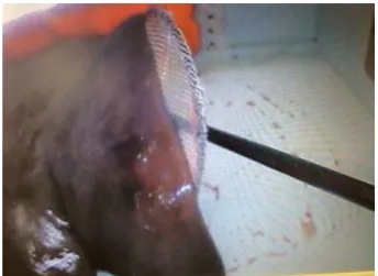
To perform the HR efficacy experiment in providing surgical field, a tape measure and ruler behind the swine liver were fixed and the highest degree of liver retraction in centimeters was measured (Figure 2). The largest measure of retraction provided by HR was tabulated as degree 1 of exposure and the lowest measure of retraction as grade 3. The intermediate measurement was tabulated as grade 2 of exposure. In case of not being able to see the tape measure due to insufficient retraction of the liver or it was not possible to maintain the maximum hepatic retraction for 10 seconds it was considered degree 4 of
Figure 2 - Moment of greatest hepatic retraction.
Macroscopic liver lesion was assessed visually by identifying lesions on hepatic surfaces which suffered the HR direct action. Hepatic lesions were classified according to the graduation scale established by the American
Association of Trauma Surgery11. The presence
of lacerations or bruises (grade I to III) was evaluated. Vascular lesions (grade IV and V) and hepatic avulsion (grade VI) do not apply to the experiment for the obvious reason of being
an ex-vivo research model. Liver photographs
Statistical analysis
Categorical quantitative results were presented as percentages and counts. The collected data were tabulated and analyzed by SPSS software, v23, IBM, Inc.
■
Results
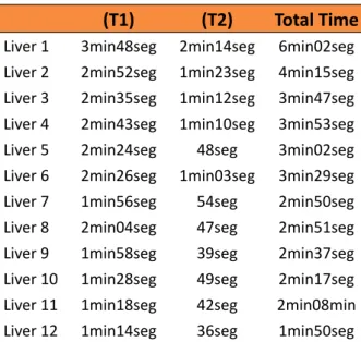
In Table 1, it was observed that the time to use the retractor was decreasing as it was being used and after 12 repetitions the evaluator took less than 2 minutes to do the HR positioning and withdrawal procedure. The average time to complete the procedure was 3.24 minutes.
Table 1 - Time for the use of HR.
(T1) (T2) Total Time
Liver 1 3min48seg 2min14seg 6min02seg Liver 2 2min52seg 1min23seg 4min15seg Liver 3 2min35seg 1min12seg 3min47seg Liver 4 2min43seg 1min10seg 3min53seg Liver 5 2min24seg 48seg 3min02seg Liver 6 2min26seg 1min03seg 3min29seg Liver 7 1min56seg 54seg 2min50seg Liver 8 2min04seg 47seg 2min51seg Liver 9 1min58seg 39seg 2min37seg Liver 10 1min28seg 49seg 2min17seg Liver 11 1min18seg 42seg 2min08min Liver 12 1min14seg 36seg 1min50seg
Table 2 shows the maximum retraction of the left hepatic lobe and the EHE grade. It was found that at the beginning of the experiment it was possible to reach maximal liver retraction and the highest EHE. In eight experiments the maximum EHE degree was obtained.
Table 2 - Expansion of the operative field with HR. Experiments Maximum
Retraction Exposition grade
1 10cm 3
2 13 cm 2
3 13 cm 2
4 15 cm 1
5 15 cm 1
6 15 cm 1
7 13 cm 2
8 15 cm 1
9 15 cm 1
10 15 cm 1
11 15 cm 1
12 15 cm 1
No macroscopic lesions were observed in any of the 12 livers used in the experiments. Table 3 shows the force applied to the liver at the time of greatest retraction. There was a slight variation in the force used that was not able to cause bruising or lacerations in any of the scenarios.
Table 3 - Strength applied to the liver by HR.
Kgf N
Liver 1 0.105 1.029
Liver 2 0.110 1.078
Liver 3 0.108 1.059
Liver 4 0.121 1.186
Liver 5 0.113 1.108
Liver 6 0.103 1.010
Liver 7 0.115 1.127
Liver 8 0.118 1.157
Liver 9 0.134 1.314
Liver 10 0.125 1.225
Liver 11 0.116 1.137
Liver 12 0.112 1.098
■
Discussion
HR is an important aspect in laparoscopic procedures of the upper abdomen. Surgeons have created several methods for HR because of limitations related to the absence of commercially available equipment. A review study analyzed the use of various hepatic retractors in the EHE and the time required for the EHE to be displaced through the analysis of surgical videos. Nasogastric tube (NG), hepatic suspension by puncture and drainage, 3 mm laparoscopic forceps, umbilical tape (sling method) and suture of the diaphragmatic pillar (crural suture) were used to mobilize the liver. The use of 3mm laparoscopic forceps achieved the best EIE grade, followed by umbilical tape and NG suspension. The use of 3mm laparoscopic forceps required less time to remove the liver (average of 2.8 minutes), followed by diaphragmatic pillar suture (average of 5.14 minutes) and NG suspension (average of 7 minutes). The average time to create liver retraction ranged from 2.8 to 8.6 minutes. The most used methods were: NG suspension (36 cases), umbilical tape (22 cases) and hepatic suspension by puncture and drain use (22 cases). It is not possible to compare
these methods with what was used in ex vivo
pig liver2.
A review study evaluated 10 instruments or techniques used for HR. Devices included were Nathanson liver retractor, liver suspension tape, V-List technique, a silicone disc with or without a snake retractor, Endoloop, Endograb, a magnetic retractor, the VaroLift, a laparoscope support and a retraction sponge. None of the instruments reported was associated with increased morbidity and conversion rates for open surgery. All articles reported that the instruments tested may save the use of the assistant during the procedure. All instruments
surgical procedure due to heterogeneity of procedures and reduced sample. The authors reported good to excellent field of vision1. One
study compared three different HR methods by laparoscopy during Roux-en-Y gastric bypass surgery. Sixty patients were randomly divided into groups. He evaluated Nathanson’s HR and V-LIST (V-LIST) methods using penrose drain and tape suspension technique using the Jackson-Pratt drain. Hepatic dysfunction related to Nathanson’s HR was observed, but there was no evidence of hepatic impairment. Postoperative pain was lower in the group that used hepatic suspension with Jackson-Pratt drain. Hepatic suspension techniques are easy to learn and do not need an extra incision for
HR placement3.
It is possible to eliminate the need for a trocarte for the use of HR by using a suture in the upper portion of the right pillar with subsequent exteriorization of the two ends of the strand by a percutaneous device. The traction of the wire well positioned in the LHL is capable of suspending it and widening the
operative field12. Another technique designed
to suspend the LHL nontraumatically, uses a Penrose drain 8 cm long and 1 cm wide. Two sutures with silk strands were performed at both ends of the penrose prior to insertion of the drain into the abdominal cavity. After opening the triangular ligament, the drain is positioned under the left lateral segment of the liver and the silk thread is passed through the opening created by the section of the triangular ligament. The silk thread of approximately 10cm at each end of the penrose is exteriorized from the abdominal cavity by improvisation of a device used for venous access puncture. After traction and fixation in the abdominal wall of the two ends of the wire a larger EHE is possible5.
the yarn is exteriorized from the abdominal cavity through the use of straight needles at the
ends of the gauze4. Moura-Júnior et al.13, used
a zero-lined silk thread in 7 cm of nelaton probe to protect the hepatic parenchyma during LHL displacement. The wire had one end anchored to the right diaphragmatic pillar and the other to the portal to the left of the surgeon. The goal was to improve the EHE during 48 laparoscopic procedures of the upper abdomen. Placement of the flexible liver retractor did not increase operative time, was easy to handle, and may improve EHE during single port and robotic surgery.
A device for internal retraction of the liver was used in fourteen surgical procedures, through laparoscopic access, of the upper abdomen. The HR was formed by a disposable telescopic rod with anchoring jaws at each end. The retractor was introduced into the peritoneal cavity through a 5 mm trocarte and needed a proper tool to attach the left end previously to the diaphragmatic origin of the right pillar. The right extremity was medial to the falciform ligament and anchored to the peritoneum located below the left border of the liver. After fixation of the retractor, the fixation tool was removed and the trocarte made available for the use of other surgical clamps. Thus, it eliminated the need for an incision for the retractor and freed one of the surgeons’ hands. The retractor was easy to use and allowed adequate exposure of the operative field in all procedures. There were no intra-operative or post-operative complications (intra-abdominal bleeding or diaphragm perforation) related to the device. The position time of the retractor improved with the experiment and reached
approximately 1 minute14.
Shibao et al.15 used a leaf-shaped
retractor and made with a rubber membrane and flexible memory structure as HR during laparoscopic surgeries of the upper abdomen. A snake retractor is needed to aid in liver
retraction. An elastic band attached to the skin was used for the external fixation of the snake retractor and release of the auxiliary surgeon’s hand. The entire procedure lasts less than 3 minutes and provides a better surgical field.
Transient elevation of hepatic enzymes has been described in the immediate post-operative period of surgeries that use some type of HR. This laboratory change is not usually related to clinical repercussions16. However,
inadequate manipulation of an HR can lead to liver damage and, consequently, increase the surgery morbidity. Avoid applying excessive force to the liver and adjust the HR during long
surgeries17. There are reports of more serious
cases, such as hepatic lobe necrosis related to
the use of HR18. The described HR format allows
a reduction of the force applied to the liver and, consequently, protection against liver damage. The greater the force required to manipulate HR, the less convenience for the surgeon and the greater possibility of loss of the surgical field. It was not possible to evaluate changes in liver enzymes because it is an ex vivo organ.
■
Conclusion
The use of the described HR can increase the operative field, without causing macroscopic hepatic lesions and prolong the surgical time. Further studies are needed to validate and perfect this new surgical instrument.
■
References
1. Vargas-Palacios A, Hulme C, Veale T, Downey CL. Systematic Review of Retraction Devices for Laparoscopic Surgery. Surg Innov. 2015 Feb;23(1):90-101. doi: 10.1177/1553350615587991.
2. Palanivelu P, Patil KP, Parthasarathi R, Viswambharan JK, Senthilnathan P, Palanivelu C. Review of various liver retraction techniques in single incision laparoscopic surgery for the exposure of hiatus. J Min Access Surg. 2015 Jul-Sep;11(3):198-202. doi: 10.4103/0972-9941.140202.
3. Goel R, Shabbir A, Tai CM, Eng A, Lin HY, Lee SL, Huang CK. Randomized controlled trial comparing three methods of liver retraction in laparoscopic Roux-en-Y gastric bypass. Surg Endosc. 2013 Feb;27(2):679-84. doi: 10.1007/s00464-012-2438-6.
4. Woo Y, Hyung WJ, Kim HI, Obama K, Son T, Noh SH. Minimizing hepatic trauma with a novel liver retraction method: a simple liver suspension using gauze suture. Surg Endosc. 2011 Dec;25(12):3939-45. doi: 10.1007/ s00464-011-1788-9.
5. Hamzaoglu I, Karahasanoglu T, Aytac E, Karatas A, Baca B. Transumbilical totally laparoscopic single-port nissen fundoplication: a new method of liver retraction: the Istanbul technique. J Gastrointest Surg. 2010 Jun;14(6):1035-9. doi: 10.1007/s11605-010-1183-1.
6. Cazarin KC, Corrêa CL, Zambrone FA. Redução, refinamento e substituição do uso de animais em estudos toxicológicos: uma abordagem atual. Rev Bras Ciênc Farm. 2004 Jul;40(3):289-98. doi: 10.1590/S1516-93322004000300004.
AR, Maddem GJ. Segmental nature of the porcine liver and its potential as a model for experimental partial hepatectomy. Br J Surg. 2003 Apr;90(4):440-4. doi: 10.1002/ bjs.4053.
8. Guimarães MV, Freire JE, Menezes LM. Utilização de animais em pesquisas: breve revisão da legislação no Brasil. Rev Bioét. 2016 May;24(2): 217-24. doi: 10.1590/1983-80422016242121.
9. Barreira MA, Siveira DG, Rocha HA, Moura-Junior LG, Mesquita CJ, Borges GC. Model for simulated training of laparoscopic gastroenterostomy. Acta Cir Bras. 2017
Jan:32(1):81-9. doi:
10.1590/s0102-865020170110.
10. Ferreira Filho F, Moura Júnior LG, Rocha HA, Rocha SG, Ferreira LF, Ferreira AF. Abdominal cavity simulator for skill progression in videolaparoscopic sutures in Brazil. Acta Cir Bras. 2018 Jan;33(1):75-85. doi: 10.1590/ s0102-865020180010000008.
11. Coccolini F, Catena F, Moore EE, Ivatury R, Biffl W, Peitzman A, Coimbra R, Rizoli S, Kluger Y, Abu-Zidan FM, Ceresoli M, Montori G, Sarteli M, Weber D, Naidoo N, Moore FA, Zanini N, Ansaloni L. WSES classification and guidelines for liver trauma. World J Emerg Surg. 2016 Oct 10;11:50. doi: 10.1186/ s13017-016-0105-2.
12. De la Torre RA, Satgunam S, Morales MP, Dwyer CL, Scott JS. Transumbilical single-port laparoscopic adjustable gastric band placement with liver suture retractor. Obes Surg. 2009 Dec;19(12):1707-10. doi: 10.1007/s11695-009-9896-5.
13. Moura-Júnior LG, Castro-Filho HF, Machado FH, Babadopulos RF, Feijó FC, Fernandes SD. Minimização de portais com miniportes e afastador flexível de fígado: alternativa ergonômica e estética ao single port em bypass gástrico laparoscópico. Arq Bras Cir Dig. 2014 Jul;27(Suppl 1):77-9. doi:10.1590/ s0102-6720201400s100019.
14. Elazary R, Kedar A, Shussman N, Abu-Gazala M, Khalaileh A, Faroja M, Rivkind AL, Mintz Y. A novel totally internal laparoscopic liver retractor. Surg Laparosc Endosc Percutan Tech. 2013 Dec;23(6):e222-4. doi: 10.1097/ SLE.0b013e31828e3fc5.
during laparoscopic surgery (with video). Surg Endosc. 2011 Aug;25(8):2733-7. doi: 10.1007/s00464-011-1614-4.
16. Morris-Stiff G, Jones R, Mitchell S, Barton K, Hassn A. Retraction transaminitis:an inevitable but benign complication of laparoscopic fundoplication. World J Surg. 2008 Dec;32(12):2650-4. doi: 10.1007/ s00268-008-9744-0.
17. Nozaki T, Kato T, Komiya A, Fuse H. Retraction-related acute liver failure after urological laparoscopic surgery. Curr Urol. 2014 Oct;7(4):199-203. doi: 10.1159/000365676. 18. Tamhankar AP, Kelty CJ, Jacob G.
Retraction-related liver lobe necrosis after laparoscopic gastric surgery. JSLS. 2011 Jan-Mar;15(1):117-21. doi: 10.4293/108680811 X13022985131651.
Correspondence:
Delano Gurgel Silveira
Avenida Beira Mar,3960/2304 60165121 Fortaleza – CE Brasil Tel.: (55 85)98822-9299
Received: July 22, 2018 Review: Sept 19, 2018 Accepted: Oct 20, 2018
Conflict of interest: none Financial source: none
1Research performed at Training Laboratory