Syntaxin-3 Binds and Regulates Both R- and
L-Type Calcium Channels in Insulin-Secreting
INS-1 832/13 Cells
Li Xie1, Subhankar Dolai1, Youhou Kang1, Tao Liang1, Huanli Xie1, Tairan Qin1, Lu Yang2, Liangyi Chen2, Herbert Y. Gaisano1*
1Department of Medicine, Faculty of Medicine, University of Toronto, Toronto, ON, Canada,2Institute of Molecular Medicine, Peking University, Beijing, China
Abstract
Syntaxin (Syn)-1A mediates exocytosis of predocked insulin-containing secretory granules (SGs) during first-phase glucose-stimulated insulin secretion (GSIS) in part via its interac-tion with plasma membrane (PM)-bound L-type voltage-gated calcium channels (Cav). In
contrast, Syn-3 mediates exocytosis of newcomer SGs that accounts for second-phase GSIS. We now hypothesize that the newcomer SG Syn-3 preferentially binds and modu-lates R-type Cavopening, which was postulated to mediate second-phase GSIS. Indeed,
glucose-stimulation of pancreatic isletβ-cell line INS-1 induced a predominant increase in interaction between Syn-3 and Cavα1 pore-forming subunits of R-type Cav2.3 and to lesser
extent L-type Cavs, while confirming the preferential interactions between Syn-1A with
L-type (Cav1.2, Cav1.3) Cavs. Consistently, direct binding studies employing heterologous
HEK cells confirmed that Syn-3 preferentially binds Cav2.3, whereas Syn-1A prefers L-type
Cavs. We then used siRNA knockdown (KD) of Syn-3 in INS-1 to study the endogenous
modulatory actions of Syn-3 on Cavchannels. Syn-3 KD enhanced Ca2+currents by 46%
attributed mostly to R- and L-type Cavs. Interestingly, while the transmembrane domain of
Syn-1A is the putative functional domain modulating Cavactivity, it is the cytoplasmic
domain of Syn-3 that appears to modulate Cavactivity. We conclude that Syn-3 may mimic
Syn-1A in the ability to bind and modulate Cavs, but preferring Cav2.3 to perhaps participate
in triggering fusion of newcomer insulin SGs during second-phase GSIS.
Introduction
SolubleN-ethylmaleimide-sensitive factor attachment protein receptor (SNARE) proteins, including target- (t-) membrane SNAREs (Syntaxins [Syn]) and synaptosomal-associated pro-teins of 25 kDa (SNAP25) and vesicle-associated membrane propro-teins (VAMPs), are the funda-mental components of the exocytotic machinery required for the docking and fusion of secretory granules (SGs) with the plasma membrane (PM), which have been well studied in neurons [1,2] and neuroendocrine cells, particularly pancreatic isletβ-cells [3–5]. t-SNAREs OPEN ACCESS
Citation:Xie L, Dolai S, Kang Y, Liang T, Xie H, Qin T, et al. (2016) Syntaxin-3 Binds and Regulates Both R- and L-Type Calcium Channels in Insulin-Secreting INS-1 832/13 Cells. PLoS ONE 11(2): e0147862. doi:10.1371/journal.pone.0147862
Editor:Alexander G Obukhov, Indiana University School of Medicine, UNITED STATES
Received:October 14, 2015
Accepted:January 8, 2016
Published:February 5, 2016
Copyright:© 2016 Xie et al. This is an open access article distributed under the terms of theCreative Commons Attribution License, which permits unrestricted use, distribution, and reproduction in any medium, provided the original author and source are credited.
Data Availability Statement:All relevant data are within the paper and its Supporting Information files.
Syn-1A and SNAP25 through their interactions with PM-bound voltage-gated calcium chan-nels (Cav), L-type inβ-cells and N-type in neurons, position the predocked SGs to the site of
maximum Ca2+influx for efficient exocytosis [6–12].
Cavs regulate secretion in neurons andβ-cells [13,14]. Cavα1 pore-forming subunits, Cav1
and Cav2, exist as heteromeric complexes by association with auxiliary subunits,βandα2δ
sub-units, which mediate trafficking of Cavs to the PM and fine-tune their biophysical properties
[13,14]. Inβ-cells, L-type Cav1s (Cav1.2 is abundant in rodents; Cav1.3 is abundant in human)
[15,16], are believed to effect first-phase GSIS by acting on the readily releasable pool (RRP) of predocked SGs [17–20]. Genetic deletion of R-type/Cav2.3 suppressed only the second-phase
GSIS from the mouse islets; and did not affect the early component of depolarization-induced exocytosis (corresponding to the RRP) in theβ-cells [21,22], leaving intact the late component, referred to as SG mobilization from the reserve pool, which corresponds to the newcomer SGs. This led us and others to hypothesize that predocked SGs mediating a major portion of first-phase GSIS and newcomer SGs accounting for all of second-first-phase GSIS are respectively medi-ated by L- and R-type Cavs.
Of the four Syns that mediate exocytosis, Syn-1A, Syn-2 and Syn-4 are present and localized predominantly to theβ-cell PM, whereas Syn-3 is more abundant in SGs [5,23–25]. Genetic deletion of Syn-1A in mice blunted first-phase GSIS, which was attributed to loss of ability of predocked insulin SGs to undergo exocytosis, without perturbation of recruitment and fusion of newcomer SGs [5]. Syn-1A binding and inhibition of L-type Cav[9,10] was demonstrated
to be via the two highly conserved cysteines, Cys271 and Cys272 at its transmembrane domain
[26–28]. In contrast, depletion of endogenous Syn-3 by RNA interference (RNAi) in INS-1
cells inhibited GSIS by impairing the recruitment and fusion of newcomer SGs affecting pre-dominantly the second-phase GSIS, without affecting predocked SGs [23]. Overexpression of Syn-3 was reported to also inhibitβ-cell L-type Cav[9]. However, it remains unclear whether
endogenous Syn-3 modulates Cavchannels inβ-cells. Furthermore, the putative Cav
-interact-ing transmembrane cysteine residues in Syn-1A are not conserved in Syn-3. Therefore, more work is required to clarify whichβ-cell Cavs Syn-3 acts on and the putative Cav-binding
domain within Syn-3.
In this work, we assessed the endogenous function of Syn-3 onβ-cell Cavactivity by siRNA
depletion and provide detailed biochemical and functional evidence for the interactions between endogenous Syn-3 and R-type (Cav2.3) and to lesser degree also L-type (Cav1.2 and
Cav1.3) Cavs.
Methods
Cell Culture
INS-1 832/13 cells and HEK293 cells lines were cultured as previously reported [29,30]. INS-1 832/13 cell line (herein called INS-1) was a gift from Christopher Newgard (Duke University, Durham, North Carolina) [31]. Syn-3 siRNA/mCherry plasmid (Dharmacon, Chicago, IL, USA) used here we previously reported to efficiently knockdown (KD) Syn-3 expression in INS-1 cells [23]. A mCherry plasmid was used as control [23]. After the cells were transfected with these plasmids for 48 h, cellular entry of the plasmids was confirmed by the mCherry expression observed by epifluorescence imaging. These mCherry-expressing cells were sub-jected to electrophysiology and TIRF imaging studies.
Immunoprecipitation
This was performed on INS-1 cells as previously reported [30,32]. INS-1 cells at 80%–85% confluence were washed with PBS (37°C) and incubated for 30 min at 37°C in Krebs–Ringer Competing Interests:The authors have declared
that no competing interests exist.
Abbreviations:Syn, syntaxin; SG, insulin-containing secretory granule; GSIS, glucose-stimulated insulin secretion; PM, plasma membrane; Cav, voltage-gated
HEPES buffer (KRB, in mM: 125 NaCl, 5.6 KCl, 1.28 CaCl2, 5 Na2CO3, 25 HEPES, pH 7.4.
with 0.1% BSA) containing basal 0.8 mM glucose concentration. Cells intended for stimulation were preincubated for 30 min with 0.8 mM glucose, and then stimulated with 16.7 mM glucose plus 10 nM glucagon-like peptide (GLP)-1 for 30 min. 1 mg protein extract of cell lysates were used for each condition. Immunoprecipitation (IP) was with 2μg Syn-1A or Syn-3 antibodies
or pre-immune IgG (as control). Precipitated proteins were immunodecorated and identified using the indicated primary antibodies, which include anti-Cav1.2, -Cav1.3, -Cav2.3, -Cav2.2,
-Cavα2δ-1, -Cavβ3, -SNAP25, -Syn-1A and–Syn-3. All Cavsubunits antibodies are from
Alo-mone Labs (Jerusalem, Israel), and SNAP25, Syn-1A and Syn-3 are from SySy (Goettingen, Germany); the specificity of these antibodies was well characterized by these companies, which been used broadly. IP experiments on HEK293 cells transfected with Syn-1A, Syn-3, Cav1.3 or
Cav2.3, were similar to those performed on INS-1 cells.
In Vitro Binding Assay and Western Blotting
In vitro binding assays were performed according to the method we previously described [30,
32,33], which also showed the specificity of the SNARE protein antibodies. Briefly, GST-Syn-1A (cytoplasmic domain a.a. 1–265), GST-Syn-3 (cytoplasmic domain a.a. 1–263) and GST (as control, 300 pM protein each, all bound to glutathione agarose beads) were incubated with HEK293 cell lysate at 4°C for 2 h with constant agitation. The beads were then washed three times with washing buffer (in mM: 20 HEPES (pH 7.4), 150 KOAC, 1 EDTA, 1 MgCl2; with
5% glycerol and 0.1% Triton X-100). Precipitated proteins were separated on 10% SDS-PAGE and identified with anti-Cav1.3 or -Cav2.3 antibody (1:200).
All of the Western blot bands were quantified using image J software (http://rsb.info.nih.
gov/ij). We employed two approaches to quantify the‘input control’and‘
co-immunoprecipi-tated (co-IPed)’blots. For quantification of‘input control’bands, we considered maximum intense band for each protein from each experiment as 100 and expressed other bands for that particular protein as % of maximum. For quantification of‘co-IPed’blots, we measured inten-sities of both‘co-IPed’and‘input control’bands that are processed in parallel. The intensity of
‘co-IPed’band was then calculated as a ratio to the corresponding‘input control’band intensity and multiplied by 5 (as‘input control’is 5% of total protein used for IP) and expressed as‘ per-centage of recovery’.
Electrophysiology
Recording pipettes were pulled from 1.5-mm borosilicate glass capillary tubes using a program-mable micropipette puller. Pipettes were heat polished to a tip resistance ranging from 2 to 3 MOhm when filled with the intracellular solution. For measurement of Cavcurrents, barium
was used as charge carrier. Pipettes were filled with the buffer containing (in mM): 120 CsCl, 20 tetraethylammonium chloride, 5 EGTA, 5 MgATP and 5 HEPES (pH 7.2 with CsOH). The external solution contained (in mM): 100 NaCl, 20 BaCl2, 20 tetraethylammonium chloride, 4
CsCl, 1 MgCl2, 10 glucose and 5 HEPES (pH 7.4 with NaOH). L-type Cavinhibitor nifedipine
(10μM), R-type Cavinhibitor SNX482 (400 nM) and N-type Cavinhibitorω-Conotoxin GVIA
(100 nM) are all from Alomone labs (Jerusalem, Israel). Cells were held at−70 mV for 2 min
Statistical Analysis
Data are presented as mean±SEM. Statistical significance was evaluated by Student’s t test or Mann-Whitney rank sum test (SigmaStat 3.11. Systat Software Inc., Chicago, IL, USA) and considered significant when P<0.05.
Results
Syn-3 Binds to Distinct
β
-cell Ca
vs than Syn-1A
We postulated that Syn-3’s actions on Cavchannels [9] may be by direct physical binding to
Cavsubunits as had been demonstrated for Syn-1A [11,12]. This was assessed with Syn-3
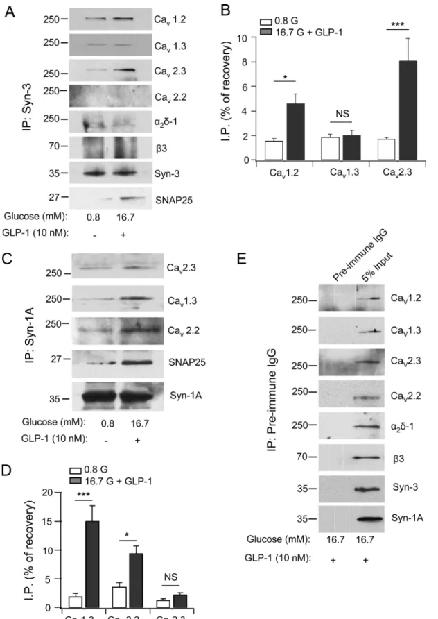
anti-body co-immunoprecipitation of INS-1 at basal (0.8 mM glucose) and maximal stimulated conditions (16.7 mM glucose plus 10 nM GLP-1).Fig 1A and 1Bshow that Syn-3 co-precipi-tated Cavα1 pore-forming subunits including Cav1.2, Cav1.3 and Cav2.3, but not Cav2.2. Syn-3
also brought down small amounts of auxiliary subunitsα2δ-1 andβ3. Remarkably, GLP-1/high glucose stimulation caused a large increase in the amount of Cav2.3 co-precipitated (from 1.7%
to 8.1%, a 4.7 fold increase; p<0.001), a more moderate increase in Cav1.2 co-precipitated (from 1.6% to 4.6%, a 2.9 fold increase; p<0.05), and no significant increase in Cav1.3 co-pre-cipitated (1.9% to 2.0%). There was no increase in the levels of co-preco-pre-cipitated auxiliary sub-unitsα2δ-1 andβ3; and there was no detectable Cav2.2 brought down. There was also an
expected large increase in the amount of SNAP25 co-precipitated (from 0.9% to 7.5%, p<0.001). These results indicate a preferential formation of the Syn-3/Cav2.3α1 complex, and the more moderate formation of the Syn-3/Cav1.2 complex could explain our previous report
on why Syn-3 overexpression also affected L-type Cavcurrent [9].
Syn-1A antibody co-precipitated Cav1.3, Cav2.2 and Cav2.3α1 subunits (Fig 1C). However,
only the amounts of Cav1.3 (from 1.9% at basal to 15% when stimulated, a 7.8 fold increase;
p<0.001; N = 3;Fig 1C and 1D) and Cav2.2 (from 3.6% to 9.4%; p<0.05; N = 3;Fig 1C and
1D) to lesser degree increased with stimulation, whereas there was no further increase in the amount of Cav2.3 co-precipitated. SNAP25 co-precipitated increased from 1.25% to 6.3%
(p<0.05; N = 3) after stimulation. The latter results confirm the previous hypothesis that Syn-1A preferentially associates with L-type Cavto form a complex exictosomes, which is
function-ally important in mediating exocytosis of predocked insulin SG [6,7]. While our results sup-port that Syn-1A could also bind R-type Cav[34], this complex is perhaps less important inβ
-cell or at least subordinate to the more abundant Syn-3-R-type Cavcomplex that formed.
Syn-1A is known to also bind N-type Cavin neurons [35] that is also found in INS-1 [34], albeit less
abundant, thus presumably less important inβ-cell. As control, at maximal stimulation with high glucose plus GLP-1, preimmune IgG did not pull down the syntaxins or any of the Cav
subunits (Fig 1E).S1 Figshows the corresponding input proteins with the Syn-3 or Syn-1A IP experiments inFig 1in both control and stimulated INS-1 cells.
Syn-3 Preferentially Binds Ca
v2.3, whereas Syn-1A Preferentially Binds
Ca
v1.2 and Ca
v1.3
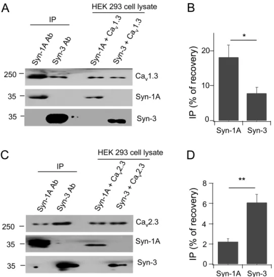
The co-IP studies of endogenously interacting proteins (Fig 1) showed a preference of Syn-3 for R-type/Cav2.3 over L-type Cavs. We therefore next critically assessed whether 3 vs
Syn-1A do indeed preferentially bind R- and L-type Cavs, respectively, by employing the HEK cell
model. HEK cells, devoid of endogenous Cavs and SNARE proteins, allow these proteins to be
individually exogenously expressed presumably in their native conformations, unperturbed by any other proteins that might affect them or their interactions as may be the case using native
Fig 1. Syn-3 co-immunoprecipitates (IP) distinct Cavs than Syn-1A in INS-1 cells.Syn-3 (A) and Syn-1A (C) interactions with the indicated Cavα1
subunits (Cav1.2, Cav1.3, Cav2.3 and Cav2.2) and auxiliary subunits (α2δ-1 andβ3) and SNAP25 in INS-1 cells. Densitometric analysis of Syn-3 co-IP (B)
Immunoprecipitation experiments were conducted in HEK cells expressing only Syn-3 or Syn-1A with Cav1.3 or Cav2.3α1 subunits. When calculated as the percentage of protein
recov-ery from total lysate input, Syn-1A co-precipitated Cav1.3 with an average (N = 3) of 18.1
±3.5% versus Syn-3 of 7.8±1.8%, which is 2.3 times higher (Fig 2A and 2B). In contrast, Syn-3 antibody co-precipitated more Cav2.3 (6.1±0.8%) than Syn-1A antibody (2.2±0.3%), which is
2.8 times higher (Fig 2C and 2D). Therefore, while there is some promiscuity in the binding of Syn-1A and Syn-3 for these Cavs, Syn-1A preferentially binds Cav1.3 and Syn-3 preferentially
binds Cav2.3. This is consistent with the current thinking that while the Syn-1A-Cav1.3
com-plex targets the PM sites of maximal Ca2+influx to where predocked insulin SGs exocytose [6,
7], we further postulate that the Syn-3-Cav2.3 complex likely targets the PM sites of Ca2+influx
where exocytosis of newcomer insulin SGs [23] would likely occur. This thinking is also consis-tent with the role of Cav2.3 in second-phase GSIS [21].
Depletion of Endogenous Syn-3 Selectively Enhances Ca
vChannels
Activity
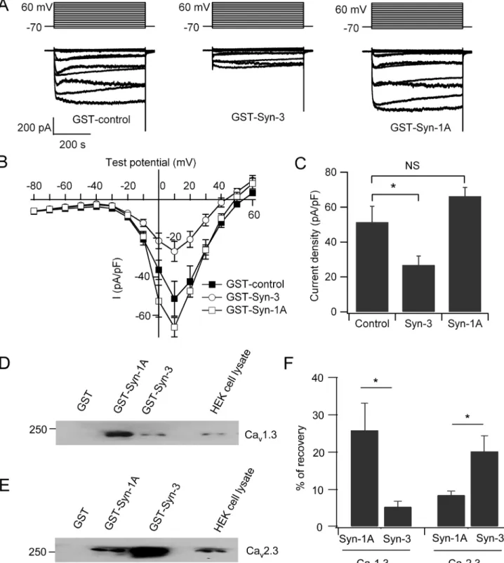
We then assessed the functional consequence of Syn-3 interactions with these Cavs by depletion
of endogenous Syn-3. Syn-3 siRNA plasmid co-expressing mCherry was used to transfect INS-1 832/13 cells, which effectively reduced Syn-3 protein expression by>70%, as shown in our previ-ous report [23]. INS-1 cells expressing mCherry would be expected to exhibit near-total depletion of Syn-3, thus ideal for single cell analysis by electrophysiology. Whole cell Cavcurrent recording
of Syn-3-depleted INS-1 cells showed the Cavcurrent amplitudes were increased by 46% (54.8
±5.6 pA/pF; n = 11;Fig 3A–3C) compared to control (mCherry transfected) cells (37.4±5.5 pA/ pF; p<0.05; n = 16). L- and R-type Cavs have been postulated to be the major Cavs in rodents to affect first- and second-phase GSIS, respectively [17–22]. N-type Cavwas reported to also
con-tribute to first-phase GSIS [36]. We thus used selective antagonists to determine the contribution of each of these Cavs to the overall Cavcurrent density in INS-1 cells (Fig 3D).Fig 3Eis the
sum-mary analysis of their Cavcurrent densities normalized to control values, showing that amounts
of Cavcurrent blocked was 52% by nifedipine (L-type antagonist) (n = 8; p<0.05), 31% (n = 10;
p<0.001) by SNX482 (R-type antagonist), and only 24% byω-Conotoxin GVIA (N-type antago-nist) (n = 9; p<0.01). This suggests that more of Cavcurrent blocked by the L- on R-type Cav antagonists, and not N-type Cavare likely attributed to the Syn-3 actions, which would be
consis-tent with our protein-binding data (Figs1and2). To confirm that L- and R-type Cavs accounted
for most of the increased Cavcurrent caused by the Syn-3 KD, we performed another set of
experiments with selective blockade with nifedipine and SNX482 on Syn-3 KD cells (Fig 3H and 3I) and Control cells (Fig 3F and 3G). Nifedipine reduced the Cavcurrent by 55%% (n = 12,
p<0.05) in Syn-3 KD cells which was slightly less than the 58% reduction (n = 14; p<0.001) in Control cells. SNX482 reduced the Cavcurrent by 28% (n = 6; p<0.05) in Syn-3 KD cells which
was slightly more than the 25% reduction (n = 10; p<0.05) in Control cells.
The Functional Domain of Syn-3 that Modulates Ca
vChannel Activity is
Different from Syn-1A
It has been well studied that the putative domain of Syn-1A that modulates Cavactivity is the
transmembrane domain (amino acid 266–288), particularly the two vicinal cysteines (C271 mM) plus GLP-1 (10 nM) increased the association of these syntaxins with the respective Cavs. Values are means±SEM, n = 3.*p<0.05,***p<0.001, NS:
not significant. As control (E) shows representative blots from five separate co-IP experiments with pre-immune IgG, which did not bring down syntaxins or Cavs (leftlanes).Right lanes show the input lysates. All five experiments probed for the Cavαsubunits andα2δ-1, whereasβ3, Syn-3 and Syn-1A were
probed on two blots from separate experiments.
and C272) [26–28]. Indeed, dialysis of the cytoplasmic domain of Syn-1A, GST-Syn-1A (a.a. 1–265), by patch pipette into INS-1 cells had no significant effects on the Cavcurrent (66.2±5.1
pA/pF; n = 8) compared to GST control (51.4±9 pA/pF; n = 9;Fig 4A–4C). Peculiarly, the two vicinal cysteines in the Syn-1A transmembrane domain are not conserved in the transmem-brane domain of Syn-3. Remarkably, dialysis of the cytoplasmic domain of Syn-3, GST-Syn-3 (a.a. 1–263) inhibited Cavcurrent by 48% compared to control cells (26.8±5.2 pA/pF; n = 9;
p<0.05;Fig 4A–4C). This result suggests that the Cav-interacting domains of 3 and Syn-1A are different. Syn-3 shares a low 64% amino acid identity to Syn-Syn-1A [37], suggesting that it may be the cytoplasmic domains in Syn-3 distinct from Syn-1A that bind the Cavs.
We next employed protein binding and pull down assays of the cytoplasmic domains of Syn-1A (a.a. 1–265) or Syn-3 (a.a. 1–263) with Cav1.3 or Cav2.3α1 subunit expressed in
HEK293 cells. As shown inFig 4D–4Fwith transfected HEK293 cells, GST-Syn-1A (a.a. 1–265) preferentially binds to Cav1.3 with an average of 25.7±7.3% versus GST-Syn-3 (a.a.
1–263) of 5.3±1.5%, which is 4.8 times higher (Fig 4D and 4F; n = 4; p<0.05). In contrast,
Fig 2. Syn-3 preferentially binds Cav2.3 while Syn-1A preferentially binds Cav1.3.Representative blots show HEK293 cells co-transfected with Cav1.3 (A) or Cav2.3 (C) with either Syn-1A or Syn-3, then subjected
to co-IP with anti-Syn-1A or Syn-3 antibody. Co-precipitated proteins were identified with the indicated antibodies. Densitometric analysis of the co-precipitated Cav1.3 (B) or Cav2.3 (D), expressed as percent
recovery of total lysate inputs. Values are means±SEM, n = 3;*p<0.05,**p<0.01.
Fig 3. Depletion of Syntaxin 3 in INS-1 cells increased voltage-gated Ca2+currents.(A) Representative traces showing Cavcurrents recorded in
whole-cell mode from control and Syn-3 KD INS-1 whole-cells. (B) Current-voltage relationship of Cavs from control (n = 16) and Syn-3 KD (n = 11) INS-1 cells. Currents
were normalized to cell capacitance to yield current density. Values are means±SEM. (C) Bar chart shows the maximum increase in current density under stimulation of 10 mV voltage.*p<0.05 for control vs Syn-3 KD (D) Representative Cavcurrents from normal INS-1 cells before and after treatment with
GST-Syn-3 binds more Cav2.3 (20.1±4.1%) than GST-Syn-1A (8.4±1.0%), which is 2.4 times
higher (Fig 4E and 4F; n = 4; p<0.05). These results indicate the following. First, whereas Syn-1A cytoplasmic domain can bind Cavα1 subunits, preferentially L-type Cavs, it seems this
bind-ing does not significantly regulate the Cavactivity. Second, the cytoplasmic domain of Syn-3
binds the Cavα1 subunits, preferring R-type Cav2.3, thus presumably influencing Cav2.3
activ-ity. More work will be required to elucidate the putative functional Cav-binding domain(s)
within the Syn-3 cytoplasmic domain.
Discussion
Taken together, these results demonstrate that Syn-3, via its cytoplasmic domain, preferentially binds and regulates R-type/Cav2.3α1 subunit. This is consistent with Syn-3’s role in mediating
fusion of newcomer SGs that account for all of second-phase GSIS [23] and Cav2.3’s role in
mediating second-phase GSIS in rodents [21]. In contrast, Syn-1A preferentially binds L-type Cav1.2 and Cav1.3 to direct the PM sites of Ca2+influx to where fusion of predocked SGs
would occur during first-phase GSIS. Nonetheless, both syntaxins are promiscuous in binding both L- and R-type Cavs and could therefore potentially assist each other in regulating various
Cavs perhaps when either syntaxin becomes deficient. In pancreatic islets of human type-2
dia-betes, Syn-1A levels are severely reduced [38] which may presumably affectβ-cell L-type Cav
function that may contribute to the reduced efficiency of exocytosis of predocked insulin SGs, with ensuing reduction to absent first phase GSIS [5]. An increased in Syn-3 expression might compensate for the Syn-1A deficiency, perhaps by forming complexes with L-type Cavs which
could affect an increase in newcomer SGs exocytosis known to also occur in first-phase GSIS [25]. This could rescue the reduced first-phase secretion in type-2 diabetes islets that has been attributed to a loss of fusion competence of predocked SGs [38].
Syn-3 on insulin SGs [23] likely acts to direct the recruitment of newcomer SGs to PM-bound Cav2.3 by forming an excitosome complex, mimicking the actions of Syn-1A-Cav1.2
and Syn-1A-Cav1.3 excitosomes on predocked SGs [6,7]. During exocytosis, we postulate that
Syn-3 would dissociate from Cav2.3, relieving the inhibition and allowing Ca2+influx to affect
fusion of the newcomer SG. Whereas both syntaxins bind these Cavs, the putative functional
domain of Syn-1A is its transmembrane domain [26,28] whereas the putative functional Cav
-binding domain of Syn-3 is within its cytoplasmic domain. It is also possible that the syntaxin-binding domain(s) in Cav2.3 [34] may be different from that reported for L-type Cavs [6,11],
called the synprint site localized to the cytosolic II-III linker connecting the second and third transmembrane domains. Much further work is required to determine the putative interacting domains between Syn-3 and Cav2.3.
Lastly, our data are consistent with newcomer SGs (containing Syn-3) being located further away from the PM-bound R-type Cav2.3 but are recruited to Cav2.3 upon stimulation where
they undergo minimal docking time at the PM before fusion. The latter would indicate more rapid priming and a high-affinity Ca2+sensor (i.e. synaptotagmins) for newcomer SGs [39]. Synaptotagmin 7 has been purported to be the major Ca2+sensor forβ-cells [40], but whether this synaptotagmin or another synaptotagmin is the Ca2+sensor for newcomer SGs remain to be elucidated. Consistent with this thinking, R-type Cavchannel has been shown to recruit
synaptotagmins to the PM to form part of the excitosome [34]. We hope that this work will (n = 6); their summary analysis (G) of the maximum increase in current densities normalized to the percentage of control (Con).***p<0.001;*p<0.05 compared to Control. (H) Representative Cavcurrents of Syn-3 KD INS-1 cells before (Syn-3 KD, n = 11) and after treatment with nifedipine (n = 12) or
SNX482 (n = 6); and their summary analysis (I) of the maximum increase in current densities normalized as percentages of the Syn-3 KD cells.*p<0.05 compared to Syn-3 KD. Here, Syn-3 KD Cavcurrents were 148% of Control cells, similar to A and B.
Fig 4. Cytoplasmic Syn-3 domain but not cytoplasmic Syn-1A domain regulates Cavcurrents.(A) GST-Syn-3 cytoplasmic domain (a.a. 1–263) or GST-Syn-1A cytoplasmic domain (a.a. 1–265) or GST (control) was dialyzed into INS-1 cells, then Cavcurrents recorded. Shown are representative traces in
the whole-cell mode with stimulation from−80–60 mV. (B) Current-voltage relationship of Cavchannels. Currents were normalized to cell capacitance to yield
current density. (C) Bar chart showing the maximum current density in INS-1 cells dialyzed with GST control (n = 9), GST-Syn-3 (n = 9), or GST-Syn-1A (n = 8). Values are means±SEM;*p<0.05; NS: no significant difference. (D and E) Representative blots show HEK293 cells transfected with Cav1.3 (D) or Cav2.3 (E) subjected to pull down with 300 pmol of GST-Syn-1A (a.a. 1–265), GST-Syn-3 (a.a. 1–263) or GST. (F) Summary analysis of four separate experiments. Data was expressed as means±SEM;*p<0.05.
trigger more future study that will lead to the full characterization of the newcomer SG excito-some as has been worked out for the predocked SG excitoexcito-some. This is of broad importance to endocrine secretory biology, as newcomer SGs likely also account for the sustained phase of secretion in other endocrine cells. While our work with the INS-1 cell line establishes proof of concept of novel Syn-3-Cavcomplexes influencing Cavactivity, humanβ-cells do not contain
R-type Cav, but rather employ P/Q-type Cav(Cav2.1) to likely mediate second-phase GSIS and
consequently newcomer SG exocytosis [41]. Therefore, more exciting work will be required to assess if Syn-3 might similarly form complexes with human P/Q-type Cav(Cav2.1) to mediate
exocytosis of newcomer SGs in humanβ-cells.
Supporting Information
S1 Fig. The corresponding input proteins with the Syn-3 or Syn-1A IP experiments inFig 1
for control and stimulated INS-1 cells. (TIF)
Author Contributions
Conceived and designed the experiments: LX SD YK HX HG. Performed the experiments: LX SD YK HX. Analyzed the data: LX SD YK. Contributed reagents/materials/analysis tools: LX SD YK TL HX TQ LY. Wrote the paper: LX HG. Critiqued the ms: LC.
References
1. Pfeffer SR. A prize for membrane magic. Cell. 2013; 155(6):1203–6. doi:10.1016/j.cell.2013.11.014 PMID:24315088
2. Sudhof TC, Rothman JE. Membrane fusion: grappling with SNARE and SM proteins. Science. 2009; 323(5913):474–7. doi:10.1126/science.1161748PMID:19164740
3. Wheeler MB, Sheu L, Ghai M, Bouquillon A, Grondin G, Weller U, et al. Characterization of SNARE pro-tein expression in cell lines and pancreatic islets. Endocrinology. 1996; 137(4):1340–8. PMID: 8625909
4. Huang XH, Pasyk EA, Kang YH, Sheu L, Wheeler MB, Trimble WS, et al. Ca2+influx and cAMP
eleva-tion overcame botulinum toxin A but not tetanus toxin inhibieleva-tion of insulin exocytosis. Am J Physiol Cell Physiol. 2001; 281(3):C740–C750. PMID:11502551
5. Ohara-Imaizumi M, Fujiwara T, Nakamichi Y, Okamura T, Akimoto Y, Kawai J, et al. Imaging analysis reveals mechanistic differences between first- and second- phase insulin exocytosis. J Cell Biol. 2007; 177(4):695–705. PMID:17502420
6. Wiser O, Trus M, Hernandez A, Renstrom E, Barg S, Rorsman P, et al. The voltage sensitive Lc-type Ca2+channel is functionally coupled to the exocytotic machinery. Proc Natl Acad Sci U S A. 1999; 96 (1):248–253. PMID:9874804
7. Yang SN, Larsson O, Bränström R, Bertorello AM, Leibiger B, Leibiger IB, et al. Syntaxin 1 interacts with the L(D) Subtype of voltage-gated Ca2+channels in pancreatic beta cells. Proc Natl Acad Sci U S A. 1999; 96(18):10164–9. PMID:10468580
8. Ji J, Yang SN, Huang X, Li X, Sheu L, Diamant N, et al. Modulation of L-type calcium channels by dis-tinct domains within SNAP-25. Diabetes. 2002; 51(5): 1425–36. PMID:11978639
9. Kang Y, Huang X, Pasyk EA, Ji J, Holz GG, Wheeler MB, et al. Syntaxins-3 and–1A inhibit L-type cal-cium channel activity, insulin biosynthesis and exocytosis in beta-cell lines. Diabetologia. 2002; 45 (2):231–41. PMID:11935155
10. Sheng ZH, Rettig J, Cook T, Catterall WA. Calcium-dependent interaction of N-type calcium channels with the synaptic core complex. Nature. 1996; 379(6564):451–4. PMID:8559250
11. Atlas D. Functional and physical coupling of voltage-sensitive calcium channels with exocytotic pro-teins: ramifications for the secretion mechanism. J Neurochem. 2001; 77(4):972–85. PMID:11359862 12. Atlas D. The voltage-gated calcium channel functions as the molecular switch of synaptic transmission.
13. Yang SN, Berggren PO. The role of voltage-gated calcium channels in pancreatic beta-cell physiology and pathophysiology. Endocr Rev. 2006; 27(6):621–76. PMID:16868246
14. Dolphin AC. Calcium channel diversity: multiple roles of calcium channel subunits. Curr Opin Neurobiol. 2009; 19(3):237–44. doi:10.1016/j.conb.2009.06.006PMID:19559597
15. Nitert MD, Nagorny CL, Wendt A, Eliasson L, Mulder H. Cav1.2 rather than Cav1.3 is coupled to
glu-cose-stimulated insulin secretion in INS-1 832/13 cells. J Mol Endocrinol. 2008; 41(1):1–11. doi:10. 1677/JME-07-0133PMID:18562674
16. Reinbothe TM, Alkayyali S, Ahlqvist E, Tuomi T, Isomaa B, Lyssenko V, et al. The human L-type cal-cium channel Cav1.3 regulates insulin release and polymorphisms in CACNA1D associate with type 2
diabetes. Diabetologia. 2013; 56(2):340–9. doi:10.1007/s00125-012-2758-zPMID:23229155 17. Davalli AM, Biancardi E, Pollo A, Socci C, Pontiroli AE, Pozza G, et al. Dihydropyridine-sensitive and
-insensitive voltage-operated calcium channels participate in the control of glucose-induced insulin release from human pancreatic beta cells. J Endocrinol. 1996; 150(2):195–203. PMID:8869586 18. Barg S, Ma X, Eliasson L, Galvanovskis J, Göpel SO, Obermuller S, et al. Fast exocytosis with few Ca2
+channels in insulin-secreting mouse pancreatic B cells. Biophys J. 2001; 81(6):3308
–23. PMID: 11720994
19. Schulla V, Renstrom E, Feil R, Feil S, Franklin I, Gjinovci A, et al. Impaired insulin secretion and glucose tolerance in beta cell-selective Cav1.2 Ca2+channel null mice. EMBO J. 2003; 22(15):3844–54. PMID: 12881419
20. Trus M, Corkey RF, Nesher R, Richard AM, Deeney JT, Corkey BE, et al. The L-type voltage-gated Ca2 +channel is the Ca2+sensor protein of stimulus-secretion coupling in pancreatic beta cells.
Biochemis-try. 2007; 46(50):14461–7. PMID:18027971
21. Jing X, Li DQ, Olofsson CS, Salehi A, Surve VV, Caballero J, et al. Cav2.3 calcium channels control
second-phase insulin release. J Clin Invest. 2005; 115(1):146–54. PMID:15630454
22. Yang SN, Berggren PO. Cav2.3 channel and PKClambda: new players in insulin secretion. J Clin
Invest. 2005; 115(1):16–20. PMID:15630435
23. Zhu D, Koo E, Kwan E, Kang Y, Park S, Xie H, et al. Syntaxin-3 regulates newcomer insulin granules and compound fusion. Diabetologia. 2013; 56(2):359–69. doi:10.1007/s00125-012-2757-0PMID: 23132338
24. Xie L, Zhu D, Dolai S, Liang T, Qin T, Kang Y, et al. Syntaxin-4 mediates exocytosis of pre-docked and newcomer insulin granules underlying biphasic glucose-stimulated insulin secretion in human pancre-atic beta cells. Diabetologia. 2015; 58(6):1250–9. doi:10.1007/s00125-015-3545-4PMID:25762204 25. Gaisano HY. Here come the newcomer granules, better late than never. Trends Endocrinol Metab.
2014; 25(8):381–8. doi:10.1016/j.tem.2014.03.005PMID:24746186
26. Trus M, Wiser O, Goodnough MC, Atlas D. The transmembrane domain of syntaxin 1A negatively regu-lates voltage-sensitive Ca2+channels. Neuroscience. 2001; 104(2):599–607. PMID:11377859 27. Arien H, Wiser O, Arkin IT, Leonov H, Atlas D. Syntaxin 1A modulates the voltage-gated L-type calcium
channel (Cav1.2) in a Cooperative Manner. J Biol Chem. 2003; 278(31):29231–9. PMID:12721298 28. Cohen R, Marom M, Atlas D. Depolarization-evoked secretion requires two vicinal transmembrane
cys-teines of syntaxin 1A. PLoS one. 2007; 2(12):e1273. PMID:18060067
29. Xie L, Zhu D, Kang Y, Liang T, He Y, Gaisano HY. Exocyst sec5 regulates exocytosis of newcomer insulin granules underlying biphasic insulin secretion. PLoS One. 2013; 8(7):e67561. doi:10.1371/ journal.pone.0067561PMID:23844030
30. Zhu D, Zhang Y, Lam PP, Dolai S, Liu Y, Cai EP, et al. Dual Role of VAMP8 in Regulating Insulin Exo-cytosis and Islet beta Cell Growth. Cell Metab. 2012; 16(2):238–49. doi:10.1016/j.cmet.2012.07.001 PMID:22841572
31. Hohmeier HE, Newgard CB. Cell lines derived from pancreatic islets. Mol Cell Endocrinol. 2004; 228 (1–2):121–8. PMID:15541576
32. Lam PP, Ohno M, Dolai S, He Y, Qin T, Liang T, et al. Munc18b is a major mediator of insulin exocytosis in rat pancreaticβ-cells. Diabetes. 2013; 62(7):2416–28. doi:10.2337/db12-1380PMID:23423569 33. Kang Y, Leung YM, Manning-Fox JE, Xia F, Xie H, Sheu L, et al. Syntaxin-1A inhibits cardiac KATP
channels by its actions on nucleotide binding folds 1 and 2 of sulfonylurea receptor 2A. J Biol Chem. 2004; 279(45):47125–31. PMID:15339904
34. Cohen R, Atlas D. R-type voltage-gated Ca2+channel interacts with synaptic proteins and recruits
synaptotagmin to the plasma membrane of Xenopus oocytes. Neuroscience. 2004; 128(4):831–41. PMID:15464290
36. Taylor JT, Huang L, Keyser BM, Zhuang H, Clarkson CW, Li M. Role of high-voltage-activated calcium channels in glucose-regulated beta-cell calcium homeostasis and insulin release. Am J Physiol Endo-crinol Metab. 2005; 289(5):E900–8. PMID:15956052
37. Bennett MK, García-Arrarás JE, Elferink LA, Peterson K, Fleming AM, Hazuka CD, et al. The syntaxin family of vesicular transport receptors. Cell. 1993; 74(5):863–73. PMID:7690687
38. Ostenson CG, Gaisano H, Sheu L, Tibell A, Bartfai T. Impaired gene and protein expression of exocy-totic soluble N-ethylmaleimide attachment protein receptor complex proteins in pancreatic islets of type 2 diabetic patients. Diabetes. 2006; 55(2):435–40. PMID:16443778
39. Pedersen MG, Sherman A. Newcomer insulin secretory granules as a highly calcium-sensitive pool. Proc Natl Acad Sci U S A. 2009; 106(18):7432–6. doi:10.1073/pnas.0901202106PMID:19372374 40. Gustavsson N, Lao Y, Maximov A, Chuang JC, Kostromina E, Repa JJ, et al. Impaired insulin secretion
and glucose intolerance in synaptotagmin-7 null mutant mice. Proc Natl Acad Sci U S A. 2008; 105 (10):3992–7. doi:10.1073/pnas.0711700105PMID:18308938