Submitted28 January 2017 Accepted 5 June 2017 Published28 June 2017
Corresponding authors Lena Eliasson,
[email protected] Jonathan Lou S. Esguerra, [email protected]
Academic editor Padmapriya Banada
Additional Information and Declarations can be found on page 8
DOI10.7717/peerj.3503
Copyright 2017 Ofori et al.
Distributed under
Creative Commons CC-BY 4.0
OPEN ACCESS
Confluence does not affect the expression
of miR-375 and its direct targets in rat
and human insulin-secreting cell lines
Jones K. Ofori1,2,*, Helena A. Malm1,2,*, Ines G. Mollet3, Lena Eliasson1,2and
Jonathan Lou S. Esguerra1,2
1Department of Clinical Sciences Malmö, Lund University, SUS-Malmö, Sweden 2Lund University Diabetes Centre, Lund University, Lund and Malmö, Sweden 3Faculdade de Ciências Médicas, Universidade Nova de Lisboa, Lisbon, Portugal
*These authors contributed equally to this work.
ABSTRACT
MicroRNAs are small non-coding RNAs, which negatively regulate the expression of target genes. They have emerged as important modulators in beta cell compensation upon increased metabolic demand, failure of which leads to reduced insulin secretion and type 2 diabetes. To elucidate the function of miRNAs in beta cells, insulin-secreting cell lines, such as the rat insulinoma INS-1 832/13 and the human EndoC-βH1, are widely used. Previous studies in the cancer field have suggested that miRNA expression is influenced by confluency of adherent cells. We therefore aimed to investigate whether one of the most enriched miRNAs in the pancreatic endocrine cells, miR-375, and two of its validated targets in mouse,Cav1andAifm1, were differentially-expressed in cell cultures with different confluences. Additionally, we measured the expression of other miRNAs, such as miR-152, miR-130a, miR-132, miR-212 and miR-200a, with known roles in beta cell function. We did not see any significant expression changes of miR-375 nor any of the two targets, in both the rat and human beta cell lines at different confluences. Interestingly, among the other miRNAs measured, the expression of miR-132 and miR-212 positively correlated with confluence, but only in the INS-1 832/13 cells. Our results show that the expression of miR-375 and other miRNAs with known roles in beta cell function is independent of, or at least minimally influenced by the density of proliferating adherent cells, especially within the confluence range optimal for functional assays to elucidate miRNA-dependent regulatory mechanisms in the beta cell.
SubjectsMolecular Biology, Diabetes and Endocrinology
Keywords microRNA, Pancreatic beta cell, Confluence, Diabetes, miR-375, Cell density
INTRODUCTION
various cellular processes within the beta cell, microRNAs (miRNAs) have been suggested to play important roles in rapid compensatory response to changing environments (Eliasson & Esguerra, 2014;Esguerra et al., 2014).
MiRNAs are small non-coding RNAs involved in the regulation of gene expression. They bind to the 3’UTR of the target mRNA leading to mRNA degradation and/or translational repression (Bartel, 2009). In diabetes, several miRNAs have been shown to be differentially expressed and have been shown to be involved in important beta cell functions and in maintaining beta cell mass (Esguerra et al., 2014;Poy et al., 2009).
The first miRNA discovered in the pancreatic islet cells was miR-375 (Poy et al., 2004), which is one of the most highly-enriched miRNAs in the pancreatic islets. Since its discovery, miR-375 has been shown to negatively regulate a plethora of genes involved in pancreatic beta cell function (Eliasson, 2017) such as in insulin secretion by regulating myotrophin (Mtpn) (Poy et al., 2004) and various voltage-gated sodium channels (SCNs) (Salunkhe et al., 2015). Knock out of miR-375 in mouse (375KO), resulted in hyperglycaemic animals with defective proliferative capacity of endocrine cells leading to decreased beta cell mass (Poy et al., 2009). Studies on islets of375KOmice reveal direct regulation of multiple genes involved in the negative control of cellular growth and proliferation such as the apoptosis-inducing factor, mitochondrion-associated 1 (Aifm1) and caveolin1 (Cav1) (Poy et al., 2009).
Another highly-enriched beta cell miRNA is miR-200a, demonstrated to be upregulated in islets of the db/db diabetic mouse model and shown to contribute in regulating pancreatic beta cell survival in T2D (Belgardt et al., 2015). There are also a number of miRNAs such as miR-132, miR-212, miR-130a and miR-152 shown to be upregulated in the pancreatic islets of the widely-studied T2D model Goto-Kakizaki rats (Esguerra et al., 2011) with active roles in beta cell stimulus-secretion coupling (Malm et al., 2016;Ofori et al., 2017).
Cell lines are commonly utilized to unravel the molecular mechanisms by which miRNAs participate in cellular processes. The ease of handling, maintenance and availability of cell line models make them indispensable tools in molecular biology investigations. Indeed, studying molecular mechanisms underlying fundamental beta cell processes such as the stimulus-secretion coupling have been made possible by tumour-derived rat insulin-secreting cell lines such as INS-1 (Asfari et al., 1992), and its more recent derivative sub-line, INS-1 832/13 cells (Hohmeier et al., 2000). Recently, the human beta cell line EndoC-βH1 has also been made available which further enabled deeper investigations of molecular mechanisms governing insulin secretion in humans (Andersson et al., 2015;Ravassard et al., 2011).
validated targets in the mouse beta cell,Aifm1andCav1, in the rat INS-1 832/13 cells and in the human EndoC-βH1 cells. We also investigated the influence of confluence on the expression levels of miR-200a, miR-130a, miR-152, miR-132 and miR-212.
MATERIALS & METHODS
ReagentsAll reagents were purchased from Sigma Aldrich (St. Louis, MO, USA) unless otherwise stated.
Cell culture, seeding and imaging
EndoC-βH1 cells (EndoCells, Paris, France) (Andersson et al., 2015;Ravassard et al., 2011) (passages between 76–80) were seeded on 24-well plates coated with Matrigel-fibronectin (100µg/mL and 2µg/mL; Sigma-Aldrich, Steinheim, Germany) at the following densities:
390,000 cells/well, 312,000 cells/well, 234,000 cells/well and 78,000 cells/well to reach 100%, 80%, 60% and 20% estimated confluence respectively after 48 h. The cells were maintained in a culture medium containing: DMEM (5.6 mM glucose), 2% BSA fraction V (Roche Diagnostics, Mannheim, Germany), 10 mM nicotinamide (Merck Millipore, Darmstadt, Germany), 50µM 2-mercaptoethanol, 5.5µg/mL transferrin, 6.7 ng/mL sodium selenite
(Sigma-Aldrich), 100 U/mL penicillin, and 100µg/mL streptomycin (PAA Laboratories,
Pasching, Austria).
Rat insulinoma INS-1 832/13 cells (passages between 50–55) (Hohmeier et al., 2000) were seeded accordingly: 300,000 cells/well, 240,000 cells/well, 180,000 cells/well and 60,000 cells/well to reach 100%, 80%, 60% and 20% estimated confluence respectively after 48 h. The growth area per well in the 24 well plate is 1.9 cm2. Cells were maintained in RPMI 1640 medium containing 11.1 mM glucose (HyClone, UT, USA) as previously described (Salunkhe et al., 2015). Both cell lines were incubated in a humidified atmosphere with 5% CO2at 37◦C for 48 h. All experiments were performed within culture passages in
which the cell lines respond robustly to glucose-stimulated insulin secretion assay. To measure cell-to-cell contact, 300 uL suspension of EndoC-βH1 cells were seeded on microscope slides with 8 chambered wells (growth area per well: 1.0 cm2) (Cat. No. 80827; ibidi Gmbh, Planegg, Germany). The corresponding seeding cell densities for each estimated harvest confluency were as follows: 20% confluence: 41,053 cells/cm2; 60% confluence: 123,158 cells/cm2; 80% confluence: 1642,10 cells/cm2; 100% confluence:
205,263 cells/cm2. The seeded cells were imaged at 40×magnification on a Zeiss LSM 510 microscope. Cell-to-cell distances between randomly selected cells were measured (n=40–90) and averaged. See Fig. S1 for representative image panels with distance measurements.
RNA extraction, RT-PCR and qPCR
RNA extraction, RT-PCR for total RNA and stem-loop RT-qPCR for microRNA was performed as previously described (Salunkhe et al., 2015) using the following TaqManR
(RT_001712), human RNU44 (RT_001094) and human RNU48 (RT_001006) for generating cDNA. The following primers from TaqManR Gene Expression and TaqManR
miRNA Assays were used for qPCR: Cav1/CAV1 (Rn00755834_m1/Hs00971716_m1), Aifm1/AIFM1 (Rn00442540_m1/ Hs00377585_m1), miR-375 (TM_ 000564), miR-200a (TM_000502), miR-130a (TM_00454), miR-152 (TM_000475), miR-132 (TM_000457) and miR-212 (TM_002551) were used for qPCR. Hprt1/HPRT1 (Rn_01527840/4333768F) and Ppia/PPIA (Rn_ 00690933/4333763F) were used as endogenous controls for mRNA expression, while rat U87 (TM_001712) or human RNU44 (TM_001094) and human RNU48 (TM_001006) were used as endogenous control for miRNA expression. The relative quantities were calculated using the11Ctmethod. The average Ctvalues of qPCR
assays for each duplicate or triplicate runs are provided inTable S1.
Statistical analysis
Differences between groups were tested using one-way ANOVA followed by Tukey’s multiple comparison test as implemented in GraphPad Prism 7. Data are presented as mean±SEM.
RESULTS & DISCUSSION
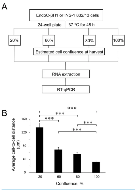
To find out the influence of cell confluence on the expression of selected miRNAs and targets, we utilized the rat (INS-1 832/13) and human (EndoC-βH1) insulin-secreting cell lines seeded at different cell densities, followed by gene expression measurements (Fig. 1A). To quantify the cell-to-cell contact, we also seeded EndoC-βH1 cells in parallel, at different densities corresponding to each confluence harvest point. We measured on average 30µm
between cells in 100% confluent plates, while the lowest 20% confluent plates contained cells with an average of 140µm cell-to-cell distance (Fig. 1BandFig. S1).
This study mainly addressed the issue whether confluence affects miR-375 expression, as it is one of the most enriched miRNAs in the pancreatic beta cells influencing diverse molecular processes, from insulin secretion to cellular growth and proliferation (Eliasson, 2017;Poy et al., 2004;Poy et al., 2009;Salunkhe et al., 2015). The genesAifm1andCav1
are among the many genes shown to be directly targeted by miR-375 in mouse beta cells. The negative effect of miR-375 on both the mRNA and protein levels of the two genes has been demonstrated, and in the islets of 375KO mice, increased expression of these targets was also detected at the mRNA level (Poy et al., 2009).Aifm1andCav1are involved in signaling mechanisms that negatively regulate cellular growth and proliferation, hence
375KOmice were found to have reduced beta cell mass and defective proliferative capacity in the pancreatic endocrine cells (Poy et al., 2009).
Figure 1 Experimental design and average cell-to-cell distance at harvest.(A) Rat (INS-1832/13) and human (EndoC-βH1) insulin-secreting cell lines were seeded at different cell densities prior to downstream assays as outlined. (B) The average distance between cells at different harvest confluences for EndoC-βH1 cells. Data are presented as mean±SEM ofN=40–90 distance measurements from three independent seedings. (∗ ∗ ∗)p<0.001; one-way ANOVA Tukey’s multiple comparison test.
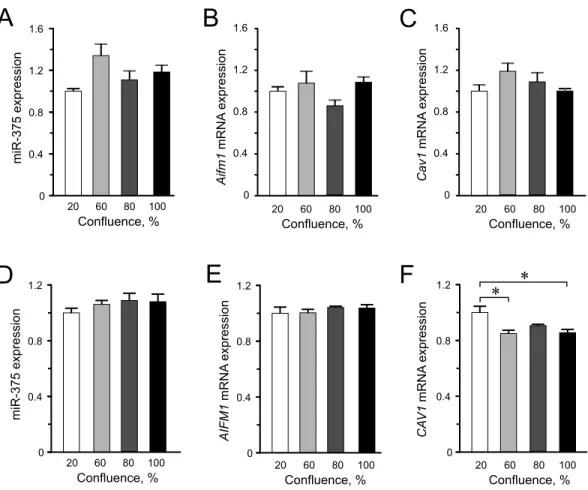
Figure 2 Expression of miR-375 and its targets in INS-1 832/13 cells (A–C) or in EndoC-βH1 cells (D– F). (A) miR-375 expression at different cell confluence of INS-1 832/13 cells. Expression was normal-ized to ratU87. (B and C)Aifm1andCav1expression in INS-1 832/13 cells, respectively. Expression was normalized toHprt1andPpia. (D) Expression of miR-375 at different cell density in EndoC-βH1 cells. Expression was normalized to humanRNU44andRNU48. (E and F)AIFM1andCAV1expression in EndoC-βH1 cells, respectively. Expression were normalized toHPRT1andPPIA. For all experiments, data are presented as mean ofN=3–4 biological replicates, (∗)p<0.05 using one-way ANOVA Tukey’s
mul-tiple comparison test.
underline the impact of species-specific effects of miRNA-mediated regulation in cellular processes.
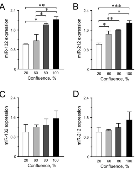
Among the other miRNAs included in this study, we observed significantly higher expression levels of miR-132 and miR-212 at higher confluences in INS-1 832/13 cells (Figs. 3A–3B) but only an increasing trend in the human EndoC-βH1 cells (Figs. 3C–
3D). For miR-200a, miR-130a and miR-152, the expression levels were found not to be influenced by cellular confluence (Fig. S2). However, although not significant, we observed a trend of increasing miRNA expression from 20% to higher confluences in the EndoC-βH1 cells (Figs. S2D–S2F). Overall, the pattern of increased miRNA expression with increasing confluence in this study, supports previous observation of increased activation of miRNA biogenesis and expression at higher cellular densities (Hwang, Wentzel & Mendell, 2009;
Figure 3 miR-132 and miR-212 expression in INS-1 832/13 cells (A–B) or in EndoC-βH1 cells (C–D) at different confluences. Expression was normalized to ratU87 or to humanRNU44andRNU48. Data are presented as mean±SEM ofN=3–4 biological replicates. (∗)p<0.05, (∗∗)p<0.01, (∗ ∗ ∗)p<0.001;
one-way ANOVA Tukey’s multiple comparison test.
CONCLUSION
We found virtually no significant differences in the expression levels of miR-375,CAV1
mRNA andAIFM1mRNA at higher confluences, from 60%–100%, either in the rat or human beta cell lines. Moreover, we did not find significant differences in the expression of the other miRNAs tested in either INS-1 832/13 or EndoC-βH1 cells between 80% and 100% confluence. These results are comforting because most functional assays employing pancreatic beta cell lines utilize these confluence levels to attain consistent results. For instance, to ensure optimal insulin secretion in cultured beta cell lines, insulin-secretion assays are commonly performed when the cell culture confluence is at least 90%.
2009;Van Rooij, 2011). One must therefore be cautious in controlling for cell densities when investigating specific miRNAs inin vitrosystems.
It has been observed that primary tissues generally exhibit higher global miRNA abundance compared to cell lines in part attributed to tighter, and greater cell-to-cell contact in three-dimensions (Lu et al., 2005). Nevertheless, it remains to be seen in the pancreatic endocrine cells how the three-dimensional organization of the cells impacts the global miRNA expression and hence, the regulation of various cellular processes.
ACKNOWLEDGEMENTS
We thank Britt-Marie Nilsson and Anna Maria V. Ramsay for the technical support. Special acknowledgment to R Scharfmann and P Ravassard, INSERM and ENDOCELL for providing us with the EndoC-βH1 human beta cell line, and C Newgard and H Mulder for providing us with the INS-1 832/13 cells.
ADDITIONAL INFORMATION AND DECLARATIONS
Funding
This work was supported by Swedish Research Council (LE; 2016-02124), Linnaeus grant to LUDC, SFO-EXODIAB, Region Skåne ALF, Swedish Foundation for Strategic Research-IRC-LUDC, The Swedish diabetes foundation (LE), Albert Påhlsson Foundation, Region Skåne ALF (LE), Magnus Bergvalls Stiftelse (JLSE), The Edla and Eric Smedberg Foundation Fund through The Royal Physiographic Society of Lund, and Crafoord Foundation (JLSE). The funders had no role in study design, data collection and analysis, decision to publish, or preparation of the manuscript.
Grant Disclosures
The following grant information was disclosed by the authors: Swedish Research Council: LE; 2016-02124.
Linnaeus grant to LUDC; SFO-EXODIAB.
Swedish Foundation for Strategic Research - IRC LUDC. Region Skåne ALF (LE) Boehringer-Ingellheim.
Swedish Diabetes Foundation (LE). Crafoord Foundation (JLSE). Albert Påhlsson Foundation.
The Edla and Eric Smedberg Foundation Fund through The Royal Physiographic Society of Lund.
Magnus Bergvalls Stiftelse (JLSE).
Competing Interests
Author Contributions
• Jones K. Ofori, Helena A. Malm and Ines G. Mollet conceived and designed the experiments, performed the experiments, analyzed the data, wrote the paper, prepared figures and/or tables, reviewed drafts of the paper.
• Lena Eliasson conceived and designed the experiments, analyzed the data, contributed reagents/materials/analysis tools, wrote the paper, prepared figures and/or tables, reviewed drafts of the paper.
• Jonathan Lou S. Esguerra conceived and designed the experiments, performed the experiments, analyzed the data, contributed reagents/materials/analysis tools, wrote the paper, prepared figures and/or tables, reviewed drafts of the paper.
Data Availability
The following information was supplied regarding data availability: The raw data has been supplied as aSupplementary File.
Supplemental Information
Supplemental information for this article can be found online athttp://dx.doi.org/10.7717/ peerj.3503#supplemental-information.
REFERENCES
Agarwal V, Bell GW, Nam JW, Bartel DP. 2015.Predicting effective microRNA target sites in mammalian mRNAs.Elife4:e05005DOI 10.7554/eLife.05005.
Andersson LE, Valtat B, Bagge A, Sharoyko VV, Nicholls DG, Ravassard P, Scharfmann R, Spegel P, Mulder H. 2015.Characterization of stimulus-secretion coupling in the human pancreatic EndoC-betaH1 beta cell line.PLOS ONE10:e0120879
DOI 10.1371/journal.pone.0120879.
Asfari M, Janjic D, Meda P, Li G, Halban PA, Wollheim CB. 1992.Establishment of 2-mercaptoethanol-dependent differentiated insulin-secreting cell lines.Endocrinology
130:167–178DOI 10.1210/endo.130.1.1370150.
Bartel DP. 2009.MicroRNAs: target recognition and regulatory functions.Cell
136:215–233DOI 10.1016/j.cell.2009.01.002.
Belgardt BF, Ahmed K, Spranger M, Latreille M, Denzler R, Kondratiuk N, Von Meyenn F, Villena FN, Herrmanns K, Bosco D, Kerr-Conte J, Pattou F, Rulicke T, Stoffel M. 2015.The microRNA-200 family regulates pancreatic beta cell survival in type 2 diabetes.Nature Medicine 21:619–627DOI 10.1038/nm.3862.
Dweep H, Gretz N. 2015.miRWalk20: a comprehensive atlas of microRNA-target interactions.Nature Methods12:697DOI 10.1038/nmeth.3485.
Eliasson L. 2017.The small RNA miR-375—a pancreatic islet abundant miRNA with multiple roles in endocrine beta cell function.Molecular and Cellular Endocrinology
DOI 10.1016/j.mce.2017.02.043.
Esguerra JL, Bolmeson C, Cilio CM, Eliasson L. 2011.Differential glucose-regulation of microRNAs in pancreatic islets of non-obese type 2 diabetes model Goto-Kakizaki rat.PLOS ONE6:e18613DOI 10.1371/journal.pone.0018613.
Esguerra JL, Mollet IG, Salunkhe VA, Wendt A, Eliasson L. 2014.Regulation of Pan-creatic Beta Cell Stimulus-Secretion Coupling by microRNAs.Genes5:1018–1031
DOI 10.3390/genes5041018.
Halban PA, Polonsky KS, Bowden DW, Hawkins MA, Ling C, Mather KJ, Powers AC, Rhodes CJ, Sussel L, Weir GC. 2014.beta-cell failure in type 2 diabetes: postulated mechanisms and prospects for prevention and treatment.Diabetes Care
37:1751–1758DOI 10.2337/dc14-0396.
Hohmeier HE, Mulder H, Chen G, Henkel-Rieger R, Prentki M, Newgard CB. 2000.
Isolation of INS-1-derived cell lines with robust ATP-sensitive K+ channel-dependent and -inchannel-dependent glucose-stimulated insulin secretion.Diabetes
49:424–430DOI 10.2337/diabetes.49.3.424.
Hwang HW, Wentzel EA, Mendell JT. 2009.Cell–cell contact globally activates mi-croRNA biogenesis.Proceedings of the National Academy of Sciences of the United States of America106:7016–7021DOI 10.1073/pnas.0811523106.
Lu J, Getz G, Miska EA, Alvarez-Saavedra E, Lamb J, Peck D, Sweet-Cordero A, Ebert BL, Mak RH, Ferrando AA, Downing JR, Jacks T, Horvitz HR, Golub TR. 2005.
MicroRNA expression profiles classify human cancers.Nature435:834–838
DOI 10.1038/nature03702.
Malm HA, Mollet IG, Berggreen C, Orho-Melander M, Esguerra JL, Goransson O, Eliasson L. 2016.Transcriptional regulation of the miR-212/miR-132 cluster in insulin-secreting beta-cells by cAMP-regulated transcriptional co-activator 1 and salt-inducible kinases.Molecular and Cellular Endocrinology424:23–33
DOI 10.1016/j.mce.2016.01.010.
Ofori JK, Salunkhe VA, Bagge A, Vishnu N, Nagao M, Mulder H, Wollheim CB, Eliasson L, Esguerra JL. 2017.Elevated miR-130a/miR130b/miR-152 expression reduces intracellular ATP levels in the pancreatic beta cell.Scientific Reports7:44986
DOI 10.1038/srep44986.
Poy MN, Eliasson L, Krutzfeldt J, Kuwajima S, Ma X, Macdonald PE, Pfeffer S, Tuschl T, Rajewsky N, Rorsman P, Stoffel M. 2004.A pancreatic islet-specific microRNA regulates insulin secretion.Nature432:226–230DOI 10.1038/nature03076.
Poy MN, Hausser J, Trajkovski M, Braun M, Collins S, Rorsman P, Zavolan M, Stoffel M. 2009.miR-375 maintains normal pancreatic alpha- and beta-cell mass.
Proceedings of the National Academy of Sciences of the United States of America
106:5813–5818DOI 10.1073/pnas.0810550106.
Prasad RB, Groop L. 2015.Genetics of type 2 diabetes-pitfalls and possibilities.Genes
6:87–123DOI 10.3390/genes6010087.
Ravassard P, Hazhouz Y, Pechberty S, Bricout-Neveu E, Armanet M, Czernichow P, Scharfmann R. 2011.A genetically engineered human pancreatic beta cell line exhibiting glucose-inducible insulin secretion.Journal of Clinical Investigation
Salunkhe VA, Esguerra JL, Ofori JK, Mollet IG, Braun M, Stoffel M, Wendt A, Eliasson L. 2015.Modulation of microRNA-375 expression alters voltage-gated Na(+) channel properties and exocytosis in insulin-secreting cells.Acta Physiologica
213:882–892DOI 10.1111/apha.12460.
Van Rooij E. 2011.The art of microRNA research.Circulation Research108:219–234