R E S E A R C H A R T I C L E
Open Access
Bovine pericardium retail preserved in
glutaraldehyde and used as a vascular patch
Wladimir F Saporito
1, Adílson C Pires
1, Sérgio H Cardoso
1, João A Correa
1, Luiz Carlos de Abreu
2*, Vitor E Valenti
2,3,
Luciano MR Miller
2and Eduardo Colombari
2Abstract
Background:In this study we evaluated the performance of bovine pericardium preserved in glutaraldehyde used as a vascular patch.
Methods:Fourteen young pigs, six females and eight males, weighting 10.3 - 18.4 kg were used in our study. We implanted three remnants in each pig, two in the abdominal aorta and one was juxtaposed to the peritoneum. The smooth face (SF) and rough face (RF) of each remnant were implanted turned to the vessel inner portion and one remnant was juxtaposed to the peritoneum. The animals were sacrificed in 4.5 8 months after surgery (75 -109 kg). The remnants were assessed for aorta wall, fibroses formation in inner apposition and calcification related to the face turned to the vessel inner portion.
Results:The rough face showed a lower dilatation level compared to the face implanted in adjacent aorta. There was no difference between intensity and/or incidence of graft calcification when the superficies were compared. The bovine pericardium preserved in glutaraldehyde did not show alterations in its structure when implanted with different faces turned to the inner portion of vessel.
Conclusion:When turned to the inner portion of the vessel, the rough face of the remnant presented a lower dilatation in relation to the adjacent aorta and a better quality of endothelium layer and did not show a difference between intensity and/or incidence of graft calcification.
Background
The use of biological tissues such as vascular substitutes dates back to early days of cardiovascular surgery, when an autologous vein was used as an arterial substitute. The use of prostheses and orthoses have become necessary to the development of cardiovascular surgery, since the cor-rection of complex congenital heart disease and valvular heart disease require the use of these materials as vascu-lar substitutes [1]. Carpentier et al [2], with the introduc-tion of glutaraldehyde at low concentraintroduc-tion for the preservation of biological tissues, started a new period by introducing the concept of bioprosthesis, which despite being a biological tissue, it loses its antigenicity, does not induce the formation of antibodies and produces the dis-ruption of intra and intermolecular protein, enhancing its
structural stability, decreasing its antigenicity and main-taining the sterile tissue.
The pericardium treated with glutaraldehyde is one of the biological materials more widely used in cardiovascu-lar surgery [3-5]. Despite the good results obtained in clinical trials, degeneration of biological tissue is fre-quently observed, especially calcification, which is the major cause of dysfunction of these tissues that may cause mechanical failure and degeneration [6-8].
Pires et al [9,10] described the profile of bovine pericar-dium preserved in glutaraldehyde, while employed as a vascular patch. They reported large functional differences between the smooth (membranous) and rough (fibrosis) face of the pericardium. These differences depended on the location. The authors suggested a possible correlation between calcification and the contact of the rough face of the pericardium with the bloodstream. Several experi-ments were conducted in order to avoid or reduce the calcification of glutaraldehyde-treated pericardium [9,10], * Correspondence: [email protected]
2
Laboratório de Escrita Científica, Departamento de Morfologia e Fisiologia, Faculdade de Medicina do ABC, Santo André, SP, Brasil
Full list of author information is available at the end of the article
but none was able to completely inhibit the calcification of the material in question.
We tried to understand the mechanisms of this dysfunc-tion, which is not fully elucidated yet. One aspect to con-sider is that the bovine pericardium presents two distinct sides, the smooth and rugged faces [11]. In clinical practice the smooth side is usually used with the smooth side facing the lumen [12]. However, no studies have described the differences between the two sides (smooth face vs. rough face). Therefore, we aimed to evaluate the perfor-mance of bovine pericardium preserved in glutaraldehyde used as vascular patch and we analyzed the possible differ-ences between smooth and rough faces in contact with the bloodstream.
Methods
We used commercially produced bovine pericardium. The material used in our study was produced by Braile
Biomédica®
industry. The remnant was made by using a metal elliptical mold, measuring 2.0 cm in the greatest diameter and 1.0 cm in the smallest diameter. The surgi-cal procedure was performed under sterile conditions in the operating room of the department of surgical techni-que of our Institution. This study was approved by the Ethics Committee in Research on Animal Experiments of our Institution (Number 1808/99).
We implanted the standardized retails of bovine peri-cardium preserved in glutaraldehyde, obtained from the Gravataí industry in 14 young Large White breed pigs, weighing between 10.3 kg and 18.4 kg, six females and eight males, We implanted three remnants in each pig, two in the abdominal aorta and one was juxtaposed to peritoneum, with a smooth face (SF), the other with the rough face (RF) facing the lumen and one remnant was juxtaposed to peritoneum (Figure 1). We followed the following standardized procedures:
Animals were kept in the immediate postoperative per-iod in the vivarium of our University, where they received antibiotic prophylaxis with intramuscular penicillin G benzathine (Sigma®) at a dose of 600,000 units, analgesia with tenoxicam (Sigma®) in the first 24 hours after sur-gery, and all the necessary postoperative care including feeding, vaccinations and other medications, supervised by a veterinarian. They remained on average until the seventh postoperative day, depending on their clinical conditions. They were then transferred to a farm specia-lizing in raising pigs where they received usual care as needed.
The animals were divided into three groups according to their postoperative period, ranging from 4.5 to 8 months, and weighing between 75 to 109 kg. They were sacrificed using a lethal dose of sodium pentobarbital (Sigma®
). Segments of the aorta with the location of the implants were removed and longitudinally opened
opposite to the implant. The aorta segment which was opened was fixed on a cork board in order to keep its shape, with the inner side facing down and fixed in
aqu-eous 10% formaldehyde (Sigma®
). We evaluated the deformation of the retail which is characterized by its expansion in relation to the adjacent aorta.
The removed parts were analyzed at the Department of Pathology of the Universidade Federal de São Paulo. The pieces were cut along the longest axis of the retail
and embedded in paraffin. Fifteen sections (8-15 μm)
were cut from each tissue sample. Slides were stained by the hematoxylin - eosin method and the method of Verhoeff.
The removed parts were macroscopically and micro-scopically evaluated. We used criteria from previous stu-dies to conduct a quantitative analysis of each slide as follows [13-15]:
Calcification: characterized by deposition of calcium in the intimacy of the retail and the adjacent aorta. The pre-sence of one to two foci was considered to be mild calci-fication (1st degree), three foci were considered moderate (2nd degree), and more than three foci or confluent areas of calcification were considered intense (3rd degree).
Integration tissue: composed of fibrous connective tis-sue responsible for the integration of the bovine pericar-dium retail to the aorta.
Internal apposition fibrosis: characterized by the pre-sence of fibrous tissue in the face of the pericardium toward the interior of the aorta. Internal apposition fibro-sis was assesses according to the extent and thickness. The extent was considered mild (1st degree) when cover-ing less than half of the retail area, moderate (2nd degree) when covering half or more of the retail area and intense (3rd degree) when covering the entire area of the retail. The thickness was considered mild (1st degree) when its thickness was lower than the retail thickness, moderate (2nd degree) when its thickness was equivalent to the retail thickness and intense (3rd degree) when its thick-ness exceeded the retail thickthick-ness.
Bone metaplasia - cartilaginous: characterized by the formation of bone tissue - cartilage in the fibrous layer of the internal apposition. It was considered mild (1st degree) in the presence of one to two foci, moderate (2nd degree) in the presence of three foci, and intense (3rd degree) in the presence of more than three foci or confluent areas.
The experiments lasted a total of 14 months and the surgical procedures were performed by the same surgi-cal team. Data were reviewed by two independent readers.
Results
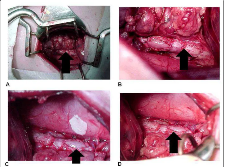
All animals progressed well in the immediate and late postoperative period, all stayed asymptomatic with good growth and adequate weight for their age; we did not observe any deaths. Only one of 14 animals (7.14%) developed wound infection, requiring prolonged hospita-lization in a vivarium, treated with antibiotics. The patches were well incorporated into the aorta of the ani-mals, keeping their limits, irrespective of the side that was facing the lumen (Figure 2).
We observed dilation of the surgical patch area in relation to the surrounding aorta in both groups. In the SF group dilation occurred in all animals and was mild in almost half. On the other hand, in the RF group we did not observe dilation in most animals (Table 1).
The calcification of the surgical patch occurred in both groups. In the SF group it was absent in more than a third of the animals, while in the RF group it was
absent in over half of the animals (Table 2). The calcifi-cation of the aorta adjacent to the vascular patch was observed in nine animals in the SF group (Table 3) and in six animals in the RF group (Table 4). We did not observe calcification in the retail implanted in the peri-toneum in approximately 40% of the animals (Tables 3 and 4).
The coaptation between the bovine pericardial patch and the aortic wall was noted by apposition or slight invagination of edges in 12 cases. In two cases one side presented a slight overlap the edges. The integration between the retail and the aortic wall was observed in a thin band of fibrous connective tissue that also involved the suture.
was moderate (2nd degree) in most animals (Table 5). In the group in which the flat edge of the retail was turned into the aorta, this phenomenon was more intense on the suture line and it occurred in both groups. In the SF group the thickness of the internal apposition fibrosis in relation to the retail area was
slight (1st degree) in most animals while in the RF group we found intense thickness in all patches (Table 6).
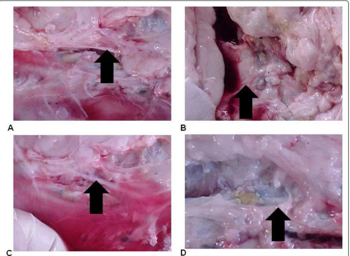
In both groups we found a fibrous layer internal appo-sition that consisted of dense fibrous tissue predomi-nantly modeled, and the cells and fibromas were compactly arranged parallel to each other, acquiring the pattern of tendons and ligaments tissue (Figure 3). Figure 2(A) Aspect of the patch at the postoperative period (animal 3). (B) Aspect of the patch at the postoperative period (animal 7). (C) Aspect of the patch at the postoperative period (animal 11). (D) Retail integrated to the aorta. Arrows indicate the position of the implants.
Table 1 Evaluation of the aortic patch and the expansion of the retail implanted in the aorta in relation to the adjacent aorta
SF RF p
Zero - 12 < 0.05
1stdegree 6 2 < 0.05
2nddegree 4 - < 0.05
3rddegree 4 - < 0.05
Statistical test: Fischer’s test. RF: Rough Face - Retail dilatation; SF: Smooth Face - Retail dilatation. (Zero) absent; (1stdegree) light, when reaching a
depth of 1.0 mm in its longest axis in relation to the adjacent aorta; (2nd
degree) moderate, when reaching a depth of 2.0 mm in its longest axis in relation to the adjacent aorta; (3rddegree) intense, when reaching a depth of
3.0 mm or more in its longest axis, in relation to the adjacent aorta.
Table 2 Evaluation of the aortic patch in relation to the calcification of the retail implanted in the aorta
SF RF P
Zero 5 8 0.1
1stdegree 4 5 0.23
2nddegree 3 1 0.12
3rddegree 2 - 0.4
Statistical test: Fischer’s test. SF: Smooth Face - Retail calcification; RF: Rough Face - Retail calcification. (1stdegree) up to 2 small foci; (2nddegree) up to 3
There was only one animal (7.1%) in the RF group that presented osteo-cartilaginous metaplasia in the internal apposition fibrous layer.
The anatomic and pathologic evaluation showed that all parts of the bovine pericardium were conserved. Moreover, we found a superposition of a fibroelastic dense connective tissue collagen fibers on the face found facing the lumen of the vessel, which was mod-eled in a compact arrangement and arranged parallel to the surface, similar to modeled dense connective tissue. There were no degenerative, inflammatory or metaplas-tic diseases.
Discussion
Our main finding was that the bovine pericardium pre-served in glutaraldehyde presented no changes in its structure when deployed with different faces to the ves-sel lumen. Its production followed a rough standardiza-tion of manufacturing and quality control. In order to reduce the variables that could influence the results, the pericardium was obtained from only one source and the patch was made with the aid of the same mold.
It is important to note the presence of an expansion of the retail in the aortic wall. There was dilation of the material with the formation of an aneurysm wall at the implant site, which was significantly higher when the smooth face was turned to the light of the aorta. Furthermore, we observed that the bovine pericardium patch was well integrated into the wall of the aorta,
confirming what Pires et al [9] observed. The structure of the patch provided by surgical retail, when implanted in the aorta of dogs, promotes clear and precise limits in preserved shape, and it is already well defined at the end of the first month after surgery [9,10]. Also, this type of procedure is vulnerable to infections [16-18].
In the group where the rough face was turned into the aorta, we observed the formation of a covering layer on the inner surface with more plasticity, modeling elastic tendon and bone formation, which provided greater ten-sion resistance to the surgical patch, preventing its expansion. Pires et al [9,10] suggested that the internal apposition fibrosis originates from the fibrous organiza-tion of blood components deposited on the surface of the retail, because this plan was not observed in any case in retail implanted in the pericardium. In the group in which the flat edge of the retail was turned into the aorta this phenomenon was more intense on the suture line and was related to the roughness of the area and local release of thromboplastin by damaged tissues. Our results suggest that the wrinkled face of the retail would facilitate the capture and adhesion of blood components across its surface, with subsequent assemblage by the release of platelet factors and secondarily by thrombo-plastin released from damaged tissues [19]. We also ver-ified that in this group there was the formation of a thin inner cover layer, resulting in dilatation of the patch area of surgery. These results may suggest that fibrosis
Table 3 Evaluation of the peritoneal retail in relation to calcification compared to the retail implanted in the aorta with smooth face
PR SF p
Zero 6 5 0.34
1stdegree 3 4 0.2
2nddegree 5 3 0.14
3rddegree - 2 0.6
Statistical test: Fischer’s test. PR: Peritoneal Retail - Calcification of the peritoneal retail; SF: Smooth Face - Calcification of the retail. (1stdegree) up
to 2 small foci; (2nddegree) up to 3 small foci; (3rddegree) more than 3 foci.
Table 4 Evaluation of the peritoneal retail in relation to calcification compared to the retail implanted in the aorta with rough face
PR RF p
Zero 6 8 0.31
1stdegree 3 5 0.54
2nddegree 5 1 0.47
3rddegree - - 0.57
Statistical test: Fischer’s test. PR: Peritoneal Retail - Calcification of the peritoneal retail; RF: Rough Face - Calcification of the retail. (1stdegree) up to
2 small foci; (2nddegree) up to 3 small foci; (3rddegree) more than 3 foci.
Evaluation of peritoneal retail regarding calcification, compared to the retail implanted in the aorta with the Rough Face faced to the light.
Table 5 Evaluation of aortic patch in relation to the coverage area of internal apposition fibrosis of the retail deployed in the aorta
SF RF p
Zero - - < 0.001
1stdegree 11 - < 0.001
2nddegree 3 10 < 0.001
3rddegree - 4 < 0.001
Statistical test: Chi Square test. SF: Smooth Face - Extension of fibrosis; RF: Rough Face - Extension of fibrosis. (Zero) Absent; (1stdegree) light, when
covering less than half of the retail area; (2nddegree) moderate, when
covering more than a half of the retail area.; (3rddegree) intense, when covering the entire area of the retail.
Table 6 Evaluation of aortic patch in relation to the thickness of the internal apposition fibrosis of the retail deployed in the aorta
SF RF p
Zero 1stdegree
-9
-< 0.001 < 0.001
2nddegree 4 - < 0.001
3rddegree 1 14 < 0.001
p < 0.001 - Chi Square test. SF: Smooth Face - Thickness of fibrosis; RF: Rough Face - Thickness of fibrosis. (Zero) Absent; (1stdegree) light, when the fibrosis
thickness was lower than the retail; (2nddegree) moderate, when the fibrosis
thickness was the same of the retail; (3rddegree) intense, when the fibrosis
internal apposition would support retail and strengthen the structure of the surgical patch. Its histological struc-ture reveals a dense fibrous connective tissue shaped pattern tendon with great tensile resistance.
Researchers have offered different hypotheses to explain why glutaraldehyde helped preserve bovine peri-cardium from changes in its structure when deployed with different faces to the vessel lumen. Ishihara et al [11] reported that bovine pericardium treated with glu-taraldehyde loses the mesothelial cells of serosal surface, exposing the submesotelial layer of connective smooth tissue, while Pires et al [9] observed that the surface of the pericardium was covered by a layer of pavement cells and fibrous connective tissue, which they called internal apposition tissue. On the other hand Schoen et al [8] reported that platelet aggregation, on the pericardial membrane, would be a factor for accelerating the calcifi-cation process. We observed that in both sides of the retail implanted in animals, an inner surface composed of
endothelium was formed. However, when the retail was implanted with the rough side facing the lumen surface, it was isolated from bovine pericardium through blood, thus reducing the intensity and incidence of aortic dilata-tion in reladilata-tion to the adjacent aorta. Without this cover-age, we would expect an increase in calcification. Additionally, we observed that this layer of tissue was dif-ferentiated from the other sides and better trained when it had the wrinkled face toward the lumen. Furthermore, modeling elastic tendon and bone formation, formed a surgical patch level with the retail bovine pericardium which had good resistance and was well integrated into the aortic wall, probably because of the uniformity result-ing from the moldresult-ing process.
[9] who described a large amount of calcium present in the inner layer of apposition. The authors hypothesized that this calcification represented an osteo-cartilaginous metaplasia and ossification of fibrous tissue covering the graft, indicating the transformation of fibroblast into osteoblasts. Nonetheless, we feel confident that our find-ings are valid. Our methodology was validated by Sucu et al [20] who demonstrated that the use of microscopic sections merged for evaluation of calcification was more precise than the use of the chemical method of extraction of calcium from the patch.
The calcification of biological tissue is the main cause of bioprostheses dysfunction. According to Schoen et al [7,8], Chanda et al [21], Vasudev et al [22] and Pires et al [9] this calcification is a multifactorial phenomenon, but it is not well defined and varies in different animal models. The authors pointed out that calcification in bovine pericardium used as a vascular patch presented a different connotation of calcification than valvular pros-theses, because it implied a reduction in leaflet mobility, resulting in dysfunction of the prosthesis, which was not observed in vascular grafts.
We observed calcification of the retail irrespective of the side facing the lumen; the same was true regarding the retail implanted in the peritoneum of the animal. However, these finding are not consistent with findings of Schoen et al [6] and Rossi et al [23] who found calci-fication of retail implanted in rats. The authors described calcification of bovine pericardium from 24 hours of implantation and progressively increased with time, but did not report the presence of internal tissue apposition. We did not find this tissue in the patches implanted in the peritoneum (without direct contact with the bloodstream). These findings are consistent with findings by Gabbay et al [24], Bortolotti et al [25] and Pires et al [10], who indicated the major influences of the implant site of bovine pericardium treated with glutaraldehyde and its direct contact with the bloodstream.
We used a bovine pericardium produced by Braile Biomedica. However, several other industries also pro-duce the same patch. Some industries use special proce-dures to reduce the prevalence of calcification. For example, Synovis, who produces Vascu-Guard [26], fol-lows Apex-Processing. Briefly, in this process, the levels of residual glutaraldehyde are below the limits of detec-tion by the sophisticated analytical methods now avail-able (< 0.5 ppm) and products undergoing Apex-Processing have levels of cellularity that are four times lower than a variety of competitive materials including products conventionally treated.
According to our data, there was no calcification in the retail implanted in the peritoneum in almost half of the animals. Peritoneal calcification is a rare condition
developed in uremic patients on continuous ambulatory peritoneal dialysis. Once peritoneal calcification is detected, it is essential to assess whether encapsulating peritoneal sclerosis develops [27].
Bovine pericardium is also implanted for the vascular reconstruction in the femoral or carotid artery as patch-plasty and venous patch angiopatch-plasty [28]. Moreover, the bovine pericardium is not only used in cardiovascular surgery but also in hernia [29] or thoracic surgery [30]. Based on our data, we confirm the use of the rough face in those types of surgery. We propose future studies to investigate this possibility.
Our investigation presents some points that should be addressed: we did not perform blood analysis, i.e., cho-lesterol, triglycerides and blood glucose levels. However, we aimed to focus only on calcification. Hematoxylin and eosin is not the best method for investigating tissue calcification and it does not quantify proteins related to fibrosis. On the other hand, this is the first study to investigate the utilization of different faces of bovine pericardium conserved in glutaraldehyde as a vascular patch. The glutaraldehyde treatment, the resulting increased stiffness of the treated tissue and the presence of fixed cellular material in the tissue are all factors con-tributing to the calcification of cardiovascular implants. We did not perform microbiological investigation of the patch. We suggest this procedure for future studies. The time the bovine patch stayed in the circulation was not constant, possibly this factor may be a bias for our findings.
Conclusion
When turned to the inner portion of the vessel, the rough face of the remnant had a lower dilatation in rela-tion to the adjacent aorta and a better quality of endothelium layer and there was no difference between intensity and/or incidence of graft calcification. There-fore, we suggest the surgeons to use the smooth face.
Acknowledgements
This study received financial support from Núcleo de Estudos, Pesquisas e Assessoria à Saúde da Faculdade de Medicina do ABC (NEPAS-FMABC) and Fundação de Amparo à Pesquisa do Estado de São Paulo (FAPESP). We thank Dr. Hani K. Atrash for his wonderful job in helping us with English Grammar and Spelling.
Author details
1Departamento de Cirurgia and Laboratório de Escrita Científica,
Departamento de Morfologia e Fisiologia, Faculdade de Medicina do ABC, Santo André, SP, Brasil.2Laboratório de Escrita Científica, Departamento de Morfologia e Fisiologia, Faculdade de Medicina do ABC, Santo André, SP, Brasil.3Departamento de Fonoaudiologia, Faculdade de Filosofia e Ciências, Universidade Estadual Paulista, UNESP, Marília, SP, Brasil.
Authors’contributions
analysis, interpreted the data and drafted the manuscript. All authors read and gave final approval for the version submitted for publication.
Competing interests
The authors declare that they have no competing interests.
Received: 3 January 2011 Accepted: 22 December 2011 Published: 22 December 2011
References
1. Nina VJ, Assef MA, Rodrigues RR, Mendes VG, Lages JS, Amorim AM, Salgado Filho N, Nina RV:Reconstruction of the chest wall with external metal brace: alternative technique in poststernotomy mediastinitis.Rev Bras Cir Cardiovasc2008,23(4):507-11.
2. Carpentier A, Lemaigre G, Robert L, Carpentier S, Dubost C:Biological factors affecting long-term results of valvular heterografts.J Thorac Cardiovasc Surg1969,58(4):467.
3. Ionescu MI, Smith DR, Sutherland TW, Wooler GH:Heart valve replacemt with aortic heterografts.The Ann of Thorac Surg1972,13(1):1-4. 4. Ionescu MI, Tandon AP, Mary DA, Abid A:Heart valve replacemt with
Ionescu - Shiley pericardial xenograft.J Thorac Cardiovasc Surg1977, 73(1):31-5.
5. Braile DM, Bilaqui A, Ardito RV:Prótese de pericárdio bovino- 44 meses em 164 pacientes.Anais do nono congresso nacional de cirurgia cardíaca 1981,4-5:44.
6. Schoen FJ, Levy RJ, Nelson AC:Onset and progression of experimental bioprosthetic heart valve calcification.Lab Invest1985,52(5):523-32. 7. Schoen FJ, Tsao JW, Levy RJ:Calcification of bovine pericardium used in
cardiac valve bioprostheses.Am J Pathol1986,123(1):134-145. 8. Schoen FJ, Harasaki H, Kim KM, Anderson HC, Levy RJ:
Biomaterial-associated calcification: Pathology, mechanisms, and strategies for prevention.J Biomed Mater Re: Appl Boimater1988,22(A1):11-36. 9. Pires AC, Saporito WF, Leão LEV, Forte V, Cardoso SH:Bovine pericardium
as a vascular patch.Rev Bras Cir Cardiovasc1997,4(3):190-4.
10. Pires A, Saporito WF, Leão LEV, Forte V, Cardoso SH:Bovine Pericardium used as a Cardiovascular Patch.The Heart Forun Surgery1999,2(1):16830-4. 11. Ishihara T, Ferrans VJ, Jones M, Boyce SW, Roberts WC:Structure and
classification of cuspal tears and perforations in porcine bioprosthetic cardiac valves implanted in patients.J Thorac Cardiovasc Surg1981, 48(3):747-57.
12. Braile DB, Bilaqui A, Ardito RV:Alargamento da raiz da aorta com“patch” de pericardio bovino preservado em glutaraldeído.Arq Bras Cardiol1983, 41(4):289-296.
13. Tzimas GN, Afshar M, Chevet E, Emadali A, Vali H, Metrakos PP:Graft calcifications and dysfunction following liver transplantation.BMC Surg 2004,4:9.
14. Neethling WM, Hodge AJ, Clode P, Glancy R:A multi-step approach in anti-calcification of glutaraldehyde-preserved bovine pericardium.J Cardiovasc Surg2006,47:711-8.
15. Kouvelos GN, Papas NK, Arnaoutoglou EM, Papadopoulos GS, Matsagkas MI: Endovascular repair of profunda femoral artery false aneurysms using covered stents.Vascular2011,19:51-4.
16. Krones CJ, Klosterhalfen B, Anurov M, Stumpf M, Klinge U, Oettinger AP, Schumpelick V:Missing effects of zinc in a porcine model of recurrent endotoxemia.BMC Surg2005,5:22.
17. Hirsch T, Spielmann M, Zuhaili B, Koehler T, Fossum M, Steinau HU, Yao F, Steinstraesser L, Onderdonk AB, Eriksson E:Enhanced susceptibility to infections in a diabetic wound healing model.BMC Surg2008,8:5. 18. Crane NJ, Pinto PA, Hale D, Gage FA, Tadaki D, Kirk AD, Levin IW, Elster EA:
Non-invasive monitoring of tissue oxygenation during laparoscopic donor nephrectomy.BMC Surg2008,8:8.
19. Bonduki CE, Lourenço DM, Motta EL, Soares JM Jr, Haidar MA, Baracat EC: Effect of estrogen-progestin hormonal replacement therapy on blood coagulation and fibrinolysis in postmenopausal women.Clinics2007, 62(5):553-60.
20. Sucu N, Tamer L, Dondas HA, Polat A, Aytacoglu B, Cimen MY:The effect of ethyenediaminetetraacetic acid on calcific degeneration in pericardium.Heart Vessels2004,19(2):89-96.
21. Chanda J, Rao SB, Mohanty M:Prevention of calcification of tissue valves. Artificial Organs1994,18(10):752-7.
22. Vasudev SC, Chandy T, Sharma CP:Glutaraldehyde treated bovine pericardium changes in calcification due to vitamins and platelet inhibitors.Artificial Organs1997,21(9):1007-1013.
23. Rossi MA, Braile DM, Teixeira MDR, Carillo SV:Calcific degeneration of pericardial valvular xenografts implanted subcutaneously in rats.Int J Cardiol1986,12(1):331-9.
24. Gabbay S, Bortolotti U, Factor S, Shore DF, Frater RWM:Calcification of implanted xenograft pericardium. Influence of site and function.J Thorac Cardiovasc Surg1984,87(5):782-7.
25. Bortolotti U, Zussa C, Factor S, Frater RWM:Glutaraldehyde treated auto, homo and xenograft pericardium in atrial, mitral, aortic and pericardial applications.Life Support Syst1986,4(2):148-50.
26. Ladowski JM, Ladowski JS:Retrospective analysis of bovine pericardium (vascu-guard) for patch closure in carotid endarterectomies.Ann Vasc Surg2011,25(5):646-50.
27. Dejima K, Mitsuhashi H, Yasuda G, Hirawa N, Ikeda Y, Umemura S: Localization and extent of peritoneal calcification in three uremic patients on continuous ambulatory peritoneal dialysis.Ther Apher Dial 2008,12:413-6.
28. Bisdas T, Pichlmaier M, Bisdas S, Haverich A, Teebken OE:Early neurologic outcome after bovine pericardium versus venous patch angioplasty in 599 patients undergoing carotid endarterectomy.Vascular2010, 18(3):147-53.
29. Limpert JN, Desai AR, Kumpf AL, Fallucco MA, Aridge DL:Repair of abdominal wall defects with bovine pericardium.Am J Surg2009,198(5): e60-5.
30. Jung TE, Baek JH, Lee DH:Pulmonary valve leaflet extension with bovine pericardium: for treatment of pulmonary insufficiency.Tex Heart Inst J 2011,38(1):68-70.
Pre-publication history
The pre-publication history for this paper can be accessed here: http://www.biomedcentral.com/1471-2482/11/37/prepub
doi:10.1186/1471-2482-11-37
Cite this article as:Saporitoet al.:Bovine pericardium retail preserved in glutaraldehyde and used as a vascular patch.BMC Surgery201111:37.
Submit your next manuscript to BioMed Central and take full advantage of:
• Convenient online submission
• Thorough peer review
• No space constraints or color figure charges
• Immediate publication on acceptance
• Inclusion in PubMed, CAS, Scopus and Google Scholar
• Research which is freely available for redistribution