Overexpression of BIRC6 Is a Predictor of
Prognosis for Colorectal Cancer
Tingting Hu1,2, Shuqiang Weng1, Wenqing Tang1, Ruyi Xue1, She Chen3, Guoxiang Cai4,5, Yu Cai1, Xizhong Shen1, Si Zhang3*, Ling Dong1*
1Department of Gastroenterology and Hepatology, Shanghai Institute of Liver Disease, Zhongshan Hospital, Fudan University, Shanghai, China,2Department of Gastroenterology and Hepatology, the First Affiliated Hospital of Wenzhou Medical University, Wenzhou, China,3Department of Biochemistry and Molecular Biology, Shanghai Medical College, Fudan University, Shanghai, China,4Department of Colorectal Surgery, Fudan University Shanghai Cancer Center, Fudan University, Shanghai, China,
5Department of Oncology, Shanghai Medical College, Fudan University, Shanghai, China
*[email protected](LD); [email protected](SZ)
Abstract
Background and Objective
Inhibitors of apoptosis proteins (IAPs) have been well investigated in human cancers, where they are frequently overexpressed and associated with poor prognosis. Here we ex-plored the role of baculoviral IAP repeat containing 6 (BIRC6), a member of IAPs, in human colorectal cancer (CRC).
Methods
We used Western blotting and immunohistochemistry to examine BIRC6 expression in 7 CRC cell lines and 126 CRC clinical samples. We determined the biological significance of BIRC6 in CRC cell lines by a lentivirus-mediated silencing method.
Results
We reported that BIRC6 was overexpressed in CRC cell lines and clinical CRC tissues. BIRC6 overexpression was correlated with tumor size and invasion depth of CRC. BIRC6 overexpression is associated with worse overall survival (OS) (P= 0.001) and shorter dis-ease-free survival (DFS) (P= 0.010). BIRC6 knockdown inhibited cell proliferation, arrested cell cycle at S phase, downregulated cyclin A2, B1, D1 and E1 levels, and sensitized CRC cells to chemotherapyin vitroandin vivo.
Conclusions
Taken together, these data suggests that BIRC6 overexpression is a predictor of poor prog-nosis in colorectal cancer and BIRC6 could be a potential target of CRC therapy.
OPEN ACCESS
Citation:Hu T, Weng S, Tang W, Xue R, Chen S, Cai G, et al. (2015) Overexpression of BIRC6 Is a Predictor of Prognosis for Colorectal Cancer. PLoS ONE 10(5): e0125281. doi:10.1371/journal. pone.0125281
Academic Editor:Ajay Goel, Baylor University Medical Center, UNITED STATES
Received:July 15, 2014
Accepted:March 23, 2015
Published:May 1, 2015
Copyright:© 2015 Hu et al. This is an open access article distributed under the terms of theCreative Commons Attribution License, which permits unrestricted use, distribution, and reproduction in any medium, provided the original author and source are credited.
Data Availability Statement:All relevant data are within the paper and its Supporting Information files.
Introduction
Colorectal cancer (CRC) is the third most frequently diagnosed malignancy and the fourth leading cause of cancer death worldwide[1]. Due to screening and removal of premalignant polyps, incidence rates have declined over the last 3 decades[2]. The use of new drugs such as oxaliplatin, bevacizumab, cetuximab and panitumumab allows patients with metastatic colo-rectal cancer survive longer[3,4,5,6]. Despite the progress in the diagnosis and treatment of CRC, the mortality from this disease remains high. Given the poor prognosis of CRC, novel prognostic markers and therapeutic strategies need to be developed to further improve the out-come of colorectal cancer.
The inhibitors of apoptosis protein (IAP) family has been demonstrated to be crucial in apo-ptosis resistance in a wide range of malignancy[7,8,9,10]. These proteins are distinguished by the presence of up to three copies of Baculoviral IAP Repeat (BIR) domain. The IAPs bind to and suppress a variety of pro-apoptotic factors, thereby effectively inhibit apoptosis in cancer cells[11]. Baculoviral inhibition of apoptosis protein repeat containing 6 (BIRC6), also known as Apollon or Bruce, is the largest member of the IAP family with a single BIR domain at its N-terminal and an active ubiquitin-conjugating (UBC) enzyme domain at its C-N-terminal[12]. In addition to its IAP activity, BIRC6 has the distinctive property of activity as a chimeric E2/E3 ubiquitin ligase in mammals[13]. With its UBC domain, BIRC6 promotes the degradation of Smac and inhibits the activity of caspase-9, which play key roles in the initiation of apoptosis [14]. While BIRC6 itself is regulated by ubiquitination and proteasomal degradation mediated by E2, UbcH5c and Nrdp1[15].
A large amount of evidence showed that BIRC6 was highly expressed in several types of can-cer. It has been reported that brain cancer cells with high levels of BIRC6 were resistant to vari-ous anticancer drugs[12]. BIRC6 overexpression was associated with unfavorable prognosis in childhoodde novoacute myeloid leukemia[16]. Moreover, it has been reported that p53 is a
downstream effector of BIRC6[17,18]. These findings suggest that BIRC6 maybe a new thera-peutic target for malignant tumor.
Qiuet al. [14] first proved that siRNAs targeting BIRC6 promoted apoptotic cell death.
Sub-sequently, several studies confirmed that silencing BIRC6 caused elevated apoptosis[17,18,19]. In addition, preliminary investigation demonstrated that BIRC6 expression was more abun-dant in CRC tissues than in non-neoplastic tissues using cDNA microarrays[20]. Similar result was obtained in the comparative proteomics study of colon cancer stem cells[21]. However, there have been no reports of its prognostic relevance based on clinical data. In the present study, we explored the prognostic significance and biologic features of BIRC6 in colorectal can-cer. Our data showed a strong correlation between the BIRC6 overexpression and the unfavor-able clinical features. In addition, BIRC6 upregulation in CRC predicted poor prognosis of patients. Furthermore, We found that BIRC6 knockdown inhibited CRC cell proliferation, ar-rested CRC cell cycle at S phase, downregulated cyclin A2, B1, D1 and E1 levels, and sensitized CRC cells to chemotherapy.
Materials and Methods
Patients, tissue specimens and follow-up
The present study was predominantly conducted by a retrospective design. CRC samples (n = 126) were obtained from patients who received surgery at the Zhongshan Hospital, Fudan University between January 2008 and August 2008. None of them received radiotherapy. Among 126 patients, 42 Patients receieved oxaliplatin-based chemotherapy (FOLFOX4; oxali-platin, 5-fluorouracil and leucovorin) after curative resection. The follow-up information of all study design, data collection and analysis, decision to
publish, or preparation of the manuscript.
participants was updated every 3 months by telephone. All patients were monitored prospec-tively by serum CEA, ultrasound examination, endoscope, computed tomography every 3–6 months. The diagnosis of recurrence was based on the imaging method and biopsy (if possi-ble). Patients were followed until death or April 1, 2013, with a mean postoperative follow-up duration of 59 months. Overall survival (OS) was defined as the time from the date of surgery to death by any cause, patients alive being censored at the last follow-up. Disease-free survival (DFS) was defined as the time elapsed from surgery to the first occurrence of any of the follow-ing events: colorectal cancer distant metastasis; recurrence of colorectal cancer; development of second non-colorectal malignancy excluding basal cell carcinomas of the skin and carcinoma in situ of the cervix; or death from any cause without documentation of a cancer-related event. Patients with TNM stage IV tumors were excluded when analyzing DFS. Thirty paired fresh re-section CRC tissues and the paired adjacent non-cancerous tissues were collected for Western blotting. The study was approved by the Research Ethics Committee of Zhongshan Hospital. All patients provided written consent forms for this study.
Microsatellite instability (MSI) analysis and KRAS mutation analysis
MSI analysis was examined using three mononucleotide repeat markers: BAT25, BAT26, and CAT25 as described previously[22]. KRAS mutation analysis was performed by PCR amplifica-tion of genomic DNA using the following primers: sense 5’ -AAGGCCTGCTGAAAAT-GACTG-3’and antisense 5’-AGAATGGTCCTGCACCAGTAA-3’.
Immunohistochemistry and evaluation
Paraffin-embedded sections of normal and tumor tissues were deparaffinized in xylene and re-hydrated in a decreasing ethanol series diluted in distilled water. Following antigen retrieval with a 10 mM citrate buffer, CRC sections were incubated overnight at 4°C with the primary anti-BIRC6 polyclonal antibody (Abcam, Cambridge, MA). Following 30 min incubation with secondary antibody against HRP-conjugated-rabbit Ig, sections were developed in 3, 3’ -diami-nobenzidine solution under microscopic observation and counterstained with hematoxylin.
The sections were observed under a light microscopy, for a histological review, to examine tumor microheterogeneity in antigen distribution. Five randomized microscopic views of 400-fold magnification of each section were observed and scored. Both the intensity of immu-nohistochemical staining (0, negative; 1, weak; 2, intermediate; 3, strong) and the percentage of positive cells (0, 0% positive cells; 1, 1–10% positive cells; 2, 11–50% positive cells; 3,>50%
positive cells) were evaluated. The final score of each sample was obtained by multiplying the scores of staining intensity and percentage of positive cells. Samples were classified as negative when the final scores were 0 to 3 and positive when 4 to 9 [23]. The BIRC6 staining was scored independently by two pathologists blinded to the clinical characteristics of the patients. The intra-observer reproducibility was tested by obtaining the widely used statisticalκ-scores that grade the strength of agreement to six categories [poor (κ-score,<0.00), slight (0.00–0.20), fair
(0.21–0.40), moderate (0.41–0.60), substantial (0.61–0.80) and almost perfect (0.81–1.00)][24].
Cell culture
were maintained in RPMI 1640 medium plus 10% fetal bovine serum. NCM460 were grown in M3:10 medium. Cells were maintained at 37°C in a humidified incubator under 5% CO2.
Establishment of stable BIRC6-knockdown clones
Lentiviral transduction was used to establish BIRC6 knockdown stable clones. A panel of shRNA (short hairpin RNA) lentiviral vectors differed in BIRC6-targeting sequences
(TRCN0000041-57~61) and pLKO.1-puro empty vector were purchased from Sigma Aldrich (St Louis, MO, USA). Cells were transfected at a MOI of 1 for 24h, screened by puromycin. The inhibition efficiency was identified by Western blotting.
Western blotting
Colorectum tissue or harvested cells were homogenized in SDS lysis buffer (40 mM Tris pH 7.4, 150 mM NaCl, 1 mM EDTA, 1% SDS, 1 mM aprotinin, 1 mM PMSF and 10 mg/ml leu-peptin) and then centrifuged for 15 min at 4°C. Total protein lysates extracted from samples were quantitated with BCA Protein Assay Kit (Pierce, Rockford, IL, USA). An aliquot of pro-tein was boiled with propro-tein loading buffer for 5 min, and was loaded on SDS polyacrylamide gel. Equal amounts of protein were separated by 8% SDS-PAGE and transferred to polyvinyli-dene difluoride membranes (Millipore, Billerica, MA, USA) using a mini trans-blot apparatus (Bio-Rad Laboratories, Hercules, CA, USA). Membranes were blocked with PBS-0.05% Tween 20 containing 5% nonfat dry milk for 1 h, followed by incubation with antibody against BIRC6 or glyceraldehyde-3-phosphate dehydrogenase antibody at 4°C overnight. The membrane was then incubated with secondary horseradish peroxidase-conjugated goat rabbit or anti-mouse antibody (Jackson Immune Research Laboratories Inc, West Grove, PA). Blots were de-veloped using an enhanced chemiluminescence kit (Tiangen, China). Each experiment was re-peated at least three times.
Cell proliferation assay
Cell proliferation was measured using the Cell Counting Kit-8 (CCK-8) (Dojindo Co., Kuma-moto, Japan) according to the instructions of the supplier. Cells were incubated with CCK-8 for 1 hour with 3 multiples and proliferation rate was assessed by measuring the absorbance at 450 nm with the Universal Micro-plate Reader. Each experiment was repeated three times[25].
Colony formation assay
Cell cycle and apoptosis assay
The effect of BIRC6 knockdown on the cell cycle was analyzed by flow cytometry. Cells were trypsinized and fixed with ice-cold 70% ethanol at 4°C overnight. For DNA content analysis, cells were incubated with 50μg/mL propidium iodide (Sigma, St. Louis, MO) in the presence
of 100μg/mL RNase and 0.2% Triton X-100 for 30 minutes at 37°C. DNA content was
deter-mined in the BD FACSAriaII. The distribution of cells in each phase of the cell cycle was calcu-lated using the Flowjo Program. Each experiment was performed in triplicate.
For apoptosis analysis by flow cytometry, cells were treated with 5-FU, CDDP, oxaliplatin or CPT-11 (All from Sigma, St. Louis, MO)[26]. Drug-induced apoptosis was analyzed using Annexin V assay kit (BD Biosciences, San Jose, CA) according to the instructions of the manufacturer.
Tumor xenograft models
Twenty male Balb/c nude mice (4 weeks of age, 12–14 g) were purchased from Experimental Animals Center of Shanghai Institute of Life Science (Shanghai, China) and were raised under specific pathogen-free conditions. All surgery was performed under anesthesia with sodium pentobarbital. DLD-1 cells (stable BIRC6 knockdown 59 and the control clones, 106) in 0.1 ml of PBS were injected subcutaneously into the right flank of each mouse. Mice were sacrificed at 4 weeks post-injection; tumors were excised and weighed. Tumor volume was calculated by the formula: 0.5 × L × W2(L = length of tumor; W = width of tumor). Animal experiments were performed according to the criteria outlined in the Guide for the Care and Use of Laboratory Animals, prepared by the National Academy of Sciences and published by the National Insti-tutes of Health, and also approved by the ethics committee of Fudan University.
Statistical analysis
Statistical analysis was performed using IBM SPSS statistical software (version 17.0). Categori-cal variables were compared byχ2test or Continuity Correction or Fisher’s exact tests as appro-priate. Continuous variables were compared using Wilcoxon ranksum test and independent two samplet-test. Univariate analysis were performed to identify the factors that affect survival
of CRC. Multivariate analysis were done using the Cox multivariate proportional hazard re-gression model. Survival curves were estimated using the Kaplan-Meier method (the log-rank test). All data were presented as mean ± standard error of the mean (SEM) from three indepen-dent experiments andP-values were determined from 2-sided tests. Statistical significance was
displayed as
,P<0.05 or,P<0.01.
Results
Enhanced BIRC6 expression in CRC cells lines and clinic CRC tissues
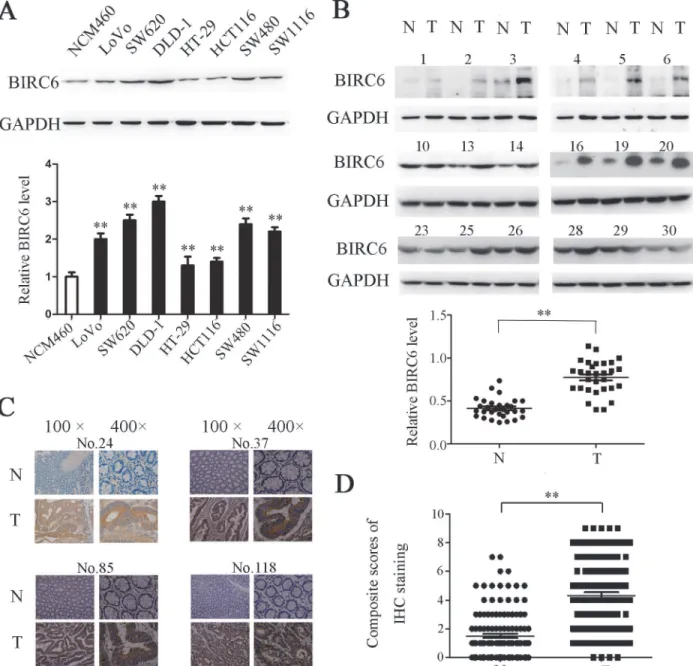
We first detected the BIRC6 expression in 7 CRC cells. Western blotting showed that BIRC6 was overexpressed in LoVo, SW620, DLD-1, HT-29, HCT116, SW480 and SW1116, whereas it was weakly detected in normal colonic epithelial cell line NCM460 (Fig 1A). We next per-formed Western blotting to examine the BIRC6 expression in 30 paired CRC tissues and adja-cent nontumorous tissues. The data implied that BIRC6 was elevated in tumor tissues (Fig 1B). We further assessed the BIRC6 expression in 126 CRC patients by immunohistochemistry. As a result, significant BIRC6 staining was detected in CRC tissues (positive, 73 of 126), whereas the staining in corresponding normal tissues was much weaker (positive, 17 of 126) (Fig 1C
and 1D). Notably, the reproducibility of our classification of BIRC6 expression was found to be
independent observers. The results above indicated that BIRC6 was significantly upregulated in CRC cells lines and clinic CRC tissues.
Correlation between BIRC6 expression and clinical pathological data
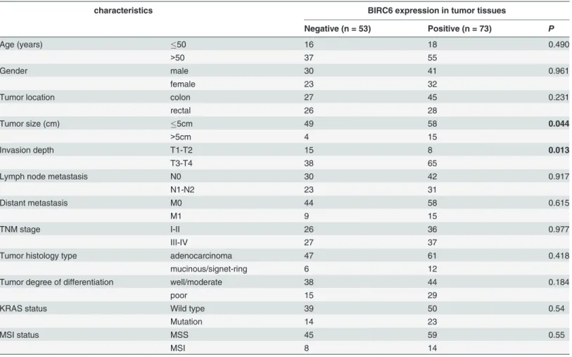
We investigated the correlation of BIRC6 expression with clinicopathologic features in 126 CRC patients. Patient clinical characteristics are listed inS1 Table. There was no significant Fig 1. BIRC6 was overexpressed in CRC cell lines and tumor tissues of CRC patients.(A) Expression of BIRC6 in 1 human colon healthy cell line and 7 CRC cell lines. Upper panel: Typical patterns of BIRC6 expression in NCM460 and CRC cell lines. Lower panel: Relative intensity of BIRC6 normalized to GAPDH. Data present Mean±SEM from three independent experiments. (B) Expression of BIRC6 in 30 paired CRC tissues (T) and adjacent nontumorous tissues (N). Upper panel: Typical patterns of BIRC6 expression in CRC tissues. Lower panel: Relative intensity of BIRC6 normalized to GAPDH. (C) Immunohistochemistry staining of BIRC6 in tumor tissues and self-paired adjacent non-tumorous tissues from four representative CRC patients. (D) Scores of immunohistochemistry staining of BIRC6 in 126 cases.**,P<0.01.
correlation between BIRC6 expression and age, gender, tumor location, lymph node metastasis (N stage), distant metastasis (M stage), histology type, degree of differentiation, KRAS status and MSI status. However, BIRC6 expression positively correlated with tumor size (P= 0.044)
and invasion depth (T stage) (P= 0.013) (Table 1).
Prognostic value of enhanced BIRC6 expression
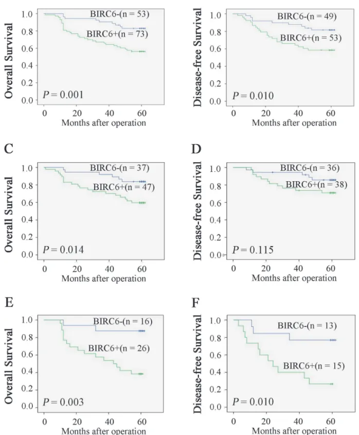
Kaplan-Meier analysis and log-rank test were used to determine the relationship between BIRC6 expression and prognosis. CRC patients with positive BIRC6 expression tended to have shorter overall survival (OS) and disease-free survival (DFS) (P= 0.001 andP= 0.010,
respec-tively) (Fig 2A and 2B). We next divided patients into two groups: no chemotherapy group and chemotherapy group. As it showed inFig 2C and 2D, BIRC6 expression was correlated with OS (P= 0.038) and DFS (P= 0.041) in no chemotherapy group. Similar results were observed
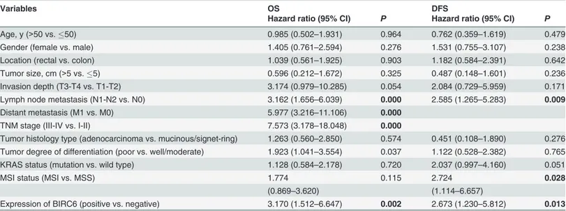
in chemotherapy group (P= 0.003 andP= 0.010) (Fig 2E and 2F). Univariate analysis
demon-strated that positive BIRC6 expression was associated with worse OS (P= 0.002) and DFS
(P= 0.013) (Table 2). Other factors correlated with OS were T stage, N stage, M stage and
tumor degree of differentiation. Factors affecting DFS included T stage, N stage, KRAS status Table 1. Correlation between BIRC6 expression and clinicopathologic characteristics.
characteristics BIRC6 expression in tumor tissues
Negative (n = 53) Positive (n = 73) P
Age (years) 50 16 18 0.490
>50 37 55
Gender male 30 41 0.961
female 23 32
Tumor location colon 27 45 0.231
rectal 26 28
Tumor size (cm) 5cm 49 58 0.044
>5cm 4 15
Invasion depth T1-T2 15 8 0.013
T3-T4 38 65
Lymph node metastasis N0 30 42 0.917
N1-N2 23 31
Distant metastasis M0 44 58 0.615
M1 9 15
TNM stage I-II 26 36 0.977
III-IV 27 37
Tumor histology type adenocarcinoma 47 61 0.418
mucinous/signet-ring 6 12
Tumor degree of differentiation well/moderate 38 44 0.184
poor 15 29
KRAS status Wild type 39 50 0.54
Mutation 14 23
MSI status MSS 45 59 0.55
MSI 8 14
Pearsonχ2 tests for all analyses. Bold items have been considered statistically significant. Abbreviations: TNM, tumor-node-metastasis; MSI, microsatellite instability; MSS, microsatellite stable.
Fig 2. High expression of BIRC6 correlated with poor survival rate.Overall survival (A) and disease-free survival (B) between patients with positive and negative expression of BIRC6 were estimated using the Kaplan-Meier method and compared by the log rank test. Overall survival (C) and disease-free survival (D) were estimated in no chemotherapy group. Overall survival (E) and disease-free survival (F) were estimated in chemotherapy group. Patients with TNM stage IV tumors were excluded when analyzing disease-free survival.
and MSI status. In addition, multivariate analysis identified enhanced BIRC6 level a risk factor for both OS (P= 0.045) and DFS (P= 0.026) (Table 3).
BIRC6 knockdown inhibited CRC cell proliferation
Since the full-length cDNA of BIRC6 extends for 14.5 kb, it is difficult to overexpress BIRC6 in a cell line. Instead, we used lentiviral transduction to establish BIRC6 knockdown stable clones in two CRC cell lines: SW480 and DLD-1. The down-regulated BIRC6 expression was observed Table 2. Univariate analysis of factors associated with survival and recurrence.
Variables OS DFS
Hazard ratio (95% CI) P Hazard ratio (95% CI) P
Age, y (>50 vs.50) 0.985 (0.502–1.931) 0.964 0.762 (0.359–1.619) 0.479
Gender (female vs. male) 1.405 (0.761–2.594) 0.276 1.531 (0.755–3.107) 0.238
Location (rectal vs. colon) 1.039 (0.561–1.925) 0.903 1.182 (0.584–2.391) 0.642
Tumor size, cm (>5 vs.5) 0.596 (0.212–1.672) 0.325 0.487 (0.148–1.601) 0.236
Invasion depth (T3-T4 vs. T1-T2) 3.174 (0.979–10.285) 0.054 2.084 (0.729–5.959) 0.171 Lymph node metastasis (N1-N2 vs. N0) 3.162 (1.656–6.039) 0.000 2.585 (1.265–5.283) 0.009
Distant metastasis (M1 vs. M0) 5.977 (3.216–11.106) 0.000
TNM stage (III-IV vs. I-II) 7.573 (3.178–18.048) 0.000
Tumor histology type (adenocarcinoma vs. mucinous/signet-ring) 1.263 (0.560–2.850) 0.574 0.451 (0.108–1.890) 0.276 Tumor degree of differentiation (poor vs. well/moderate) 1.923 (1.041–3.554) 0.037 1.122 (0.528–2.382) 0.765 KRAS status (mutation vs. wild type) 1.128 (0.584–2.178) 0.720 2.037 (0.997–4.160) 0.051
MSI status (MSI vs. MSS) 1.774 0.115 2.724 0.028
(0.869–3.620) (1.114–6.657)
Expression of BIRC6 (positive vs. negative) 3.170 (1.512–6.647) 0.002 2.673 (1.230–5.812) 0.013
Bold items have been considered statistically significant. Cox proportional hazards regression model was used in univariate analysis. Patients with TNM stage IV tumors were excluded when analyzing disease-free survival. Abbreviations: 95% CI, 95% confidence interval; OS, overall survival; DFS, disease-free survival; TNM, tumor-node-metastasis; MSI, microsatellite instability; MSS, microsatellite stable.
doi:10.1371/journal.pone.0125281.t002
Table 3. Multivariate analysis of factors associated with OS and DFS.
Hazard ratio (95% CI) P
OS
Invasion depth (T3-T4 vs. T1-T2) 0.965 (0.270–3.451) 0.956
Lymph node metastasis (N1-N2 vs. N0) 2.406 (1.234–4.692) 0.010
Distant metastasis (M1 vs. M0) 4.288 (2.243–8.196) 0.000
Tumor degree of differentiation (poor vs. well/moderate) 1.171 (0.621–2.208) 0.625
Expression of BIRC6 (positive vs. negative) 2.214 (1.019–4.810) 0.045
DFS
Invasion depth (T3-T4 vs. T1-T2) 1.028 (0.324–3.260) 0.963
Lymph node metastasis (N1-N2 vs. N0) KRAS status (mutation vs. wild type) MSI status (MSI vs. MSS)
2.198 (1.036–4.666) 1.743 (0.840–3.619) 3.138 (1.259–7.825)
0.0400.1360.014
Expression of BIRC6 (positive vs. negative) 2.564 (1.117–5.886) 0.026
Bold items have been considered statistically significant. Multivariate analysis and Cox proportional hazards regression model were used. Variables were adopted for their prognostic significance by univariate analysis (P<0.2). Abbreviations: 95% CI, 95% confidence interval; OS, overall survival; DFS, disease-free survival; MSI, microsatellite instability; MSS, microsatellite stable.
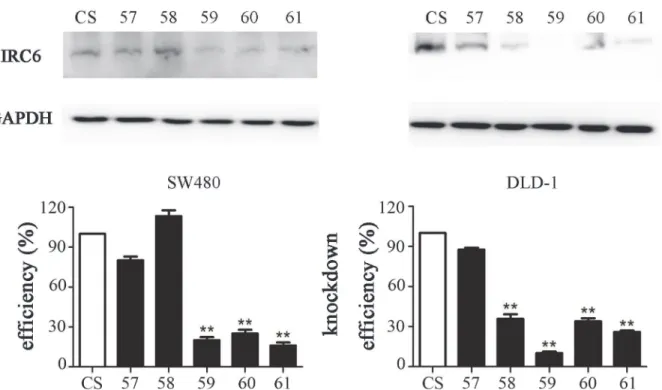
significantly in two BIRC6-knockdown cell lines (59 and 61), as shown by Western blotting
(Fig 3). These two clones were used in the subsequent analysis.
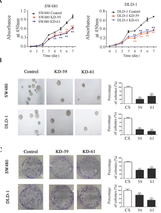
CCK-8 assay showed that BIRC6 knockdown significantly inhibited the proliferation of SW480 and DLD-1 cells in a time-dependent mannerin vitro(Fig 4A). Colony formation
assay showed that BIRC6 knockdown reduced colony formation in agar in SW480 and DLD-1 cells (Fig 4B). Similar results were observed in colony formation in plates (Fig 4C).
BIRC6 knockdown induced cell cycle arrest at S phase and
downregulated cyclin A2, B1, D1 and E1 levels in CRC cells
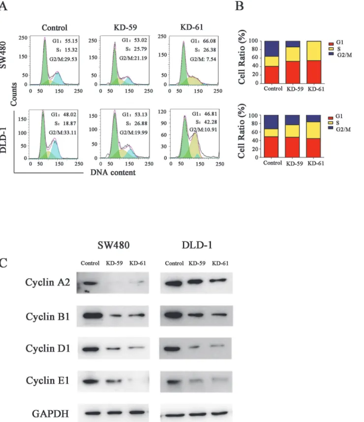
Cell cycle analysis showed that the percentage of cells in S phase increased while the percentage of cells in G2/M phase decreased after BIRC6 knockdown in both SW480 cells and DLD-1 cells
(Fig 5A and 5B), which indicated that BIRC6 knockdown prevented the CRC cells from
enter-ing the mitotic phase. To check whether BIRC6 could regulate the genes involved in cell-cycle control, we detected the effect of BIRC6 on the expression of several cyclins. Western blotting indicated that BIRC6 knockdown decreased the levels of cyclin A2, B1, D1 and E1 in both SW480 cells and DLD-1 cells (Fig 5C).
BIRC6 knockdown sensitized CRC cells to chemotherapy
in vitro
and
in
vivo
We investigate the role of BIRC6 in chmosensitivity of ESCC cells. Though BIRC6 knockdown alone did not induce apoptosis in SW480 cells and DLD-1 cells, BIRC6 knockdown sensitized Fig 3. BIRC6 knockdown stable clones were established by lentiviral transduction.(A) BIRC6 knockdown efficiency was testified by Western blotting. (B) Relative expression of BIRC6 normalized to GAPDH was calculated.**,P<0.01 vs. control.
Fig 4. BIRC6 knockdown inhibited CRC cell proliferation.(A) Cell-growth curves of control (CS) and two BIRC6 knockdown clones (59 and 61). (B) Representative images of soft agar colony formation assay and relative levels of colonies in BIRC6 knockdown clones normalized to the control. (C) Representative images of plate colony formation assay and relative levels of colonies in BIRC6 knockdown clones normalized to the control. Data present Mean±SEM from three independent experiments.**,P<0.01 vs. control.
Fig 5. BIRC6 knockdown induced cell cycle arrest at S phase.(A) Representative images of cell cycle assessed by PI staining. (B) Quantification of the cell cycle distribution. Control cells were cells transfected with pLKO.1-puro empty vector; KD-59 and KD-61 were cells transfected with shRNA lentiviral vectors differed in BIRC6-targeting sequences. (C) Expression of cyclin A2, B1, D1 and E1 were performed by Western blotting in the control and two knockdown clones.
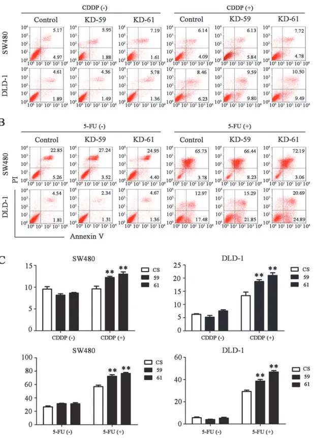
SW480 cells and DLD-1 cells to chemo-induced apoptosis including 5-FU, CDDP, oxaliplatin and CPT-11 (Figs6A and 6BandS2). The finding that BIRC6 knockdown inhibited cell growth and sensitized CRC cells to chemo-induced apoptosisin vitroprompted us to
deter-mine whether it exerts a similar effectin vivo. Control or BIRC6 stable knockdown clones of
DLD-1 were subcutaneously inoculated into nude mice. When all animals had established tu-mors averaging from 50 to 100 mm3, tumorbearing mice were treated with CDDP (4 mg/kg; i. p., twice weekly)[27]. The antitumor efficacy was measured by monitoring the tumor volume and weight after treatment. BIRC6 knockdown slightly inhibited tumor growth. Interestingly, combination of BIRC6 knockdown and CDDP treatment significantly inhibited the tumor growth compared with either BIRC6 knockdown or CDDP treatment alone (Fig 7A and 7B).
Discussion
Acquired resistance to apoptosis is one of the defining hallmarks of cancer[28]. IAPs, a group of anti-apoptosis proteins that are highly evolutionarily conserved across several species, are key regulators of apoptosis[29]. BIRC6, also named as Apollon, is the largest member of the IAP family with 4830 amino acid residues[12]. It is also the only known IAP member which is membrane associated and localizes to the Golgi compartment and the vesicular system[30]. BIRC6 is highly conserved in different species, suggesting a common biological function. Se-quence analysis showed that BIRC6 shared about 92% amino acid identity with Bruce, a mem-ber of IAP family from mouse[12]. In addition, there exists the Drosophila homolog of BIRC6 called dBruce[31]. Unlike most of the other mammalian IAPs, BIRC6 have been shown to be essential for viability. Complete inactivation of the Bruce gene led to perinatal lethality and growth retardation in mice embryos after embryonic day 14[32]. Likewise, deletion of the C-terminal half of Bruce caused activation of caspases and apoptosis in the placenta and yolk sac, and resulted in embryonic lethality[17]. Moreover, plenty of evidence indicated that BIRC6 was overexpressed in a variety of cancers including glioma, childhoodde novoacute
myeloid leukemia, breast cancer, neuroblastoma, prostate cancer and non-small-cell lung can-cer, and overexpression of BIRC6 was correlated with carcinogenesis, progression and poor prognosis of malignant tumor[12,16,18,33,34,35]. Although the previous study demonstrated that BIRC6 was up-regulated in colorectal cancer using cDNA microarray and qRT-PCR anal-ysis, the relationship between BIRC6 expression and colorectal cancer progression has not been extensively studied.
In the present study, we observed that the expression of BIRC6 could be detected in both colorectal cancer and adjacent tissues but with different percentage. The level of BIRC6 was obviously upregulated in CRC tissues compared with adjacent nontumorous tissues. We also demonstrated that high expression of BIRC6 was associated with adverse clinical and patholog-ic features in colorectal cancer. Moreover, patients with positive BIRC6 expression tended to survive shorter and had a higher risk of recurrence than those with negative BIRC6 expression. Univariate and multivariate analysis showed that BIRC6 expression was significantly correlated with OS and DFS, indicating that BIRC6 was a predictor of prognosis.
Fig 6. BIRC6 knockdown sensitized CRC cells to cisplatin (CDDP) or 5-fluorouracil (5-FU)-induced apoptosis.(A) Representative images of cell apoptosis assessed by Annexin V/PI staining in the control (CS) and BIRC6 knockdown cells treated with CDDP (40μM for SW480 or 100μM for DLD-1) for
24 h or not. (B) Representative images of cell apoptosis for the control and BIRC6 knockdown cells treated with 5-FU (75μg/mL for SW480 or 50μg/mL for
DLD-1) for 48 h or not. (C) Quantification of the apoptosis rate. Data present Mean±SEM from three independent experiments.**,P<0.01 vs. control.
which consist of hinokitiol decreased the levels of cyclin A and cyclin E and led to S phase ar-rest in HCT116 and SW620 cells. Joeet al. [37] reported that resveratrol decreased the levels of
cyclins D1, A, and B1 and resulted in S phase arrest in SW480 cells. However, unlike our re-sults, Kikuchiet al. [38] found that BIRC6 ubiquitylated cyclin A and BIRC6 deficiency
accu-mulated more cyclin A in 293T cells. Accordingly, the effect of BIRC6 on cyclins appears to depend on cell types and target proteins. Finally, we found that BIRC6 knockdown sensitized CRC cells to chemotherapy bothin vitroandin vivo, which provides a rationale for combining
BIRC6 antagonism with chemotherapy to treat CRC.
The present study has certain limitations. For example, the correlation between BIRC6 and prognosis was determined in a limited number of CRC patients (n = 126), and this correlation required confirmation on a larger-scale basis. In the same sense, the fact that we did not ob-serve a significant correlation between BIRC6 expression and MSI status needs further investi-gation in larger samples before firmer conclusions can be drawn. It's worth noting that we found that BIRC6 regulated cell cycle progression via modulation of cell-cycle-regulation pro-teins. While increasing evidence have suggested a biological correlation between MSI and alter-ations in cell-cycle-regulation proteins in cancer[39,40].
In conclusion, the present study provides evidence that BIRC6 functions as a prognostic fac-tor of human CRC. Moreover, our results suggest that BIRC6 knockdown in combination with chemotherapy may have therapeutic potential in the treatment of human CRC.
Supporting Information
S1 Dataset. Data for all Western blotting, immunohistochemistry and survival. (DOCX)
S2 Dataset. Data for cell proliferation assay, colony formation assay, cell cycle and apopto-sis assay and tumor xenograft models.
(DOCX)
Fig 7. BIRC6 knockdown sensitized CRC cells to chemotherapy in a xenograft nude mouse model.DLD-1 cells stably transfected with control shRNA and BIRC6 knockdown shRNA (59) were injected subcutaneously into nude mice. Tumorbearing mice were treated with CDDP (4 mg/kg; i.p., twice weekly). (A) The tumor size was monitored. (B) Total tumor weight of each group of mice was recorded. Data present Mean±SEM of 5 mice of each group.*,
P<0.05;**,P<0.01.
S1 Fig. Typical patterns of different staining intensity in tumor tissues and peritumoral tis-sues.Representative images of immunohistochemistry.
(TIF)
S2 Fig. BIRC6 knockdown sensitized CRC cells to oxaliplatin or CPT-11-induced apopto-sis.(A) Representative images of cell apoptosis assessed by Annexin V/PI staining in the con-trol (CS) and BIRC6 knockdown cells treated with oxaliplatin (10μM for SW480 and DLD-1)
for 48 h or not. (B) Representative images of cell apoptosis for the control and BIRC6 knock-down cells treated with CPT-11 (4μM for SW480 and DLD-1) for 24 h or not. (C)
Quantifica-tion of the apoptosis rate. Data present Mean ± SEM from three independent experiments.,
P<0.05;,P<0.01 vs. control.
(TIF)
S1 Table. Characteristics of 126 CRC patients. (DOCX)
Author Contributions
Conceived and designed the experiments: LD SZ XZS. Performed the experiments: TTH SQW WQT. Analyzed the data: TTH WQT RYX. Contributed reagents/materials/analysis tools: SQW SC GXC YC. Wrote the paper: TTH SZ.
References
1. Jemal A, Bray F, Center MM, Ferlay J, Ward E, Forman D (2011) Global cancer statistics. CA Cancer J Clin 61: 69–90. doi:10.3322/caac.20107PMID:21296855
2. Akin O, Brennan SB, Dershaw DD, Ginsberg MS, Gollub MJ, Schöder H, et al. (2012) Advances in on-cologic imaging: update on 5 common cancers. CA Cancer J Clin 62: 364–393. doi:10.3322/caac. 21156PMID:23070605
3. Grothey A, Sargent D, Goldberg RM, Schmoll HJ (2004) Survival of patients with advanced colorectal cancer improves with the availability of fluorouracil-leucovorin, irinotecan, and oxaliplatin in the course of treatment. J Clin Oncol 22: 1209–1214. PMID:15051767
4. Fuchs CS, Marshall J, Barrueco J (2008) Randomized, controlled trial of irinotecan plus infusional, bolus, or oral fluoropyrimidines in first-line treatment of metastatic colorectal cancer: updated results from the BICC-C study. J Clin Oncol 26: 689–690. doi:10.1200/JCO.2007.15.5390PMID:18235136 5. Bokemeyer C, Bondarenko I, Makhson A, Hartmann JT, Aparicio J, Braud FD, et al. (2009) Fluorouracil,
leucovorin, and oxaliplatin with and without cetuximab in the first-line treatment of metastatic colorectal cancer. J Clin Oncol 27: 663–671. doi:10.1200/JCO.2008.20.8397PMID:19114683
6. Douillard JY, Siena S, Cassidy J, Tabernero J, Burkes R, Barugel M, et al. (2010) Randomized, phase III trial of panitumumab with infusional fluorouracil, leucovorin, and oxaliplatin (FOLFOX4) versus FOL-FOX4 alone as first-line treatment in patients with previously untreated metastatic colorectal cancer: the PRIME study. J Clin Oncol 28: 4697–4705. doi:10.1200/JCO.2009.27.4860PMID:20921465 7. Srinivasula SM, Ashwell JD (2008) IAPs: what's in a name? Mol Cell 30: 123–135. doi:10.1016/j.
molcel.2008.03.008PMID:18439892
8. Athanasoula K, Gogas H, Polonifi K, Vaiopoulos AG, Polyzos A, Mantzourani M (2014) Survivin beyond physiology: orchestration of multistep carcinogenesis and therapeutic potentials. Cancer Lett 347: 175–182. doi:10.1016/j.canlet.2014.02.014PMID:24560928
9. Fulda S (2014) Inhibitor of Apoptosis (IAP) proteins in hematological malignancies: molecular mecha-nisms and therapeutic opportunities. Leukemia 28: 1414–1422. doi:10.1038/leu.2014.56PMID: 24487414
10. Guicciardi ME, Werneburg NW, Bronk SF, Franke A, Yagita H, Thomas G, et al. (2014) Cellular inhibitor of apoptosis (cIAP)-mediated ubiquitination of phosphofurin acidic cluster sorting protein 2 (PACS-2) negatively regulates tumor necrosis factor-related apoptosis-inducing ligand (TRAIL) cytotoxicity. PLOS One 9: e92124. doi:10.1371/journal.pone.0092124PMID:24633224
12. Chen Z, Naito M, Hori S, Mashima T, Yamori T, Tsuruo T (1999) A human IAP-family gene, apollon, ex-pressed in human brain cancer cells. Biochem Biophys Res Commun 264: 847–854. PMID:10544019 13. Bartke T, Pohl C, Pyrowolakis G, Jentsch S (2004) Dual role of BRUCE as an antiapoptotic IAP and a
chimeric E2/E3 ubiquitin ligase. Mol Cell 14: 801–811. PMID:15200957
14. Qiu XB, Goldberg AL (2005) The membrane-associated inhibitor of apoptosis protein, BRUCE/Apollon, antagonizes both the precursor and mature forms of Smac and caspase-9. J Biol Chem 280: 174–182. PMID:15507451
15. Qiu XB, Markant SL, Yuan J, Goldberg AL (2004) Nrdp1-mediated degradation of the gigantic IAP, BRUCE, is a novel pathway for triggering apoptosis. EMBO J 23: 800–810. PMID:14765125
16. Sung KW, Choi J, Hwang YK, Lee SJ, Kim HJ, Lee SH, et al. (2007) Overexpression of Apollon, an anti-apoptotic protein, is associated with poor prognosis in childhood de novo acute myeloid leukemia. Clin Cancer Res 13: 5109–5114. PMID:17785565
17. Ren J, Shi M, Liu R, Yang QH, Johnson T, Skarnes WC, et al. (2005) The Birc6 (Bruce) gene regulates p53 and the mitochondrial pathway of apoptosis and is essential for mouse embryonic development. Proc Natl Acad Sci U S A 102: 565–570. PMID:15640352
18. Lopergolo A, Pennati M, Gandellini P, Orlotti NI, Poma P, Daidone MG, et al. (2009) Apollon gene si-lencing induces apoptosis in breast cancer cells through p53 stabilisation and caspase-3 activation. Br J Cancer 100: 739–746. doi:10.1038/sj.bjc.6604927PMID:19223905
19. Chu L, Gu J, Sun L, Qian Q, Qian C, Liu X (2008) Oncolytic adenovirus-mediated shRNA against Apol-lon inhibits tumor cell growth and enhances antitumor effect of 5-fluorouracil. Gene Ther 15: 484–494. doi:10.1038/gt.2008.6PMID:18239605
20. Bianchini M, Levy E, Zucchini C, Pinski V, Macagno C, Sanctis P, et al. (2006) Comparative study of gene expression by cDNA microarray in human colorectal cancer tissues and normal mucosa. Int J Oncol 29: 83–94. PMID:16773188
21. Van Houdt WJ, Emmink BL, Pham TV, Piersma SR, Verheem A, Vries RG, et al. (2011) Comparative proteomics of colon cancer stem cells and differentiated tumor cells identifies BIRC6 as a potential ther-apeutic target. Mol Cell Proteomics 10: M111–M11353.
22. Findeisen P, Kloor M, Merx S, Sutter C, Woerner SM, Dostmann N, et al. (2005) T25 repeat in the 3' un-translated region of the CASP2 gene: a sensitive and specific marker for microsatellite instability in co-lorectal cancer. Cancer Res 18:8072–8078. PMID:16166278
23. Yang XR, Xu Y, Yu B, Zhou J, Li JC, Qiu SJ, et al. (2009) CD24 is a novel predictor for poor prognosis of hepatocellular carcinoma after surgery. Clin Cancer Res 15: 5518–5527. doi:10.1158/1078-0432. CCR-09-0151PMID:19706825
24. Landis JR, Koch GG (1977) The measurement of observer agreement for categorical data. Biometrics 33: 159–174. PMID:843571
25. Zhao N, Wang X, Zhang Y, Gu Q, Huang F, Zheng W, et al. (2013) Gestational zinc deficiency impairs humoral and cellular immune responses to hepatitis B vaccination in offspring mice. PLOS One 8: e73461. doi:10.1371/journal.pone.0073461PMID:24069198
26. Akiyoshi T, Nakamura M, Yanai K, Nagai S, Wada J, Koga K, et al. (2008) Gamma-secretase inhibitors enhance taxane-induced mitotic arrest and apoptosis in colon cancer cells. Gastroenterology 134: 131–144. doi:10.1053/j.gastro.2007.10.008PMID:18166351
27. Suzuki K, Kokuryo T, Senga T, Yokoyama Y, Nagino M, Hamaguchi M (2010) Novel combination treat-ment for colorectal cancer using Nek2 siRNA and cisplatin. Cancer Sci 101: 1163–1169. doi:10.1111/ j.1349-7006.2010.01504.xPMID:20345485
28. Hanahan D, Weinberg RA (2000) The hallmarks of cancer. Cell 100: 57–70. PMID:10647931 29. LaCasse EC, Mahoney DJ, Cheung HH, Plenchette S, Baird S, Korneluk RG (2008) IAP-targeted
ther-apies for cancer. Oncogene 27: 6252–6275. doi:10.1038/onc.2008.302PMID:18931692
30. Hauser HP, Bardroff M, Pyrowolakis G, Jentsch S (1998) A giant ubiquitin-conjugating enzyme related to IAP apoptosis inhibitors. J Cell Biol 141: 1415–1422. PMID:9628897
31. Vernooy SY, Chow V, Su J, Verbrugghe K, Yang J, Cole S, et al. (2002) Drosophila Bruce can potently suppress Rpr- and Grim-dependent but not Hid-dependent cell death. Curr Biol 12: 1164–1168. PMID: 12121627
32. Lotz K, Pyrowolakis G, Jentsch S (2004) BRUCE, a giant E2/E3 ubiquitin ligase and inhibitor of apopto-sis protein of the trans-Golgi network, is required for normal placenta development and mouse survival. Mol Cell Biol 24: 9339–9350. PMID:15485903
34. Low CG, Luk IS, Lin D, Fazli L, Yang K, Xu Y, et al. (2013) BIRC6 protein, an inhibitor of apoptosis: role in survival of human prostate cancer cells. PLOS One 8: e55837. doi:10.1371/journal.pone.0055837 PMID:23409057
35. Dong X, Lin D, Low C, Vucic EA, English JC, Yee J, et al. (2013) Elevated expression of BIRC6 protein in non-small-cell lung cancers is associated with cancer recurrence and chemoresistance. J Thorac Oncol 8: 161–170. doi:10.1097/JTO.0b013e31827d5237PMID:23287853
36. Lee YS, Choi KM, Kim W, Jeon YS, Lee YM, Hong JT, et al. (2013) Hinokitiol inhibits cell growth through induction of S-phase arrest and apoptosis in human colon cancer cells and suppresses tumor growth in a mouse xenograft experiment. J Nat Prod 76: 2195–2202. doi:10.1021/np4005135PMID: 24308647
37. Joe AK, Liu H, Suzui M, Vural ME, Xiao D, Weinstein IB (2002) Resveratrol induces growth inhibition, S-phase arrest, apoptosis, and changes in biomarker expression in several human cancer cell lines. Clin Cancer Res 8: 893–903. PMID:11895924
38. Kikuchi R, Ohata H, Ohoka N, Kawabata A, Naito M (2014) APOLLON protein promotes early mitotic CYCLIN A degradation independent of the spindle assembly checkpoint. J Biol Chem 289: 3457– 3467. doi:10.1074/jbc.M113.514430PMID:24302728
39. Morikawa T, Kuchiba A, Liao X, Imamura Y, Yamauchi M, Qian ZR, et al. (2012) Tumor TP53 expres-sion status, body mass index and prognosis in colorectal cancer. Int J Cancer 131: 1169–1178. doi: 10.1002/ijc.26495PMID:22038927