*Corresponding author: Carlos Rogério Figueiredo, Tel: + 55 (11) 5084-2991, Fax: + 55 (11) 5571-5877, Email: c.figueiredo@unifesp.br ©
2014 The Authors. This is an Open Access article distributed under the terms of the Creative Commons Attribution (CC BY), which permits Adv Pharm Bull, 2014, 4(Suppl 1), 429-436
doi: 10.5681/apb.2014.063 http://apb.tbzmed.ac.ir
Advanced
Pharmaceutical
Bulletin
Antitumor Activity of Kielmeyera Coriacea Leaf Constituents in
Experimental Melanoma, Tested in Vitro and in Vivo in Syngeneic Mice
Carlos Rogério Figueiredo1*, Alisson Leonardo Matsuo1, Mariana Hiromi Massaoka1, Natalia Girola1, Ricardo Alexandre Azevedo1, Aline Nogueira Rabaça1, Camyla Fernandes Farias1, Felipe Valença Pereira1, Natalia Silva Matias2, Luciana Pereira Silva2, Elaine Guadelupe Rodrigues1, João Henrique Guilardi Lago3, Luiz Rodolpho Travassos1, Regildo Márcio Gonçalves Silva2
1
Department of Microbiology, Immunology and Parasitology, Experimental Oncology Unit (UNONEX), Federal University of São Paulo (UNIFESP), São Paulo, Brazil.
2 Department of Biological Sciences – Laboratory of Herbal Medicines, Universidade Estadual Paulista (UNESP- FLC/Assis), São
Paulo, Brazil.
3
Institute of Environmental, Chemical and Pharmaceutical Sciences, Federal University of São Paulo (UNIFESP), Diadema, São Paulo, SP, Brazil.
Introduction
Plant-derived compounds have received considerable attention in recent years because of their pharmacological properties, including cytotoxicity and chemotherapeutic activities in cancer. Brazilian Cerrado, the second largest biome in Brazil,1 is the source of many species containing bioactive compounds assayed in different experimental models.2,3 The Cerrado is one of the 25 important biodiversity hotspots in the World, with an exceptional concentration of rare endemic species.4 Recent studies have described the biological activities of several plant extracts or isolated compounds from this tropical savanna ecoregion.5-8
Kielmeyera coriacea Mart. & Zucc. (Clusiaceae) is a medicinal plant derived from the Brazilian Cerrado, used by the native population in the treatment of several tropical diseases, including schistosomiasis, leishmaniasis, malaria, fungal and bacterial infections.9 Recent studies have reported on the cytotoxic activity of K. coriacea Root
and Bark extracts in tumor cells, such as HCT-8 (human colon carcinoma), HL-60 (human leukemia), SF-295 (glioblastoma) and MDA-MB-435 (melanoma).8 Previous studies have shown the low toxicity of a dichlorometane extract of K. coriacea stems, suggesting a margin of safety for in vivo therapeutic doses.10 It has also been reported that the dichloromethane fraction from stems of K. coriacea can be an important therapeutic alternative in the treatment of anxiety disorders by induction of antidepressant response in rats.11,12
We evaluated the in vitro and in vivo antitumor effects of different leaf extracts of K. coriacea on murine melanoma B16F10-Nex2. We also evaluated K. coriacea cytotoxicity in several human cancer cells. Our results indicate that the chloroform extract (CE) from leaves of K. coriacea inhibited murine melanoma B16F10-Nex2 cell growth with cell cycle arrest, cell migration in vitro and tumor-cell lung colonization in vivo. Analysis of CE using 3H- Research Article
Article History: Received: 4 January 2014 Revised: 7 February 2014 Accepted: 19 February 2014 ePublished: 25 August 2014
Keywords:
Cancer
Cell cycle arrest
Cell migration
Cerrado
Anti-tumor
Cytotoxic
Abstract
Purpose: The antitumor activity of Kielmeyera coriacea (Clusiaceae), a medicinal plant
used in the treatment of parasitic, as well as fungal and bacterial infections by the Brazilian Cerrado population, was investigated.
Methods: A chloroform extract (CE) of K. coriacea was tested in the murine melanoma cell line (B16F10-Nex2) and a panel of human tumor cell lines. Tumor cell migration was determined by the wound-healing assay and the in vivo antitumor activity of CE was investigated in a melanoma cell metastatic model. 1H NMR and GC/MS were used to determine CE chemical composition.
Results: We found that CE exhibited strong cytotoxic activity against murine melanoma cells and a panel of human tumor cell lines in vitro. CE also inhibited growth of B16F10-Nex2 cells at sub lethal concentrations, inducing cell cycle arrest at S phase, and inhibition of tumor cell migration. Most importantly, administration of CE significantly reduced the number of melanoma metastatic nodules in vivo. Chemical analysis of CE indicated the presence of the long chain fatty compounds, 1-eicosanol, 1-docosanol, and 2-nonadecanone as main constituents.
Figueiredo et al.
NMR (Nuclear magnetic resonance spectroscopy) as well as GC/MS (gas chromatography–mass spectrometry) indicated the presence of long-chain fatty alcohols (C20, C22, C26), ketones (C17, C19) and an alkene (C22) as the main compounds. The length of the alkyl chains may play an important role in eliciting the biological activity of these molecules.13 Their unsuspected antitumor activity adds to several other properties of these compounds as insect repellents (USPatent 4774082), antiviral agents, neurotrophic factors, waxes for thermal insulation.14-16
Materials and Methods Plant materials and extraction
Leaves of K. coriacea were collected in the Cerrado area in the city of Patos de Minas-MG, Brazil (17°30.27'34''S and 45°31.21'17''W) in August 2008, and 2009. The plant was identified by MSc. Alice F. Amaral and a voucher specimen was deposited at the Mandevilla Herbarium, Centro Universitario de Patos de Minas (UNIPAM), No. MGHM0632-7. Hydroalcoholic (HA) and ethyl acetate (EA) extracts were obtained from 50 g of powdered leaves macerated in 250 mL of ethanol (EtOH): H2O 7:3 (v/v) for 3 h at 45°C. Chloroform (CE), hexane (HE) and heptane (HP) were used to obtain extracts from 20 g of powdered leaves. Triplicate extractions were carried out with 200-mL of each solvent with stirring for 2 h at room temperature. Stock solutions were prepared with the dry residues diluted in dimethylsulfoxide at 10 mg/ml. The yield of extraction of leaf plant compounds with chloroform was higher than with dichloromethane, therefore chloroform was chosen in the subsequent extractions.
Chemical analysis 1
H-NMR spectra were recorded at 300 MHz in a Bruker DPX300 spectrometer, using CDCl3 as solvent and TMS (tetramethylsilane) as internal standard. GC–MS analysis was performed at 70 eV in a INCOS 50 Finnigan-Mat-quadrupole spectrometer, using a capillary column (DB-5) coated with crosslinked methyl silicone gum (50 m, 0.20
program was 100C isothermal for 1 min, then 100–280C at 10C/min, and isothermal at 280°C for 20 min. The temperatures of injection and detection were 250 and 280C, respectively.
Cell lineages and mice
The following cell lineages were used: murine melanoma (B16F10-Nex2), a subclone of B16F10, deposited at BCRJ no. 0342; human colon carcinoma (HCT); human cervical cancer (Siha); human melanoma (A2058, SKmel28 and MeWO) all obtained from the Ludwig Institute for Cancer Research (LICR), São Paulo branch. Cells were cultivated as previously described.17
In vitro cytotoxicity and proliferation assays
K. coriacea extracts and specifically CE, diluted in RPMI medium with 1% DMSO (10 to 40 µg/ ml) were incubated with B16F10-Nex2 cells or human tumor cells (104 cells)
in 96-well plates in a final volume of 100 µL for 24 h for cell cytotoxicity assay. For cell proliferation assay, 5 x 10³ B16F10-Nex2 cells were incubated with 20 and 10 µg/ml of CE in a final volume of 100 µL for 24 h, 48 h and 72 h. Positive controls were carried out with doxorubicin, and negative controls with 1% DMSO supplemented RPMI medium. Cell viability was quantified using the MTT-based Cell Proliferation Kit I (SIGMA). Readings were made in a plate reader at 570 nm. Alternatively, the Trypan Blue exclusion method was also used. All experiments were performed in triplicate.
Wound healing assay
B16F10-Nex2 cells (3x10⁵) were seeded in 12-well plates and incubated over-night with a sublethal dose of CE (10 µg/ml). Wounds were made on tumor cell monolayers with a pipette tip, and images were captured at 0, 3, 8 and 24 h during cell migration and gap filling. Images of CE treated cells were compared with control cell images for quantification of cell migration ratio.
Cell cycle analysis
Tumor cells (1x10⁶) were incubated with 20 µg/ml of CE for 24 hours. Cells were trypsinized and centrifuged at 1000 rpm for 5 minutes. Pellets were suspended in 1 ml of ethanol 70% for fixation and incubated in ice for 15 minutes. Cells were centrifuged at 1000 rpm for 5 min and suspended in 500 µL of PI (propidium iodide) solution containing 50 µg/ml PI, 0.1 mg/ml RNase A and 0.05% Triton X-100, for 40 min at 4 °C. Cells were pelleted and suspended in 500 µl phosphate-buffered saline (PBS) for analysis in a Facs CantoII flow cytometer (BD Biosciences). Data were analyzed with FlowJo software (Tree Star, Inc. Ashland, OR).
Melanoma metastasis assay
Mice were endovenously injected with 5x10⁵ B16F10-Nex2 cells in 100 µl of RPMI without bovine fetal serum (SFB) in the tail veins of syngeneic C57Bl6 mice. Three groups of 5 animals were challenged with tumor cells and daily treated with intraperitoneal (i.p.) doses of CE (0.1mg and 0.5 mg/100µl 1% DMSO-PBS) during fourteen consecutive days. After 15 days, the lungs were removed and inspected for metastatic colonization and their masses quantified.
Ethics statement
All necessary permits were obtained for the described field studies, granted by the State of São Paulo Research Support Foundation (FAPESP), Brazil. The Ethics Committee of Federal University of São Paulo approved the main Project submitted by the Experimental Oncology Unit, CEP 1234/2011.
Statistical analysis
Antitumor activity of Kielmeyera coriacea
using GraphPad Prism 4.0 software (La Jolla, CA). p<0.05 was considered significant difference.
Results
Cytotoxicity study
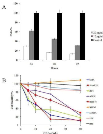
The different K coriacea leaf organic extracts were tested in the various tumor cells. The chloroform extract (CE) was the most cytotoxic in B16F10-Nex2 cells with IC50 of 10.26 µg/ml (Table 1). CE cytotoxicity was also evaluated in different tumor cells and non- tumorigenic cell lines (Figure 1A), and IC50 values are shown in Table 2 after 24 h of incubation. The positive control was run with doxorubicin at IC500.03 μg/ml in B16F10-Nex2 cells. We observed that CE induced tumor cell death at 40 µg/ml, as shown by Trypan Blue staining, suggesting a cytostatic effect of CE at concentrations lower than 40 µg/ml. B16F10-Nex2 cells incubated with subdoses of CE at 10 and 20 µg/ml, showed growth inhibition after 48 h and 72 h (Figure 1B), but the cells which showed morphological alterations did not stain with Trypan blue.
Table 1. Cytotoxic activity of different K.coriacea leaf extracts in
B16F10-Nex2 cells in vitro.
K.coriacea leaf extracts IC50 (μg/ml)
HA > 100
EA > 100
CE 10.26
HE 95.33
HP > 100
Doxorubicin 0.03
HA – Hydroalcoholic extract / EA – Ethyl acetate extract / CE – Chloroform extract
HE – Hexane extract / HP – Heptane extract
CE interferes in the cell cycle of murine melanoma cells The CE prolonged the S phase delaying the cell cycle kinetics in melanoma cells. Treated cells showed increased cytoplasmic area (Figure 2). This experiment was run with 1x10⁶ B16F10-Nex2 cells incubated with 20 µg/ml of CE for 24 h and cells were processed as described in methods. Interference in the cell cycle at sublethal concentrations explains the growth inhibition shown in Figure 1.
CE inhibits migration of murine melanoma cells The migration of B16F10-Nex2 cells using the wound-healing assay was significantly inhibited after 8 and 24 h of incubation with 10 µg/ml of CE (p < 0.05) (Figure 3).
CE anti-metastatic activity in a syngeneic melanoma system
C57Bl6 mice were endovenously challenged with 1x10⁶ B16F10-Nex2 cells and intraperitoneally (i.p) treated with a daily dose of CE during 14 days. We observed that CE induced significant protection in mice treated with 0.5 mg (p < 0.05) rather than with 0.1 mg doses of CE (Figure 4A). The tumor mass values were calculated by subtracting the average mass value (20 mg) of normal
lung from the lung mass values of treated animals (Figure 4B). Treated animals had no weight loss or other signs of toxicity during the experiment.
Figure 1.Cytotoxic effects of K. coriacea chloroform extract
(CE). (A) B16F10-Nex2 cell growth after incubation with low
concentrations of CE (10 and 20 µg/ml) compared to the untreated control, for 24, 48 and 72 hours;(B) Cytotoxic activity of CE in different cell lines. CE was incubated with 104 viable cells at concentrations ranging from 0 to 40 µg/ml for 24 h. Cell viability was determined by the MTT method.
Table 2. IC50 values obtained for CE in different cancer cell
lines and non-tumorigenic cell lines.
Cell line - IC50
(µg/ml) SD
Siha Human cervix carcinoma 6.90 1.21
HCT Human colon carcinoma 6.88 1.42
SKMel 28 Human melanoma 42.05 0.53
MeWo Human melanoma 34.26 3.56
A2058 Human melanoma 26.80 3.54
B16F10-Nex2 Murine melanoma 10.26 0.68
3T3 Murine embryo fibroblast > 100 2.90
Melan A Murine melanocyte > 100 3.28
NHEM Normal human epidermal
melanocyte > 100 3.26
HFF Human foreskin fibroblast > 100 2,79
Figueiredo et al.
Figure 2. CE effects on melanoma cell cycle. (A) Cell cycle
analysis of B16F10-Nex2 cells incubated with 20 μg/ml of CE for 24 h.; (B) Representative images of tumor cell morphology following CE treatment for 24h. Magnification, x200.
Figure 3. B16F10-Nex2 cell migration during incubation with
CE at 10 µg/ml (A) Migrationof tumor cells for 24 h. Statistical
analysis was performed and data was plotted as the mean ± standard deviation (SD) (*p < 0.05 vs. control); (B) Migration of CE-treated tumor cells. Magnification, x100.
Figure 4. In vivo protection of CE against metastatic
melanoma. (A) Representativemouse lungs after treatment with
CE; (B) Tumor mass from mice treated with 0.1 mg and 0.5 mg of CE and vehicle (1% dimethylsulfoxide - DMSO in phosphate-buffered saline - PBS) intraperitoneally during 14 consecutive days (*p < 0.05 vs. control).
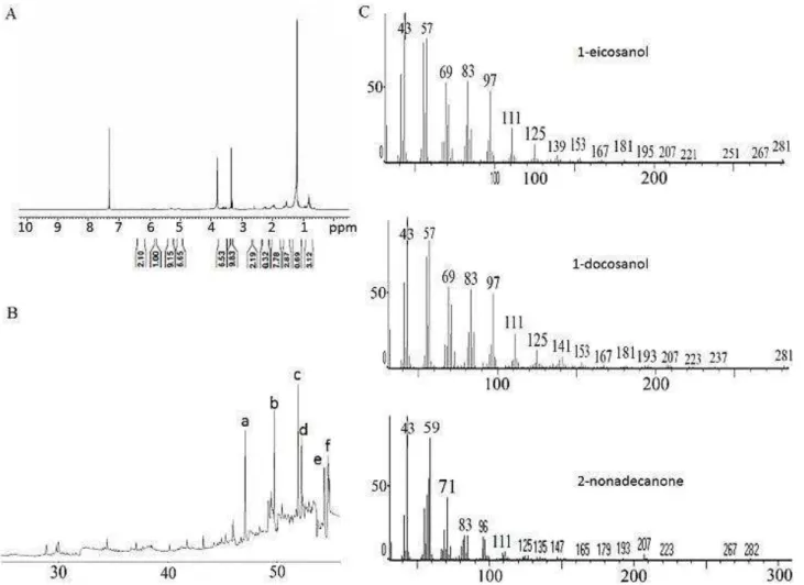
Chemical analysis of CE extract
The 1H NMR spectrum of crude CE extract from K. coriacea showed an intense broad singlet at δ 1.2 and a deformed triplet at δ 0.8 (J = 7.0 Hz). These signals, associated with the presence of multiplets at δ 5- 6 ppm, indicated the occurrence of long side chain unsaturated hydrocarbon derivatives as major derivatives in this active group.18 Additionally, several signals at δ 3-4 ppm as well as at δ 2-3 ppm were detected, which suggested the occurrence of alcohol and ketone derivatives as well. Aiming at the identification of these components, the crude extract was analyzed by GC-MS, which allowed the identification of six main fatty compounds (Table 3). 1H NMR spectrum, GC and MS spectra of CE main
compounds are shown in Figure 5. Compounds are grouped in three different classes: alkene: 1-docosene (16.59%); alcohols: 1-eicosanol (18.46%), 1-docosanol (19.80%), and 1-hexacosanol (13.38%); and ketones: 2-heptadecanone (11.63%) and 2-nonadecanone (20.12%). The characterization of these compounds was based on the mass spectra as well as on the retention times in a DB-5 column.
Discussion
Antitumor activity of Kielmeyera coriacea
extract from the root bark of K. coriacea, particularly against MDA-MB-435 melanoma cells.7-9 Our present data demonstrate the cytotoxic activity of K coriacea leaf
extracts, showing that the chloroform extract (CE) is the most active in vivo and in vitro against melanoma and other human tumor cell lines.
Figure 5. Analyses of CE chemical constituents. (A) 1H NMR spectrum depicted an intense broad singlet at δ 1.2 and a deformed
triplet at δ 0.8 ppm; (B) GC of CE chloroform extract showing peaks a: 1-docosene, b: 1-eicosanol, c: 1-docosanol, d: 2-heptadecanone,
e: 2-nonadecanone, f: 1-hexacosanol); (C) MS spectra of the main compounds.
Table 3. Fatty compounds identified in CE.
Rt / min Molecular formula Relative amount (%)
47.1 (1-docosene) 16.59
49.8 (1-eicosanol) 18.46
51.9 (1-docosanol) 19.80
52.3 (2-heptadecanone) 11.63
54.4 (2-nonadecanone) 20.12
54.7 (1-hexacosanol) 13.4
High concentrations of CE (40 µg/ml) caused cytoplasmic swelling in B16F10-Nex2 melanoma cells, bleb formation and cell lysis thus suggesting both apoptosis and necrosis.20 The genomic DNA in CE treated cells showed degradation in a ladder pattern (data
Figueiredo et al.
present work, led to proliferation inhibition and apoptosis, as previously reported 21-24
Cancer cells lack the growth control of normal cells, exhibiting unlimited self-sufficient replication.25,26 For therapeutic effectiveness, drugs are being developed that act as biological modifiers, regulating the cell cycle and promoting cell death.20 Plant derived compounds have been reported to induce cell cycle arrest and cell death in many tumor cell lines.27-32 The present study reports on the delay of the cell cycle kinetics at the S phase, significantly reducing the G2/M phase, by K. coriacea leaf chloroform extract.
K. coriacea CE inhibited B16F10-Nex2 cell migration in vitro and protected against lung metastasis in vivo. Migration and invasion are essential steps in cancer cell metastasis.33 Similar effects have been reported in many plant derived extracts and purified compounds that inhibit cancer cell migration.34-38
The previous analysis of hexane extracts from root and bark of K. coriacea described δ-tocotrienol, its dimeric derivative,3 and xanthones in the dichloromethane fraction.12 K.coriacea xanthones were shown to exert antimicrobial activities against Staphylococcus aureus at 50 µg/ml,39 the plant pathogenic fungus Cladosporium cucumerinum and also Candida albicans.40 A trypanocidal activity has also been described for K.coriacea xanthones.41
Presently, we show that the chloroform extract from leaves of K. coriacea have long-chain fatty compounds. The alkene docosene, the alcohols eicosanol, 1-docosanol and 1-hexacosanol, and the ketones 2-heptadecanone and 2-nonadecanone were identified. With chain lengths of C20 to C36,42 these compounds constitute leaf cuticular waxes that may differ widely among species.43,44 Genetic studies in Arabidopsis clarified the fatty acid elongation steps and the subsequent modification of the elongated products into primary alcohols, wax esters, secondary alcohols, and ketones, disclosing the enzymes involved in these pathways.45 Fatty acid derivatives can act as signaling molecules, modulating normal and disease-related phenotypes in animals,46 and display antimicrobial and anticarcinogenic activity 47. One of these long chain hydrocarbon derivatives has already been characterized in plant extracts with antitumor activity, the 1-eicosanol, a component of the acetate fraction of Leea indica that inhibits growth of various cancer cell lines.48
Regarding ketones (C17, C19) and C22 alkene found in CE, there are still no data on the mechanism of action of these compounds against tumor cells. Apparently, the length of the alkyl group13 and the hydrophobicity are related to their biological activities.49 Previous work has shown that some long-chain fatty alcohols and their derivatives may act on mitochondria, inhibiting both tumor cell growth in vitro and the growth of B16 melanoma in vivo. 50-54 Regarding the long-chain fatty alcohols found in CE (eicosanol, docosanol and hexacosanol), they were found in plant extracts and fractions cytotoxic to tumor cell lines in vitro. 55
Recently, the activity of mixtures of long-chain alcohols (C26-C32), such as octacosanol, hexacosanol, heptacosanol, eicosanol and many others, derived most commonly from the wax of natural sources56 and are similar to CE long-chain alcohols mixture has been studied. Antitumor properties have been described, such as inhibition of angiogenesis and metastasis in vitro and in vivo, by inhibition of matrix metalloproteinases activity (MMPs) and translocation of Nf-kB to nucleus.57 Further studies are needed to clarify the mechanism of action of these compounds on tumor cells.
Conclusion
In the present work we describe the in vitro and in vivo antitumor activity of the chloroform extract (CE) of leaves from K. coriacea containing long-chain fatty alcohols, ketones and an alkene. The CE delayed the melanoma cell cycle with morphological alterations and inhibited tumor cell migration in vitro. CE growth inhibition in vitro was shown in murine melanoma B16F10-Nex2 and a few human tumor cell lineages. CE exerted in vivo protection effect using a syngeneic metastatic melanoma model with a significant reduction in the number of lung tumor nodules. It is still unclear whether any single fatty compound in the mixture, or a combination of constituents may reproduce the antitumor effects of the CE extract.
Acknowledgements
The authors thank FAPESP and CNPq for the financial support of this work.
Conflict of Interest
There is no conflict of interest to be reported.
References
1. Araujo JF, De Castro AP, Costa MM, Togawa RC, Junior GJ, Quirino BF, et al. Characterization of soil bacterial assemblies in Brazilian savanna-like vegetation reveals acidobacteria dominance. Microb Ecol 2012;64(3):760-70.
2. Badisa RB, Chaudhuri SK, Pilarinou E, Rutkoski NJ, Hare J, Levenson CW. Licania michauxii Prance root extract induces hsp 70 mRNA and necrotic cell death in cultured human hepatoma and colon carcinoma cell lines. Cancer Lett 2000;149(1-2):61-8. 3. De Mesquita ML, Araujo RM, Bezerra DP, Filho RB, De Paula JE, Silveira ER, et al. Cytotoxicity of delta-tocotrienols from Kielmeyera coriacea against cancer cell lines. Bioorg Med Chem 2011;19(1):623-30.
4. Myers N, Mittermeier RA, Mittermeier CG, Da Fonseca GA, Kent J. Biodiversity hotspots for conservation priorities. Nature 2000;403(6772):853-8. 5. Rios JL, Recio MC. Medicinal plants and
antimicrobial activity. J Ethnopharmacol 2005;100(1-2):80-4.
Antitumor activity of Kielmeyera coriacea
antifungal activity of plants used in traditional medicine in Brazil. J Ethnopharmacol 2007;111(2):396-402.
7. Hiruma-Lima CA, Santos LC, Kushima H, Pellizzon CH, Silveira GG, Vasconcelos PC, et al. Qualea grandiflora, a Brazilian "Cerrado" medicinal plant presents an important antiulcer activity. J Ethnopharmacol 2006;104(1-2):207-14.
8. De Mesquita ML, De Paula JE, Pessoa C, De Moraes MO, Costa-Lotufo LV, Grougnet R, et al. Cytotoxic activity of Brazilian Cerrado plants used in traditional medicine against cancer cell lines. J Ethnopharmacol 2009;123(3):439-45.
9. Alves TM, Silva AF, Brandao M, Grandi TS, Smania E, Smania Junior A, et al. Biological screening of Brazilian medicinal plants. Mem Inst Oswaldo Cruz 2000;95(3):367-73.
10. Obici S, Otobone FJ, Da Silva Sela VR, Ishida K, Da Silva JC, Nakamura CV, et al. Preliminary toxicity study of dichloromethane extract of Kielmeyera coriacea stems in mice and rats. J Ethnopharmacol 2008;115(1):131-9.
11. Sela VR, Hattanda I, Albrecht CM, De Almeida CB, Obici S, Cortez DA, et al. Effect of xanthone from Kielmeyera coriacea stems on serotonergic neurons of the median raphe nucleus. Phytomedicine 2010;17(3-4):274-8.
12. Biesdorf C, Cortez DA, Audi EA. Assessment of anxiolytic and panicolytic effects of dichloromethane fraction from stems of Kielmeyera coriacea. Phytomedicine 2012;19(3-4):374-7. 13. Kubo I, Fujita K, Nihei K. Anti-Salmonella activity
of alkyl gallates. J Agric Food Chem 2002;50(23):6692-6.
14. Treister NS, Woo SB. Topical n-docosanol for management of recurrent herpes labialis. Expert Opin Pharmacother 2010;11(5):853-60.
15. Borg J. The neurotrophic factor, n-hexacosanol, reduces the neuronal damage induced by the neurotoxin, kainic acid. J Neurosci Res 1991;29(1):62-7.
16. Greer S, Wen M, Bird D, Wu X, Samuels L, Kunst L, et al. The cytochrome P450 enzyme CYP96A15 is the midchain alkane hydroxylase responsible for formation of secondary alcohols and ketones in stem cuticular wax of Arabidopsis. Plant Physiol 2007;145(3):653-67.
17. Matsuo AL, Figueiredo CR, Arruda DC, Pereira FV, Scutti JA, Massaoka MH, et al. alpha-Pinene isolated from Schinus terebinthifolius Raddi (Anacardiaceae) induces apoptosis and confers antimetastatic protection in a melanoma model. Biochem Biophys Res Commun 2011;411(2):449-54. 18. Moreira IC, Roque NF, Contini K, Lago JHG. Sesquiterpenos e hidrocarbonetos dos frutos de Xylopia emarginata (Annonaceae). Rev Bras Farmacognosia 2007;17(1):55-8.
19. Suffness M, Pezzuto JM. Assays related to cancer drug discovery. In: Hostettmann K, editor. Methods
in plant biochemistry: assays for bioactivity. London: Aacademic press; 1991.
20. Kroemer G, Galluzzi L, Vandenabeele P, Abrams J, Alnemri ES, Baehrecke EH, et al. Classification of cell death: recommendations of the Nomenclature Committee on Cell Death 2009. Cell Death Differ 2009;16(1):3-11.
21. Li L, Chen DB, Lin C, Cao K, Wan Y, Zhao XY, et al. hPNAS-4 inhibits proliferation through S phase arrest and apoptosis: underlying action mechanism in ovarian cancer cells. Apoptosis 2013;18(4):467-79.
22. Chen T, Wong YS. Selenocystine induces S-phase arrest and apoptosis in human breast adenocarcinoma MCF-7 cells by modulating ERK and Akt phosphorylation. J Agric Food Chem 2008;56(22):10574-81.
23. Yu YH, Kuo HP, Hsieh HH, Li JW, Hsu WH, Chen SJ, et al. Ganoderma tsugae Induces S Phase Arrest and Apoptosis in Doxorubicin-Resistant Lung Adenocarcinoma H23/0.3 Cells via Modulation of the PI3K/Akt Signaling Pathway. Evid Based Complement Alternat Med 2012;2012:371286. 24. Joe AK, Liu H, Suzui M, Vural ME, Xiao D,
Weinstein IB. Resveratrol induces growth inhibition, S-phase arrest, apoptosis, and changes in biomarker expression in several human cancer cell lines. Clin Cancer Res 2002;8(3):893-903.
25. Hartwell LH, Kastan MB. Cell cycle control and cancer. Science 1994;266(5192):1821-8.
26. Vermeulen K, Van Bockstaele DR, Berneman ZN. The cell cycle: a review of regulation, deregulation and therapeutic targets in cancer. Cell Prolif 2003;36(3):131-49.
27. Zhang Y, Li Q, Ge Y, Chen Y, Chen J, Dong Y, et al. Silibinin triggers apoptosis and cell-cycle arrest of SGC7901 cells. Phytother Res 2013;27(3):397-403.
28. Geethangili M, Rao YK, Fang SH, Tzeng YM. Cytotoxic constituents from Andrographis paniculata induce cell cycle arrest in jurkat cells. Phytother Res 2008;22(10):1336-41.
29. Nadova S, Miadokova E, Mucaji P, Grancai D, Cipak L. Growth inhibitory effect of ethyl acetate-soluble fraction of Cynara cardunculus L. in leukemia cells involves cell cycle arrest, cytochrome c release and activation of caspases. Phytother Res 2008;22(2):165-8.
30. Du B, Zhong X, Liao X, Xu W, Zhou X, Xu S. A new antitumor arabinopyranoside from Laurencia majuscula induces G2/M cell cycle arrest. Phytother Res 2010;24(10):1447-50.
Figueiredo et al.
32. Lee EJ, Kim WJ, Moon SK. Cordycepin suppresses TNF-alpha-induced invasion, migration and matrix metalloproteinase-9 expression in human bladder cancer cells. Phytother Res 2010;24(12):1755-61. 33. Friedl P, Wolf K. Tumour-cell invasion and
migration: diversity and escape mechanisms. Nat Rev Cancer 2003;3(5):362-74.
34. Lee SJ, Park K, Ha SD, Kim WJ, Moon SK. Gleditsia sinensis thorn extract inhibits human colon cancer cells: the role of ERK1/2, G2/M-phase cell cycle arrest and p53 expression. Phytother Res 2010;24(12):1870-6.
35. Wang HM, Chiu CC, Wu PF, Chen CY. Subamolide E from Cinnamomum subavenium induces sub-G1 cell-cycle arrest and caspase-dependent apoptosis and reduces the migration ability of human melanoma cells. J Agric Food Chem 2011;59(15):8187-92.
36. Yang EJ, Lee JS, Yun CY, Ryang YS, Kim JB, Kim IS. Suppression of ovalbumin-induced airway inflammatory responses in a mouse model of asthma by Mimosa pudica extract. Phytother Res 2011;25(1):59-66.
37. Mojzisova G, Mojzis J, Pilatova M, Varinska L, Ivanova L, Strojny L, et al. Antiproliferative and antiangiogenic properties of horse chestnut extract. Phytother Res 2013;27(2):159-65.
38. Kim EJ, Hong JE, Lim SS, Kwon GT, Kim J, Kim JS, et al. The hexane extract of Saussurea lappa and its active principle, dehydrocostus lactone, inhibit prostate cancer cell migration. J Med Food 2012;15(1):24-32.
39. Cortez DAG, Filho BAA, Nakamura CV, Filho BPD, Marston A and Hostettman K. Antibacterial Activity of a Biphenyl and Xanthones from Kielmeyera coriacea. Pharma Biol 2002;40(7):485-489
40. Cortez DAG, Young MCM, Marston A, Wolfender JL, Hostettmann K. Xanthones, triterpenes and a biphenyl from Kielmeyera coriacea. Phytochemistry 1998;47(7):1367–1374.
41. Caleare Ade O, Lazarin-Bidoia D, Cortez DA, Ueda-Nakamura T, Dias Filho BP, Silva Sde O, et al. Trypanocidal activity of 1,3,7-trihydroxy-2-(3-methylbut-2-enyl)-xanthone isolated from Kielmeyera coriacea. Parasitol Int 2013;62(5):405-11.
42. Pacini E, Guarnieri M, Nepi M. Pollen carbohydrates and water content during development, presentation, and dispersal: a short review. Protoplasma 2006;228(1-3):73-7.
43. Post-Beittenmiller D. Biochemistry and Molecular Biology of Wax Production in Plants. Annu Rev Plant Physiol Plant Mol Biol 1996;47:405-30. 44. Millar AA, Clemens S, Zachgo S, Giblin EM,
Taylor DC, Kunst L. CUT1, an Arabidopsis gene required for cuticular wax biosynthesis and pollen
fertility, encodes a very-long-chain fatty acid condensing enzyme. Plant Cell 1999;11(5):825-38. 45. Samuels L, Kunst L, Jetter R. Sealing plant surfaces:
cuticular wax formation by epidermal cells. Annu Rev Plant Biol 2008;59:683-707.
46. Cury-Boaventura MF, Curi R. Regulation of reactive oxygen species (ROS) production by C18 fatty acids in Jurkat and Raji cells. Clin Sci (Lond) 2005;108(3):245-53.
47. Dembitsky VM. Natural neo acids and neo alkanes: their analogs and derivatives. Lipids 2006;41(4):309-40.
48. Yau Hsiung W, Abdul Kadir H. Leea indica Ethyl Acetate Fraction Induces Growth-Inhibitory Effect in Various Cancer Cell Lines and Apoptosis in Ca Ski Human Cervical Epidermoid Carcinoma Cells. Evid Based Complement Alternat Med 2011;2011:293060.
49. Hansch C, Dunn WJ, 3rd. Linear relationships between lipophilic character and biological activity of drugs. J Pharm Sci 1972;61(1):1-19.
50. Scolaro MJ, Gunnill LB, Pope LE, Khalil MH, Katz DH, Berg JE. The antiviral drug docosanol as a treatment for Kaposi's sarcoma lesions in HIV type 1-infected patients: a pilot clinical study. AIDS Res Hum Retroviruses 2001;17(1):35-43.
51. Holliday MW, Jr., Cox SB, Kang MH, Maurer BJ. C22:0- and C24:0-dihydroceramides confer mixed cytotoxicity in T-cell acute lymphoblastic leukemia cell lines. PLoS One 2013;8(9):e74768.
52. Setzer WN, Vogler B, Schmidt JM, Petty JL, Haber WA. Isolation of cupanioside, a novel cytotoxic and antibacterial long-chain fatty alcohol glycoside from the bark of Cupania glabra. Planta Med 2005;71(7):686-8.
53. Voutquenne L, Lavaud C, Massiot G, Sevenet T, Hadi HA. Cytotoxic polyisoprenes and glycosides of long-chain fatty alcohols from Dimocarpus fumatus. Phytochemistry 1999;50(1):63-9.
54. Matsunaga H, Saita T, Nagumo F, Mori M, Katano M. A possible mechanism for the cytotoxicity of a polyacetylenic alcohol, panaxytriol: inhibition of mitochondrial respiration. Cancer Chemother Pharmacol 1995;35(4):291-6.
55. Piovano M, Chamy MC, Garbarino JA, Tita B, Vitalone A, Di Fabio A, et al. Cytotoxic activity of the root extract from Myoschilos oblongum. Fitoterapia 2003;74(5):497-500.
56. Banerjee S, Ghoshal S, Porter TD. Activation of AMP-kinase by policosanol requires peroxisomal metabolism. Lipids 2011;46(4):311-21.