Reassortant Mammalian Orthoreovirus from
Least Horseshoe Bat in China
Lihua Wang1,2, Shihong Fu1,2, Lei Cao1,2, Wenwen Lei1,2, Yuxi Cao1,2, Jingdong Song1,2, Qing Tang1,2, Hailin Zhang3, Yun Feng3, Weihong Yang3, Guodong Liang1,2*
1State Key Laboratory for Infectious Disease Prevention and Control, Key Laboratory for Medical Virology, National Institute for Viral Disease Control and Prevention, Chinese Center for Disease Control and Prevention, Beijing, China,2Collaborative Innovation Center for Diagnosis and Treatment of Infectious Diseases, Hangzhou, China,3Yunnan Institute of Endemic Diseases Control and Prevention, Yunnan, China
*gdliang@hotmail.com
Abstract
Background
Mammalian orthoreoviruses (MRVs) have a wide geographic distribution and can infect vir-tually all mammals. Infections in humans may be either symptomatic or asymptomatic. This study describes the isolation and identification of a natural reassortant MRV from least horseshoe bats (Rhinolophus pusillu) in China, referred to as RpMRV-YN2012.
Methods and Results
The RpMRV-YN2012 was obtained from urine samples ofRhinolophus pusillusby cell culture. Negative-staining electron microscopy revealed that RpMRV-YN2012 was a non-enveloped icosahedral virus with*75 nm in diameter. Polyacrylamide gel
electro-phoresis (PAGE) migration patterns of the genome segments showed that RpMRV-YN2012 contained 10 segments in a 3:3:4 arrangement. The whole genome sequence of RpMRV2012 was determined. The consensus terminal sequences of all segments of 5’ -GCUAh. . .yUCAUC-3’(h = A, U or C; y = C or U) were similar to the MRV species within the genusOrthoreovirus. Its evolution and evidence of genetic reassortment were ana-lyzed by sequence comparison and phylogenetic analysis. The results showed that RpMRV-YN2012 is a novel serotype 2 MRV that may have originated from reassortment among bat, human, and/or pig MRV strains which associated with diarrhea, acute gastro-enteritis and necrotizing encephalopathy in animals and humans.
Conclusions
RpMRV-YN2012 is a novel bat reassortant MRV, which may have resulted from a reassort-ment involving MRVs known to infect humans and animals. It is necessary to identify wheth-er RpMRV-YN2012 is associated with diarrhea, acute gastroentwheth-eritis and necrotizing OPEN ACCESS
Citation:Wang L, Fu S, Cao L, Lei W, Cao Y, Song J, et al. (2015) Isolation and Identification of a Natural Reassortant Mammalian Orthoreovirus from Least Horseshoe Bat in China. PLoS ONE 10(3): e0118598. doi:10.1371/journal.pone.0118598
Academic Editor:Jonas Waldenström, Linneaus University, SWEDEN
Received:September 3, 2014
Accepted:January 21, 2015
Published:March 17, 2015
Copyright:© 2015 Wang et al. This is an open access article distributed under the terms of the
Creative Commons Attribution License, which permits unrestricted use, distribution, and reproduction in any medium, provided the original author and source are credited.
Data Availability Statement:All relevant data are within the paper.
encephalopathy in clinical patients. In addition, we should carefully monitor its evolution and virulence in real time.
Introduction
Mammalian orthoreoviruses (MRVs), prototypes of the genusOrthoreovirus(family
Reoviri-dae), are non-enveloped viruses with a segmented double-stranded RNA (dsRNA) genome
(*23,500 bp) [1]. MRVs have four major serotypes (type 1 Lang, type 2 Jones, type 3 Dearing,
and type 4 Ndelle) [1,2]. Each MRV particle contains 10 genome segments divided into three
size classes based upon their characteristic mobility during gel electrophoresis: three large (L1, L2 and L3) segments, three medium segments (M1, M2 and M3), and four small segments (S1,
S2, S3 and S4) [1–3]. The virions have an average diameter of 70–80 nm with a typical
icosahe-dral, double-layered protein capsid structure [1–3].
Although MRVs had been assumed to cause mild respiratory or gastrointestinal diseases, re-cent studies have shown that they can cause severe illnesses in humans and other mammals,
in-cluding upper respiratory tract infections, encephalitis, and diarrhea [4,5]. MRVs have been
isolated from many mammalian species, including humans and bats [4,5]; however, the
natu-ral reservoirs or direct progenitors remain unclear. The significance of bats as a source of emerging infectious diseases has been recognized. Bats also are being increasingly recognized as reservoir hosts for viruses which can cross species to infect humans and other domestic and
wild mammals [6–8]. Indeed, many recent outbreaks of emerging viruses, such as the Hendra
virus, Nipah virus, Ebola virus and severe acute respiratory syndrome coronavirus (SARS-like
CoVs), have been associated with bat transmission events [9–13].
Here, we describe the first isolation of a novel natural reassortant MRV strain, named
RpMRV-YN2012, from the least horseshoe bat (Rhinolophus pusillus) in China. The whole
ge-nome sequence of RpMRV2012 was determined. Its evolution and evidence of genetic reassort-ment were analyzed by sequence comparison and phylogenetic analysis.
Materials and Methods
Ethics Statement
Bats were treated according to the guidelines of Regulations for the Administration of Labora-tory Animals (Decree No. 2 of the State Science and Technology Commission of the People's Republic of China, 1988). The sampling was approved by the Ethics Committee of Institute for Viral Disease Control and Prevention, Chinese Center for Disease Control and Prevention.
Sample collection
Clean plastic sheets measuring 2.0 by 2.0m were placed under bat roosting sites at approxi-mately 17:00. The fresh urine and fecal samples were collected at the following morning (ap-proximately 7:30 to 8:00). Pharyngeal swab and anal swab samples from captured bats were immersed into maintenance media in a virus sampling tube (Yocon, China). After collecting the swab samples, all bats were released at their capture site. The samples were transported to
the laboratory under chilled conditions and stored at−80°C until being processed.
Cell culture and virus isolation
Cell lines used in this study were BHK-21(ATCC CCL-10) and Tb1Lu (ATCC CCL88). Cells
were grown in Dulbeco’s Modified Eagle Medium (DMEM) containing high glucose
(Invitro-gen, Breda, The Netherlands), supplemented with penicillin, streptomycin and 10% fetal
bo-vine serum (FBS) (Invitrogen, Breda, The Netherlands) at 37°C in the presence of 5% CO2.
The urine and fecal samples were thawed at 4°C and centrifuged at 16,000xg for 5 min to
pellet debris. Supernatant was filtered through a 0.45-μm filter (Millipore) to remove
bacteri-um-sized particles, and then was diluted 1:10 in cell culture media. Two aliquots of 200μl
dilut-ed supernatant were adddilut-ed to monolayer BHK-21and Tb1Lu cells in a 24-well plate separately. After rocked for 2 h at 37°C, 1ml of fresh cell culture media was added and then incubated for 7 days at 37°C. The flasks were observed daily for toxicity, contamination, or viral cytopathic effect (CPE). By CPE occurrence in the third subcultivation, the supernatant was passed to an
80–90% confluent 175 cm2flask of fresh cells and incubated at cultivation conditions for four
days. To harvest virus particles, cells were homogenized by three freeze-thaw cycles and the re-sulting suspension was purified from cell debris by low-speed centrifugation. Aliquots were
used as viral stocks and stored at−80°C.
Virus cloning
Viral supernatants were applied to six-well plates (Corning, USA) of confluent BHK-21 cells with serial dilution and incubated for one hour. Plates were first overlaid with medium con-taining 75% agarose and then with medium concon-taining neutral red vital stain after three days
incubation at 37°C in a 5% CO2incubator. Plaques of different sizes and shape were shattered
in 500 ul MEM medium after being picked out using a sterile pipette tip. As described
previ-ously [14], this process was repeated until a single plaque shaped virus was obtained.
Electron microscopy
When 80% BHK-21 cells showed CPE, the supernatant of the medium containing viral parti-cles were concentrated at 40,000 rpm for 25 min in a Hitachi centrifuge (Hitachi, Japan). For negative staining, one drop of culture supernatant was adsorbed on Formvar carbon coated grid (1 min), stained with 3% phosphotungstic acid (pH 6.3) (1 min), and inactivated with ul-traviolet irradiation before examination. The infected cells were physically detached by cell scraper. After 3000xg for 15 min, the cells were fixed 2.5% glutaraldehyde in 0.1 mol/L phos-phate buffer (pH 7.4) overnight at 4°C to make ultrathin sections. The viral particles were ob-served using transmission electron microscopy (TECNAI 12, FEI, Blackwood, NJ) with an acceleration voltage of 80 kV.
Migration of genome segments
Virion from culture supernatant was harvested and viral RNA was extracted using the QIamp viral RNA kit (Qiagen, Germany). Fifteen ul of viral RNA was run on a 10% SDS polyacryl-amide/Bis gel under denaturing and reducing conditions at 150 V for 4 hrs at room tempera-ture. The gel was washed with distilled water, stained by Fast Silver Stain Kit (Beyotime, China) according to the manufacturer's protocol before the photo was taken.
Complete genome sequencing including 5
’
- and 3
’
-untranslated regions
Whole genome sequences of 10 segments (L1–L3, M1–M3, S1–S4) of the isolate were
deter-mined by DNase-sequence-independent single primer amplification (DNase-SISPA) and the
sequences of all segments were reconfirmed by overlapping nested PCRs with specific primers (Table 1) derived from the completed viral genomes.
Sequence analysis and phylogenetic comparisons
The nucleotide (nt) sequences and deduced amino acid (aa) products were analyzed and as-sembled using the DNASTAR program (Lasergene). BLAST searches were carried out using
the NCBI server (www.ncbi.nlm.nih.gov) with all available databases. An ORF search was
per-formed with the ORF Finder of the NCBI. Sequences of other Orthoreoviruses were down-loaded from GenBank and used to build the phylogenetic trees. Sequences were aligned using the CLUSTAL_X, version 2.0. Calculations of nucleic acid and amino acid sequence identities
were performed using the MEGA v6.06 software with the default settings (www.Megasoftware.
net) [16]. Maximum-likelihood trees of of L, M and S segments were also generated in MEGA,
using the p-distance and the Poisson correction algorithms. The robustness of the branching was evaluated by bootstrapping using 1,000 replications.
Prevalence study
The throat and anal swabs were thawed at 4°C and centrifuged at 16,000xg for 5 min to pellet
debris. Supernatant was filtered through a 0.45-μm filter (Millipore) to remove
bacterium-sized particles, and aliquot of 200μl used for RNA extraction using the QIAamp Viral RNA
Mini kit (Qiagen) according to the manufacturer's protocol. Extracted RNA was converted to double-stranded cDNA with random hexamers. The existence of viral RNA in the collected throat and anal swabs were detected by a reported RT-PCR procedure targeting the conserved
regions of the L1 genome segment of MRV was used [17].
Results
Isolation and preliminary identification of RpMRV-YN2012
Fifteen pool urine samples and fifteen pool feces samples were collected from six bat roosts in southwestern Yunnan Province during July and August, 2012. Roosts were occupied mainly by
Rhinolophus luctus,Rhinolophus affinis,Rhinolophus pusillus, andMyotis daubentonii. Isola-tion from pool urine and pool feces samples was attempted in BHK-21 and Tb1Lu cell lines.
One isolate was obtained from one pool urine samples ofRhinolophus pusillus, designated
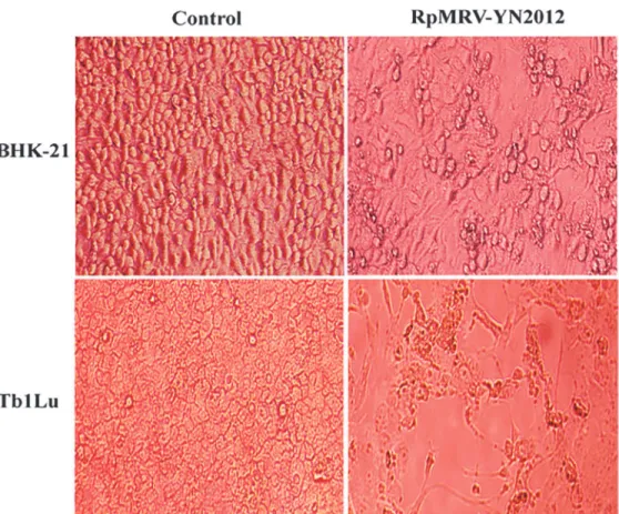
RpMRV-YN2012. The isolate caused cytopathogenic effects (CPE) 3–4 days post-inoculation
in both cell lines. These CPE included granulating, shrinking, rounding, seining, and falling off (Fig. 1). The isolate was plaque-purified and visualized by negative-staining electron microsco-py (Fig. 2A), revealing the presence of many non-enveloped icosahedral virus particles of*75
nm in diameter, morphologically related to reoviruses. Ultra-thin sections of infected BHK-21 cells displayed typical electron-dense virus particles, organized in a paracrystalline pattern
within the cytoplasm (Fig. 2B). Polyacrylamide gel electrophoresis (PAGE) migration patterns
of the genome segments showed that RpMRV-YN2012 contained 10 segments in a 3:3:4
ar-rangement, typical of reoviruses (Fig. 2C).
Genome organization and characteristics of RpMRV-YN2012
By DNase-SISPA and RACE) methods, the whole genome sequences of 10 segments (L1–L3,
M1–M3, S1–S4) of RpMRV-YN2012 were determined. The genome sequences of all segments
were reconfirmed by overlapping nested PCRs with specific primers (Table 1) derived from the
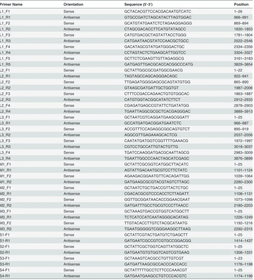
Table 1. Primers used for overlapping RT-PCR confirmation of RpMRV-YN2012.
Primer Name Orientation Sequence (5'-3') Position
L1_F1 Sense GCTACACGTTCCACGACAATGTCATC 1–26
L1_R1 Antisense GTGCCGATCTAGCATACTTAGTGGAC 966–991
L1_F2 Sense GCATGTATGAATCTCTAGAAGGAGGG 869–894
L1_R2 Antisense CTAGCGACACCTTCATGTATAGCC 1830–1853
L1_F3 Sense CATGTGACGCTAGTATTACCTGGG 1781–1804
L1_R3 Antisense CATGAATAACGTATCCAACGCTGCC 2522–2546
L1_F4 Sense GACATAGCGTATGATGGGACTGC 2334–2356
L1_R4 Antisense CCTAGTACTCTGAAGCATTGGTCC 3304–3327
L1_F5 Sense GCTTCTCGAAGTTGTTAGAGGCG 3161–3183
L1_R5 Antisense GATGAGTTGACGCACCACGGCCCATG 3829–3854
L2_F1 Sense GCTATTGGCGCGATGGCGAACG 1–22
L2_R1 Antisense TAGTAGCCAGCAGGGACAGC 922–941
L2_F2 Sense TTGAGATGGGGAGCGCAGTATGTGG 865–890
L2_R2 Antisense GTAAGCGATGATTGCTGGTGT 1987–2006
L2_F3 Sense CTTTCCGACCAGAACTGTGTGGCAC 1863–1887
L2_R3 Antisense CATGTGGTACGGCATATCTTCT 2912–2933
L2_F4 Sense CGAGATGAGCCGTATTCTGATATGG 2878–2903
L2_R4 Antisense TGAATTAGGCGCGCTCACGAGGGAC 3889–3913
L3_F1 Sense GCTAATCGTCAGGATGAAGCGGATT 1–25
L3_R1 Antisense GCCATGATGACGGATGAATCTC 966–987
L3_F2 Sense ACCGTTTCCAGAGGCGGCAGTGTCT 895–919
L3_R2 Antisense AGGCGTTGAGAAAGCACTCG 2037–2056
L3_F3 Sense CAATATGATGGTCGGTTTTGAAACG 1972–1997
L3_R3 Antisense CGTCCTGCCATTGTACTGTTG 3018–3037
L3_F4 Sense TGATCCAAGGATGACGCAATTAGCG 2983–3009
L3_R4 Antisense TGAATTGGCCCAACTAGCATCGAGC 3876–3899
M1_F1 Sense GCTATTCGCGGTCATGGCTTACATC 1–25
M1_R1 Antisense AGTATTGACAATGCGTCCTTCTATC 1101–1124
M1_F2 Sense AGAAGACGGAATGTTCACAGATTGG 1039–1064
M1_R2 Antisense GATGAAGCGCGTACGTAGTCTTAGC 2280–2300
M2_F1 Sense GCTAATCTGCTGACCGTTACTCTGC 1–25
M2_R1 Antisense CGACACGCGTCCCACCTCTTAGATT 1108–1131
M2_F2 Sense GGTTGCGGATAACACCGGAACGAAT 1073–1098
M2_R2 Antisense GATGATTTGCCTGCGTCCCTTAACC 2180–2203
M3_F1 Sense GCTAAAGTGACCGTGGTCATGGCTT 1–25
M3_R1 Antisense TCTCATCCATCAATAGGGCACATAG 1226–1249
M3_F2 Sense TTGTACACCTTGTCTACGCATAATG 1192–1216
M3_R2 Antisense TGAATGGGGGTCGGGAAGGCTTAAG 2292–2315
S1-F1 Sense GCTATTCGTACTGATGTCTGAGCTT 1–25
S1-R1 Antisense GATGAATCGCCGTCGTGCCGGACGG 1414–1437
S2-F1 Sense GCTATTCGCTGGTCAGTTATGGCTC 1–25
S2-R1 Antisense GATGAATGTGTGGTCAGTCGTGAAG 1308–1331
S3-F1 Sense GCTAAAGTCACGCCTGTTGTCGT 1–23
S3-R1 Antisense GATGATTAAGCGCCACCCACCACC 1176–1198
S4-F1 Sense GCTATTTTTGCCTCTTCCCAAACGT 1–25
S4-R1 Antisense GATGAATGAAGCCTGTCCCACGTC 1174–1196
Fig 1. Cytopathic effects of RpMRV-YN2012 on BHK-21 and Tb1Lu cells after three days of infection (200X).
doi:10.1371/journal.pone.0118598.g001
Fig 2. Electron micrographs and genome segment profile of RpMRV-YN2012. (A)Negative-stained RpMRV-YN2012. The black arrow indicates an intact particle; the white arrow indicates an empty particle (scale bar: 200 nm).(B)Image of an ultrathin section of RpMRV-YN2012-infected BHK-21 cell. N, nucleus; black arrows, paracrystalline viral arrays (scale bar: 0.5μm).(C)Genome segment profile of RpMRV-YN2012. The genome segments were separated on a 10% SDS-polyacrylamide gel. The classes of genome segments (L, M, and S) are labeled on the right. The asterisk (*) indicates co-migrating bands.
The complete genome of RpMRV-YN2012 was 23,578 nt in length, including segments L1 to L3, M1 to M3, and S1 to S4 (3,854 nt, 3,915 nt, 3,900 nt, 2,304 nt, 2,203 nt, 2,240 nt, 1,437 nt,
1,331 nt, 1,198 nt, and 1,196 nt, respectively), encoding proteinsλ1(1,275 aa),λ2 (1,289 aa),λ3
(1,267 aa),μNS (721aa),μ1 (708 aa),μ2 (736 aa),δ1 (455 aa),δ1s (114 aa),δ2 (418 aa),δNS
(366 aa), andδ3 (365 aa) (Table 2). The consensus terminal sequences of all segments of 5’
-GCUAh. . .yUCAUC-3’(h = A, U or C; y = C or U) (Table 2) were similar to the MRV species
within the genusOrthoreovirus(8), revealing the MRV character of RpMRV-YN2012.
Pair-wise nucleotide (nt) and deduced amino acid (aa) comparisons between RpMRV-YN2012 and other orthoreoviruses, including the prototype MRVs, were performed for all ten
segments (Table 3). The results showed that six segments (L1–L3, M2, M3, S1) of
RpMRV-YN2012 were closely related to those of human reoviruses (SI-MRV01 and tou05), which iso-lated from patients with acute gastroenteritis and acute necrotizing encephalopathy recently
in Slovenia and France [5,18]. Interestingly, M1 segment of RpMRV-YN2012 is closest to that
of pig diarrhea associated reoviruses (GD-1, SC-A and SHR-A) isolated in China recently
Table 2. Lengths of the coding and untranslated regions of each of the 10 genomic segments of RpMRV-YN2012.
50UTR 30UTR
Segment Length (bp) Protein (aa) Length (bp) Terminal sequence Length (bp) Terminal sequence
L1 3854 1267 18 50-GCUAC— 32 -CUCAUC-30
L2 3915 1289 12 50-GCUAU— 33 -UUCAUC-30
L3 3900 1275 13 50-GCUAA— 59 -UUCAUC-30
M1 2304 736 13 50-GCUAU— 80 -UUCAUC-30
M2 2203 708 29 50-GCUAA— 47 -AUCAUC-30
M3 2240 721 18 50-GCUAA— 56 -CUCAUC-30
S1 1437 455 13 50-GCUAU— 57 -UUCAUC-30
S2 1331 418 18 50-GCUAU— 56 -UUCAUC-30
S3 1198 366 27 50-GCUAA— 70 -AUCAUC-30
S4 1196 365 32 50-GCUAU— 66 -UUCAUC-30
doi:10.1371/journal.pone.0118598.t002
Table 3. Nucleotide and amino acid identities for segments of novel bat orthoreovirus RpMRV-YN2012, China.
RpMRV-YN2012
MRV prototype strains Bat reovirus Human reoviruses Pig reoviruses
T1L T2J T3D T4N 342/08 SI-MRV01 MRV2tou05 GD-1 SC-A SHR-A
L1 89.9/98.6 75.2/92.4 90.0/99.0 90.7/97.9 90.3/98.7 91.2/99.1 90.0/98.8 90.4/98.2 90.6/98.5 91.1/99.2 L2 76.3/92.5 73.1/80.7 86.6/97.3 NA 86.6/97.7 86.7/97.8 76.4/93.1 77.0/93.1 76.7/92.9 76.0/91.5 L3 87.9/98.6 77.0/95.9 87.9/98.5 NA 94.4/99.3 94.6/99.4 84.6/98.5 84.2/98.1 84.6/97.9 87.5/98.2 M1 94.5/97.4 70.3/79.7 93.9/97.6 NA 86.0/95.3 86.3/95.7 88.7/96.6 94.5/98.3 94.3/97.1 94.6/97.9
M2 80.7/97.3 78.5/96.3 83.8/62.4 82.7/97.3 86.9/97.4 87.3/97.7 84.8/98.0 84.3/97.4 84.6/97.7 84.2/96.8 M3 82.3/95.8 69.3/83.4 82.3/95.9 NA 87.6/97.7 87.6/97.6 89.4/97.7 81.3/94.5 89.0/97.6 82.5/96.2 S1 57.6/43.9 62.8/62.9 40.4/24.6 40.2/23.3 40.8/23.8 41.1/23.6 84.9/91.1 40.2/24.0 40.6/24.4 56.1/53.4 S2 87.2/77.8 77.0/93.7 84.4/98.8 84.8/97.3 98.1/99.2 93.5/98.3 87.3/98.5 87.0/97.8 95.8/99.2 87.5/99.0 S3 93.4/97.5 72.7/85.7 85.6/97.5 NA 94.9/98.3 94.7/98.9 88.6/98.6 94.1/98.0 88.4/97.8 87.9/97.5 S4 87.5/96.1 78.3/92.0 88.2/96.7 89.7/93.6 97.0/98.9 96.8/99.1 88.7/98.0 78.0/86.3 92.8/98.6 78.0/87.1
T1L, type 1 Lang; T2J, type 2 Jones; T3D, type 3 Dearing; T4N, type 4 Ndelle; L, large segment; M, medium segment; S, small segment; NA, not available; Boldface indicates high sequence identity.
[19,20], and is very distinct from MRVs originated human or other animals. These pig strains
have higher sequence identity (94.9–96.2%/96.8–97.5%, nt/aa identity). Identities of nt and aa
sequences of M1 between RpMRV-YN2012 and the pig strains ranged from 93.9 to 96.2% and from 96.8 to 97.9%, respectively. The three other segments (S2-S4) shared high sequence
simi-larity with bat-originated MRV strain 342/08, isolated in Germany in 2008 [21] (nt and aa
identities from 94.9 to 98.1% and from 98.3 to 99.2%, respectively).
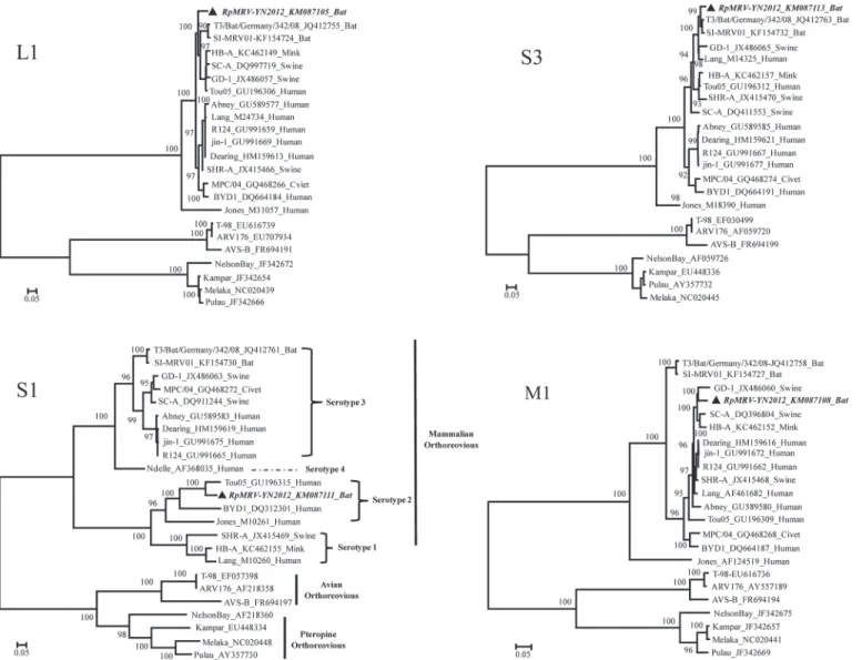
Phylogenetic classification
Whole genome sequences of 10 segments (L1–L3, M1–M3, S1–S4) of RpMRV-YN2012 were
aligned with those ofOrthoreovirusstrains available in GenBank. Maximum likelihood trees
were reconstructed based on the nucleotide sequence of the whole genome sequences. The to-pology of the phylogenetic trees confirmed the relationship of RpMRV-YN2012 with these bat,
Fig 3. Phylogenetic analysis of the L1, M1, S1, and S3 genome segments for the RpMRV-YN2012 strain and most related whole-genome strains from GenBank.Phylogenetic analyses were performed by the Maximum-likelihood method using MEGA (ver. 6.06,www.megasoftware.net). The scale bar, indicates the number of nucleotide substitutions per site. Bootstrap percentages (1000 replicates) above 50% are shown at nodes.The sequence of RpMRV-YN2012 from this study is inbold italictypeface.
pig, and human MRV strains (Fig. 3). Although all the segments of RpMRV-YN2012 were clustered within the MRVs, they separated in two serotypes (T2 and T3). Commonly,
orthoreo-viruses are classified taxonomically on the basis of the S1 segment [2]. The topology of a
phylo-genetic tree based on the nucleotide sequence of the complete S1 segment assigned RpMRV-YN2012 to the human-associated serotype 2 MRVs and was most divergent from the serotype 3 MRVs, providing phylogenetic evidence that RpMRV-YN2012 was a serotype 2 strain (Fig. 3). However, based on the other segments, the reconstructed tree sorted RpMRV-YN2012
to pig- or bat-associated serotype 3 MRVs (Fig. 3).
Prevalence of RpMRV-YN2012
The RpMRV-YN2012 prevalence study was performed in pharyngeal swabs (n= 106) and anal
swabs (n= 106) of bats (36Rhinolophus luctus, 27Rhinolophus affinis, 25Rhinolophus pusillus,
5Penthetor lucasian, and 13Myotis daubentonii) captured in the six roosts where the urine and feces samples were collected. Total RNA from all collected samples was isolated and screened by a reported RT-PCR procedure, targeting the conserved regions of the L1 genome
segment [17]. Four additional anal swab samples fromRhinolophus pusilluswere found to
har-bor strain RpMRV-YN2012, whereas other sampleswere negative.
Discussion
Bats, as the most abundant, diverse, and geographically dispersed vertebrates on earth, have been shown recently to be the reservoir hosts of a variety of zoonotic viruses responsible for
se-vere human disease outbreaks, some with very high mortality [6]. However, reports on the
iso-lation oforthoreovirusfrom bats are limited. Nelson Bay virus (NBV) was the first bat-origin
orthoreovirus isolated in 1968 from the heart blood of a flying fox (Pteropus poliocephalus) in
New South Wales, Australia [22]. In 1999, the second bat-borne orthoreovirus, called Pulau
virus (PulV), was isolated from fruit bat urine collected on Tioman Island, Malaysia [23]. In
2007, a novel bat-origin orthoreovirus, Melaka virus (MelV), was isolated from a patient with acute respiratory disease in Melaka, Malaysia, and the data provided suggested that this new orthoreovirus is capable of human-to-human transmission; this was the first report of an
orthoreovirus in association with acute human respiratory diseases [24]. In 2008, the fourth
member of the NBV species group, Kampar virus (KamV), was isolated from a human patient with fever and acute respiratory illness, and was suggested to be a bat-borne orthoreovirus by
epidemiological data [25]. Since then, bat-borne orthoreoviruses have received much attention,
and five additional orthoreoviruses (Xi-River, Kampar, Sikamat, HK23629/07 and Broome
vi-ruses) have been isolated from fruit bats, or humans with assumed contact with bats [26–29].
MRV infection in humans has been shown to be fairly common and the infections are often shown to be asymptomatic or associated with mild, self-limiting respiratory or gastrointestinal
illness in infants and children [3]. Recent studies have shown that MRV can cause severe
ill-nesses in humans and other mammals, including upper respiratory tract infections,
encephali-tis, and diarrhea[4,5,18]. In this study, we describe the first isolation of a novel natural
reassortant MRV strain, named RpMRV-YN2012, from the least horseshoe bat (Rhinolophus
pusillus) during a viral carriage study of bats in southwestern cities of Yunnan Province, China. The results provided evidence that least horseshoe bats carry MRVs and may act as a natural reservoir of MRVs.
The S1 gene segment of mammalian reovirus is bicistronic, encoding both the viral
attach-ment protein sigma-1 (δ1) and the non-structural protein (δ1 s) from overlapping open
read-ing frames [1,3]. The viralδ1 protein is unique to each prototype of mammalian reovirus and
mice [1–3]. The S1 gene of RpMRV-YN2012 showed far greater similarity to serotype 2 tou05
(84.9%/91.1%, nt/aa identity) than it did to serotype 1 and 3 MRVs S1 genes (Table 3). The
phylogenetic tree based on the nucleotide sequence of the complete S1 segment showed RpMRV-YN2012 closely related to serotype 2 MRVs and was most divergent from the serotype 1 and 3 MRVs. However, based on the other segments, the reconstructed tree sorted
RpMRV-YN2012 to serotype 3 MRVs (Fig. 3). These data confirm that RpMRV-YN2012 is a novel
sero-type 2 MRV that may have originated from reassortment among serosero-type 2 and 3 MRVs. Until
now, most of the orthoreoviruses identified in bats belong to thePteropine orthoreovirus
spe-cies or serotype 3 MRVs [18,21,30]. RpMRV-YN2012 is the first serotype 2 MRV isolated
from bats.
Considering RpMRV-YN2012 may have resulted from a reassortment of bat, pig, and/or human MRV strains, which can cause severe human or animal diseases (diarrhea, acute gastro-enteritis and necrotizing encephalopathy), it is necessary to identify whether RpMRV-YN2012 is associated with diarrhea, acute gastroenteritis and necrotizing encephalopathy in clinical pa-tients. In addition, we should carefully monitor its evolution and virulence in real time.
Acknowledgments
We thank Dr. Linfa Wang for his constructive comments and colleagues in local CDCs for help with field investigations and sample collection.
Author Contributions
Conceived and designed the experiments: LHW GDL. Performed the experiments: LHW SHF LC WWL JDS. Analyzed the data: LHW QT GDL. Wrote the paper: LHW GDL. Samples col-lection: LHW YXC HLZ YF WHY.
References
1. Attoui H, Mertens PPC, Becnel J, Belaganahalli S, Bergoin M, Brussaard CP, et al. Ninth Report of the International Committee on Taxonomy of Viruses. In: Andrew MQ King, Michael J Adams, Eric B Car-stens, Elliot J Lefkowitz, editors. Family: Reoviridae. London: Elsevier/Academic Press; 2011. pp. 541–603.
2. Day JM. The diversity of the orthoreoviruses: molecular taxonomy and phylogenetic divides. Infect Genet Evol. 2009; 9:390–400. doi:10.1016/j.meegid.2009.01.011PMID:19460305
3. Schriff LA, Nibert ML, Tyler KL. Orthoreoviruses and their replication. In: Knipe DM, Griffin DE, Lamb RA, Straus SE, Howley PM, et al., editors. Fields Virology. Philadelphia: Lippincott Williams & Wilkins; 2007; pp. 1853–1915.
4. Tyler KL, Barton ES, Ibach ML, Robinson C, Campbell JA, O'Donnell SM, et al. Isolation and molecular characterization of a novel type 3 reovirus from a child with meningitis. J Infect Dis. 2004; 189:1664– 1675. PMID:15116303
5. Ouattara LA, Barin F, Barthez MA, Bonnaud B, Roingeard P, Goudeau A, et al. Novel human reovirus isolated from children with acute necrotizing encephalopathy. Emerg Infect Dis. 2011; 17:1436–1444. doi:10.3201/eid1708.101528PMID:21801621
6. Calisher CH, Childs JE, Field HE, Holmes KV, Schountz T. Bats: Important reservoir hosts of emerging viruses. Clin Microbiol Rev. 2009; 19:531–545.
7. Kuzmin Ivan V, Bozick Brooke, Guagliardo Sarah A, Tong S, et al. Bats, emerging infectious diseases, and the rabies paradigm revisited. Emerg Health Threats J. 2011; 4: 7159. doi:10.3402/ehtj.v4i0.7159 PMID:24149032
8. Parrish CR, Holmes EC, Morens DM, Park EC, Burke DS, Calisher CH, et al. Cross-species virus trans-mission and the emergence of new epidemic diseases. Microbiol Mol Biol Rev. 2008; 72:457–470. doi: 10.1128/MMBR.00004-08PMID:18772285
10. Omatsu T, Watanabe S, Akashi H, Yoshikawa Y. Biological characters of bats in relation to natural res-ervoir of emerging viruses. Comp Immunol Microbiol Infect Dis. 2007; 30:357–374. PMID:17706776
11. van der Poel WH, Lina PH, Kramps JA. Public health awareness of emerging zoonotic viruses of bats: a European perspective. Vector Borne Zoonotic Dis. 2006; 6:315–324. PMID:17187565
12. Wong S, Lau S, Woo P, Yuen KY. Bats as a continuing source of emerging infections in humans. Rev Med Virol. 2007; 17:67–91. PMID:17042030
13. Ge XY, Li JL, Yang XL, Chmura AA, Zhu GJ, Epstein JH, et al. Isolation and characterization of a bat SARS-like coronavirus that uses the ACE2 receptor. Nature. 2013; 503:535–538. doi:10.1038/ nature12711PMID:24172901
14. Keller BC, Fredericksen BL, Samuel MA, Mock RE, Mason PW, Diamond MS, et al. Resistance to alpha/beta interferon is a determinant of West Nile virus replication fitness and virulence. J Virol. 2006; 80:9424–9434. PMID:16973548
15. Wang L, Lv X, Zhai Y, Fu S, Wang D, Rayner S, et al. Genomic Characterization of a Novel Virus of the Family Tymoviridae Isolated from Mosquitoes. PLoS One. 2012; 7(7):e39845. doi:10.1371/journal. pone.0039845PMID:22848363
16. Tamura K, Peterson D, Peterson N, Stecher G, Nei M, Kumar S. MEGA5: molecular evolutionary genet-ics analysis using maximum likelihood; evolutionary distance; and maximum parsimony methods. Mol Biol Evol. 2011; 28:2731–2739. doi:10.1093/molbev/msr121PMID:21546353
17. Leary TP, Erker JC, Chalmers ML, Wetzel JD, Desai SM, Mushahwar IK, et al. Detection of reovirus by reverse transcription-polymerase chain reaction using primers corresponding to conserved regions of the viral L1 genome segment. J Virol Methods. 2002; 104:161–165. PMID:12088825
18. Steyer A, Gutiérrez-Aguire I, Kolenc M, Koren S, Kutnjak D, Pokorn M, et al. High similarity of novel orthoreovirus detected in a child hospitalized with acute gastroenteritis to mammalian orthoreoviruses found in bats in Europe. J Clin Microbiol. 2013; 51:3818–3825. doi:10.1128/JCM.01531-13PMID: 24025904
19. Dai Y, Zhou Q, Zhang C, Song Y, Tian X, Zhang X, et al. Complete Genome Sequence of a Porcine Orthoreovirus from Southern China. J Virol. 2012; 86:12456. doi:10.1128/JVI.02254-12PMID: 23087117
20. Zhang C, Liu L, Wang P, Liu S, Lin W, Hu F, et al. A potentially novel reovirus isolated from swine. Virus Genes. 2011; 43:342–349. doi:10.1007/s11262-011-0642-4PMID:21761235
21. Kohl C, Lesnik R, Brinkmann A, Ebinger A, RadonićA, Nitsche A, et al. Isolation and characterization of three mammalian orthoreoviruses from European bats. PLoS ONE. 2012; 7:e43106. doi:10.1371/ journal.pone.0043106PMID:22905211
22. Gard GP, Marshall ID. Nelson Bay virus. A novel reovirus. Arch Gesamte Virus forsch. 1973; 43: 34– 42. PMID:4367379
23. Chua KB. A novel approach for collecting samples from fruit bats for isolation of infectious agents. Mi-crobes Infect. 2003; 5: 487–490. PMID:12758277
24. Chua KB, Crameri G, Hyatt A, Yu M, Tompang MR, Rosli J, et al. A previously unknown reovirus of bat origin is associated with an acute respiratory disease in humans. Proc Natl Acad Sci USA. 2007; 104:11424–11429. PMID:17592121
25. Chua KB, Voon K, Crameri G, Tan HS, Rosli J, McEachern JA, et al. Identification and characterization of a new orthoreovirus from patients with acute respiratory infections. PLoS One. 2008; 3:e3803. doi: 10.1371/journal.pone.0003803PMID:19030226
26. Du L, Lu Z, Fan Y, Meng K, Jiang Y, Zhu Y, et al. Xi River virus, a new bat reovirus isolated in southern China. Arch Virol. 2010; 155: 1295–1299. doi:10.1007/s00705-010-0690-4PMID:20495835
27. Thalmann CM, Cummins DM, Yu M, Lunt R, Pritchard LI, Hansson E, et al.Broome virus, a new fuso-genicOrthoreovirusspecies isolated from an Australian fruit bat. Virology. 2010; 402: 26–40. doi:10. 1016/j.virol.2009.11.048PMID:20350736
28. Cheng P, Lau CS, Lai A, Ho E, Leung P, Chan F, et al. A novel reovirus isolated from a patient with acute respiratory disease. J Clin Virol. 2009; 45:79–80. doi:10.1016/j.jcv.2009.03.001PMID: 19356975
29. Chua KB, Voon K, Yu M, Keniscope C, Abdul Rasid K, Wang LF. Investigation of a potential zoonotic transmission of orthoreovirus associated with acute influenza-like illness in an adult patient. PLoS ONE. 2011; 6: e25434. doi:10.1371/journal.pone.0025434PMID:22022394