JOCLEITA PERUZZO FERRAREZE
EFFECT OF POSTHARVEST JASMONIC ACID TREATMENT ON STORAGE DISEASES, BIOCHEMICAL AND MOLECULAR ANALYSES OF INDUCED
DISEASE RESISTANCE IN SUGARBEET (BETA VULGARIS L.)
Tese apresentada à Universidade Federal de Viçosa, como parte das exigências do Programa de Pós-Graduação em Fisiologia Vegetal, para obtenção do título de Doctor Scientiae.
VIÇOSA
Ficha catalográfica preparada pela Seção de Catalogação e Classificação da Biblioteca Central da UFV
T
Ferrareze, Jocleita Peruzzo, 1983-
F374e Effect of postharvest jasmonic acid treatment on storage 2012 diseases, biochemical and molecular analyses of induced disease resistance in sugarbeet (Beta vulgaris L.) / Jocleita
Peruzzo Ferrareze. – Viçosa, MG, 2012. x, 68f. : il. (algumas col.) ; 29cm.
Inclui anexo.
Orientador: Fernando Luiz Finger.
Tese (doutorado) - Universidade Federal de Viçosa. Inclui bibliografia.
1. Beterraba açucareira. 2. Beterraba açucareira - Fisiologia pós-colheita. 3. Regulação de expressão gênica. 4. Beterraba açucareira - Resistência a doenças e pragas. 5. Hormônios
vegetais. 6. Ácido jasmônico. 7. Enzimas. 8. Fungos patogênicos. 9. Raízes (Botânica). 10. Beta vulgaris. I. Universidade Federal de Viçosa. II. Título.
JOCLEITA PERUZZO FERRAREZE
EFFECT OF POSTHARVEST JASMONIC ACID TREATMENT ON STORAGE DISEASES, BIOCHEMICAL AND MOLECULAR ANALYSES OF INDUCED
DISEASE RESISTANCE IN SUGARBEET (BETA VULGARIS L.)
Tese apresentada à Universidade Federal de Viçosa, como parte das exigências do Programa de Pós-Graduação em Fisiologia Vegetal, para obtenção do título de Doctor Scientiae.
APROVADA: 16 de Março de 2012.
______________________________
Karen Klotz Fugate ________________________________ Daniela Vieira Chaves
______________________________
Raimundo Santos Barros ________________________________ Edward Deckard
______________________________ Fernando Luiz Finger
iii
AGRADECIMENTOS
Gostaria primeiramente de agradecer ao Professor Fernando Finger, meu querido orientador e amigo, por sempre ter o conselho certo na hora certa.
Aos Professores Fábio da Mata e Raimundo Barros, pela amizade, inspiração e pelas aulas maravilhosas.
Aos meus queridos amigos Daniela Vieira Chaves e Fábio Zanata. Que sorte conhecer vocês! Obrigada por tudo.
A minha amiga Juliane Karsten, pelo apoio nas disciplinas e pelos maravilhosos doces caseiros.
A Gládis e Alice, pelos ótimos momentos.
A Clarice Megguer, pelo apoio em todo o processo da bolsa sanduíche e pela recepção nos Estados Unidos.
A André Brandt, por estar comigo e ser comigo.
A todos os amigos que fizeram parte da minha vida nesse período incrível e que jamais serão esquecidos, muito obrigada.
Aos meus pais pelo incentivo desde sempre, amor e carinho.
Ao CNPq pela concessão da bolsa de doutorado e de doutorado sanduíche.
Ao programa de Pós-Graduação em Fisiologia Vegetal pela excelente formação proporcionada aos seus estudantes.
ACKNOWLEDGEMENTS
To Karen Fugate, for the generosity sharing her knowledge and for all the patience to understanding and teaching me despite my difficulties. Thanks for be such a good friend, an inspiration for life. Thanks for the great experience I had in US.
To Professor Ed Deckard for the help.
To Melvin Bolton for the help with the research.
To John Eide, for all the help in the lab and for the great time working together in the lab.
v
SUMÁRIO
PÁGINA
RESUMO... vii
ABSTRACT... ix
1 INTRODUÇÃO GERAL ... 1
2 REFERÊNCIAS ... 4
3 ARTIGO 1 ... 8
1 INTRODUCTION ... 10
2 MATERIALS AND METHODS ... 12
2.1 Plant material and postharvest treatments ... 12
2.2 Storage rot resistance assays ... 12
2.3 Statistical analysis... 13
3 RESULTS AND DISCUSSION ... 14
4 CONCLUSIONS ... 18
5 REFERENCES ... 19
4 ARTIGO 2 ... 23
1 INTRODUCTION ... 25
2 MATERIAL AND METHODS ... 27
2.1 Plant material and JA treatment ... 27
2.2 Protein extraction and enzyme activity assays ... 27
2.3 Total phenolic compounds concentration and antioxidant capacity .. 29
2.4 Statistical analysis... 29
3 RESULTS ... 30
3.1 ROS-scavenging enzyme activities... 30
3.2 Pathogen-related defense enzyme activities ... 32
3.3 Total phenolic compounds (TPC) concentration and antioxidant capacity (AOC) ... 34
4 DISCUSSION ... 35
5 REFERENCES ... 38
5 ARTIGO 3 ... 43
1 INTRODUCTION ... 46
2 MATERIAL AND METHODS ... 48
2.1 Plant material and JA treatment ... 48
2.3 Reference transcriptome generation ... 50
2.4 Unigene Annotation ... 50
2.5 RNA-sequencing of JA treated samples ... 51
3 RESULTS ... 52
3.1 Defense-related genes expression ... 52
3.2 Gene ontology and clusters of orthologous groups of the RNA library ... 54
3.3 JA effect on sugarbeet transcriptome ... 56
4 DISCUSSION ... 60
5 REFERENCES ... 62
6 CONCLUSÃO GERAL ... 66
vii
RESUMO
FERRAREZE, Jocleita Peruzzo, D.Sc., Universidade Federal de Viçosa, março de 2012. Efeito do tratamento com ácido jasmônico nas doenças pós-colheita e análises bioquímicas e moleculares da resistência induzida em beterraba-açucareira (Beta vulgaris L.). Orientador: Fernando Luiz Finger.
Ácido jasmônico (AJ) e seus derivativos são conhecidos por ativar os mecanismos de defesa das plantas e fornecer proteção contra fungos causadores de podridões em várias espécies de vegetais. No presente trabalho testou-se a eficiência do ácido jasmônico em proteger raízes de beterraba-açucareira (Beta vulgaris L.) contra os três principais patógenos causadores de podridões pós-colheita. AJ reduziu a podridão causada por Botrytis cinerea e Phoma betae em uma média de 51 e 71% respectivamente e em concentrações de 0.01–10 µM AJ reduziu podridão causada por Penicillium claviforme em uma média de 44%, enquanto 100 µM reduziu a podridão em 65%. Tendo-se em vista os resultados mostrando a habilidade do AJ em proteger raízes de beterraba-açucareira contra os patógenos citados, os mecanismos responsáveis pela indução da resistência por jasmonatos foram estudados. A atividade de enzimas com função antioxidante como ascorbato peroxidase (APX), catalase (CAT), peroxidase (POD) e superóxido dismutase (SOD), e metabólitos com propriedades antioxidantes foram analisados. A
atividade de enzimas relacionadas com a defesa de plantas como β
-1,3-glucanase (β-Gluc), quitinase, polifenol oxidase (PPO) e fenilalanina
ix
ABSTRACT
FERRAREZE, Jocleita Peruzzo, D.Sc., Universidade Federal de Viçosa, March, 2012. Effect of postharvest jasmonic acid treatment on storage diseases, biochemical and molecular analyses of induced disease resistance in sugarbeet (Beta vulgaris L.). Adviser: Fernando Luiz Finger.
1
1 INTRODUÇÃO GERAL
Patógenos pós-colheita são controlados por técnicas de manejo das pilhas de armazenamento de beterraba-açucareira. Baixas temperaturas reduzem o índice de crescimento de muitos organismos causadores de podridões, diminuindo a temperatura das pilhas e removendo ‘hotspots’ (locais específicos na pilha onde a temperatura é elevada) e reduz a ocorrência de perdas na pós-colheita. O manejo das pilhas, no entanto, requer condições ambientes favoráveis de armazenamento, monitoramento contínuo e ainda assim são limitados em controlar fungos causadores de podridões. Resistência genética e fungicidas químicos podem ser usados para reduzir as perdas (Miles et al., 1977; Bugbee & Cole, 1979), mas nenhum desses mecanismos de controle é utilizado porque a introdução de características adicionais em programas de melhoramento reduz a rapidez de progresso desses programas em obter outras características desejáveis. Fungicidas geralmente produzem efeitos negativos nas propriedades de armazenamento das beterrabas mesmo sem doenças (Akeson et al., 1979; Campbell, 2005).
Em grande número de produtos hortícolas, aplicações de jasmonatos proporcionam proteção contra fungos pós-colheita (Tripathi and Dubey, 2004; Rohwer and Erwin, 2008). Jasmonatos são hormônios vegetais conhecidos por ativarem os mecanismos de defesas das plantas (Ballaré, 2011). Quando aplicado, jasmonatos reduzem doenças causadas por uma variedade de fungos, bactérias e vírus patogênicos (Thaler et al., 2004; Rohwer and Erwin, 2008; Haggag et al., 2010) por reduzirem a severidade da doença, na maioria dos casos, ou por reduzirem a incidência em outros (Yao and Tian, 2005b; Cao et al., 2008; Zhang et al., 2009).
Seliskar, 1952; Campbell and Klotz, 2006; Fugate and Campbell, 2009). P. claviforme é geralmente menos agressivo que B. cinerea, produz corpos de frutificação em formato de haste com massas de esporos verdes e prevalece nas pilhas de armazenamento dos EUA (Bugbee, 1975; Bugbee and Cole, 1976; Fugate and Campbell, 2009). P. betae causa uma podridão que inicia no tecido da medula central e se desenvolve para baixo e para fora no interior da beterraba e circulando o tecido da coroa (Bugbee and Cole, 1976). Infecção devido a P. betae tipicamente se desenvolve após um período de armazenamento de 80 dias, porém o organismo é capaz de infectar o tecido em qualquer momento na pós-colheita (Bugbee, 1982).
Os mecanismos responsáveis pela indução da resistência em plantas por jasmonatos não são bem estabelecidos. Alterações na atividade de enzimas antioxidantes como ascorbato peroxidase (APX), catalase (CAT), peroxidase (POD) e superóxido dismutase (SOD), e aumentos nas concentrações de metabolitos com propriedades antioxidantes, no entanto, tem sido relacionadas com resistência a doenças induzida por jasmonatos em várias espécies de plantas (Yao and Tian, 2005a; Cao et al., 2008; Wang et al., 2009a). Aumentos nas atividades de enzimas relacionadas com a defesa de
plantas como β-1,3-glucanase (β-Gluc), quitinase, polifenol oxidase (PPO) e
fenilalanina amônia-liase (PAL) também tem sido associadas com resistências induzidas por jasmonatos em muitas plantas (Yao and Tian, 2005a; Haggag et al., 2010). Essas enzimas presumivelmente aumentam a resistência a doenças pela sua atividade antimicrobiana direta ou seu envolvimento na síntese de compostos antimicrobianos.
Alteração na expressão de genes relacionados à defesa é um elemento chave nos mecanismos de defesa induzida em plantas (Edreva, 2005). Alterações na expressão gênica de fenilalanina amônia-liase (PAL), peroxidase (POD), genes relacionados à patogênese (PR1), e quitinases são comumente relacionadas com defesas induzidas por jasmonatos nas plantas. (Sharan et al., 1998; Curtis et al., 1997; Raymond and Farmer 1998; Ding et al., 2002).
3 informação sobre milhões de pares de bases em uma única corrida. O grande número de sequencias geradas fornece valiosas informações em nível de transcriptoma para descoberta de novos genes ou investigação de mecanismos moleculares (Wang et al., 2009b; Wei et al., 2011).
2 REFERÊNCIAS
Akeson, W.R., Yun, Y.M., Sullivan, E.F., 1979. Effect of chemicals on sucrose loss in sugarbeets during storage. J. Am. Soc. Sugar Beet Technol. 20, 255-268.
Ballaré, C.L., 2011. Jasmonate-induced defenses: a tale of intelligence, collaborators and rascals. Trends Plant Sci. 16, 249-257.
Bugbee, W.M., 1975. Penicillium claviforme and Penicillium variabile: pathogens of stored sugar beets. Phytopathology 65, 926–927.
Bugbee, W.M., 1982. Storage rot of sugar beet. Plant Dis. 66, 871–873.
Bugbee, W.M., 1986. Storage rot of sugar beet. In: Whitney, E.D., Duffus, J.E. (Eds.), Compendium of Beet Diseases and Insects. APS Press, St. Paul, MN, USA, pp. 37–39.
Bugbee, W.M., Cole, D.F., 1976. Sugarbeet storage rot in the Red River Valley, 1974–1975. J. Am. Soc. Sugar Beet Technol. 19, 19–24.
Bugbee, W.M., Cole, D.F., 1979. Comparison of thiabendazole and genetic resistance for control of sugar beet storage rot. Phytopathology 69, 1230-1232.
Campbell, L.G., 2005. Post-harvest storage traits. In: Biancardi, E., Campbell, L.G., De Biaggi, M., Skaracis, G.N. (eds.) Sugar Beet Breeding and Genetics. Science Publishers, Inc, Enfield, NH. pp. 122-126.
5 Cao, S., Zheng, Y., Yang, Z., Tang, S., Jin, P., Wang, K., Wang, X., 2008. Effect of methyl jasmonate on the inhibition of Colletotrichum acutatum infection in loquat fruit and the possible mechanisms. Postharvest Biol. Technol. 49, 301-307.
Curtis, M. D., Rae, A. L., Rusu, A. G., Harrison, S. J., Manners, J. M., 1997. A Peroxidase Gene Promoter Induced by Phytopathogens and Methyl Jasmonate in Transgenic Plants. MPMI. 10, 326-338.
Ding, C., Wang, C. Y., Gross, K, C., Smith, D. L., 2002. Jasmonate and salicylate induce the expression of pathogenesis-related-protein genes and increase resistance to chilling injury in tomato fruit. Planta. 214, 895–901.
Edreva, A., 2005. Pathogenesis-related proteins: research progress in the last 15 years. General and Applied Plant Physiology, 31, 105-124.
Fugate, K., Campbell, L., 2009. Postharvest deterioration of sugar beet. In: Harveson, R.M., Hanson, L.E., Hein, G.L. (Eds.), Compendium of Beet Diseases and Pests. , 2nd ed. APS Press, St. Paul, MN, USA, pp. 92–94.
Gaskill, J.O., Seliskar, C.E., 1952. Effect of temperature on rate of rotting of sugarbeet tissue by two storage pathogens. Proc. Am. Soc. Sugar Beet Technol. 7, 571–574.
Haggag, W.M., Mahmoud, Y.S., Farag, E.M., 2010. Signaling necessities and function of polyamines/jasmonate-dependent induced resistance in sugar beet against beet mosaic virus (BtMV) infection. New York Sci. J. 3, 95-103.
Miles, W.G., Shaker, F.M., Nielson, A.K., Ames, R.R., 1977. A laboratory study of the ability of fungicides to control beet rotting fungi. J. Am. Soc. Sugar Beet Technol. 19, 288-293.
Rohwer, C.L., Erwin, J.E., 2008. Horticultural applications of jasmonates: a review. J. Hortic. Sci. Biotech. 83, 283-304.
Sharan, M., Taguchi, M., Gonda, K., Jouke, T., Shimosaka, M., Hayashida, N., Okazaki, M., 1998. Effects of methyl jasmonate and elicitor on the activation of phenylalanine ammonia-lyase and the accumulation of scopoletin and scopolin in tobacco cell cultures. Plant Science, 132, 13-19.
Thaler, J.S., Owen, B., Higgins, V.J., 2004. The role of the jasmonate response in plant susceptibility to diverse pathogens with a range of lifestyles. Plant Physiol. 135, 530-538.
Tripathi, P., Dubey, N.K., 2004. Exploitation of natural products as an alternative strategy to control postharvest fungal rotting of fruit and vegetables. Postharvest Biol. Technol. 32, 235-245.
Wang, K., Jin, P., Cao, S., Shang, H., Yang, Z., Zheng, Y., 2009a. Methyl jasmonate reduces decay and enhances antioxidant capacity in chinese bayberries. J. Agric. Food Chem. 57, 5809-5815.
Wang, Z., Gerstein, M., Snyder, M., 2009b. RNA-Seq: a revolutionary tool for transcriptomics Nature Reviews Genetics. 10, 57-63.
Wei, W., Qi, X., Wang, L., Zhang, Y., Hua, W., Li, D., Lv, H., Zhang, X., 2011. Characterization of the sesame (Sesamum indicum L.) global transcriptome using Illumina paired-end sequencing and development of EST-SSR markers. BMC Genomics. 12, 451
7 Yao, H., Tian, S., 2005b. Effects of pre- and post-harvest application of salicylic acid or methyl jasmonate on inducing disease resistance of sweet cherry fruit in storage. Postharvest Biol. Technol. 35, 253-262.
3 ARTIGO 1
doi:10.1016/j.postharvbio.2011.10.005
*Corresponding author: Karen Klotz Fugate USDA-ARS, Northern Crop Science Laboratory 1605 Albrecht Blvd., N.
Fargo, ND 58102-2765, USA telephone: (701) 239-1356 fax: (701) 239-1349
9
ABSTRACT
Although jasmonic acid (JA) and JA derivatives are known to activate plant defense mechanisms and provide protection against postharvest fungal
diseases for several horticultural crops, JA’s ability to protect sugarbeet (Beta
1 INTRODUCTION
Storage rots of sugarbeet root consume sucrose and produce sucrose- and cell wall-degrading enzymes whose reaction products impair juice purification and sucrose crystallization operations and decrease sucrose recovery during processing (Buchholz et al., 1998; Tungland et al., 1998; Dutton and Huijbregts, 2006). Storage rots also increase root respiration rate, a process that metabolizes sucrose and generates heat, creating additional storage losses and contributing to the warming of storage piles (Mumford and Wyse, 1976; Kays and Paull, 2004). As storage piles warm, the respiration of both healthy and diseased roots and the incidence and severity of storage rots increase (Campbell and Klotz, 2006). Storage rots, therefore, increase storage and processing losses and initiate events that escalate the rate of storage losses.
Botrytis cinerea Pers. ex Fr. (teleomorph: Botryotinia fuckeliana [de Bary] Whetz.), Penicillium claviforme Bainier and Phoma betae Frank (teleomorph: Pleospora bjorlingii Byford) are known storage rot pathogens of sugarbeet (Bugbee, 1986). B. cinerea is an aggressive rot-causing organism that is characterized by gray spore masses, active over a wide range of temperatures, and widely distributed in sugarbeet growing regions throughout the world (Gaskill and Seliskar, 1952; Campbell and Klotz, 2006; Fugate and Campbell, 2009). P. claviforme is generally less aggressive than B. cinerea, produces stalked fruiting bodies with green spore masses, and is prevalent in U.S. storage piles (Bugbee, 1975; Bugbee and Cole, 1976; Fugate and Campbell, 2009). P. betae causes a rot that begins in the central pith tissue of the root crown and develops downward and outward into the taproot and surrounding crown tissue (Bugbee and Cole, 1976). Rot due to P. betae typically develops after roots have been stored in excess of 80 d, although the organism is capable of rotting tissue at any time after harvest (Bugbee, 1982).
11 piles in which sugarbeet roots are stored are cooled using ambient winter air. The control of storage rots, therefore, is dependent on weather conditions and is limitedly effective against fungal organisms that are capable of growth at low temperatures (Miles et al., 1977). Although chemical fungicides can reduce the incidence and severity of storage rots, fungicides are not used by the industry since they generally have deleterious effects on sugarbeet root storage properties in the absence of disease (Miles et al., 1977; Akeson et al., 1979). Germplasm with genetic resistance against some of the major storage rot-causing fungi has been developed (Bugbee and Cole, 1979), but has not been incorporated into current commercial hybrids.
2 MATERIALS AND METHODS
2.1 Plant material and postharvest treatments
Sugarbeet hybrid VDH66156 (SESVanderHave, Tienen, Belgium) was greenhouse grown in Sunshine Mix #1 (Sun Gro Horticultural Products, Seba Beach, Alberta, Canada) in 15-L pots with supplemental light under a 16 h light/8 h dark regime, and watered as needed. Taproots were harvested 16-18 weeks after planting and washed to remove adhering soil. Seed was treated with N-(2,3-dimethylphenyl)-N-(methoxyacetyl)-alanine methyl ester (Apron-metalaxyl) and tetramethylithiuram disulfide (Thiram) to prevent fungal seedling diseases. No symptoms of root disease were evident at any time during production or at harvest. Jasmonic acid treatments were administered by submerging roots in 0, 0.01, 0.1, 1, 10, or 100 µM JA (Cayman Chemical, Ann Arbor, MI) for 1 h at room temperature, using at least 7 roots per treatment. Roots were incubated at 20 °C and 90% relative humidity for 3 d post-treatment (Yao and Tian, 2005b) to allow for induction of defense mechanisms prior to inoculation with a storage rot-causing pathogen. All experiments were repeated at least once.
2.2 Storage rot resistance assays
13 (diameter × depth) holes into each root with a hand-held drill, with holes located on opposite sides of the root in the region where root girth was greatest. Roots were inoculated by inserting a 10 mm diameter plug obtained from fungal-covered PDA plates into each hole. After inoculation, roots were incubated at 20 °C and 90% relative humidity until severe disease symptoms were visually evident on control roots, approximately 20, 30, and 50 d for B. cinerea, P. claviforme, and P. betae, respectively. Root rot severity was evaluated by excising and weighing the rotted, discolored tissue from each root.
2.3 Statistical analysis
3 RESULTS AND DISCUSSION
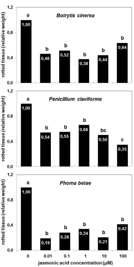
Postharvest jasmonic acid treatments of 0.01-100 µM significantly (P < 0.001) reduced the severity of rot due to Botrytis cinerea in stored sugarbeet roots (Fig. 1A). Roots treated with 0.01, 0.1, 1, 10, and 100 µM JA had 36-62% less rot than water-treated controls after inoculation with the pathogen and incubation under environmental conditions that favor disease development. Although JA treatments varied in concentration by 10,000-fold, all treatments reduced rot severity to a statistically similar extent. This suggests that JA protected sugarbeet roots against B. cinerea at an extremely low concentration (0.01 µM) with no further benefit or detriment provided by additional JA. On average, JA reduced the weight of rotted tissue by 51%.
Postharvest jasmonic acid treatments of 0.01 to 100 µM also significantly (P < 0.001) reduced the severity of rot due to Penicillium claviforme in stored sugarbeet roots (Fig. 1B). Roots treated with 0.01, 0.1, 1, 10, and 100 µM JA had 34-65% less rot than water-treated controls after inoculation with the pathogen and incubation under disease-promoting environmental conditions. JA concentrations of 0.01-10 µM caused a statistically similar reduction of rot due to P. claviforme and reduced the weight of rotted tissue by an average of 44%. An increase in JA concentration to 100 µM provided additional protection against P. claviforme and reduced the weight of rotted tissue by 65% relative to the water-treated control.
15
Fig. 1. Relative weight of rotted tissue in jasmonic acid-treated roots after inoculation with Botrytis cinerea (A), Penicillium claviforme (B), or Phoma betae (C) and incubation at 20 °C and 90% relative humidity until severe disease symptoms were evident on control roots (0 µM JA). Harvested roots were treated for 1 h at room temperature and incubated for 3 d at 20 °C and 90% relative humidity prior to inoculation. Weight of rotted tissue is expressed as a fraction of the weight of rotted tissue of the control. Experimental repetitions were not statistically different by ANOVA (α = 0.05), and data are the combined results of two experimental repetitions, with at least 7 replicates per repetition. Treatments with different letters are statistically different based
Exogenous application of jasmonates has previously been shown to protect various plants against B. cinerea (Moline et al., 1997; Thomma et al., 1998; Yu et al., 2009) and several species of Penicillium, including P. digitatum, P. expansum, and P. citrinum (Yao and Tian, 2005a; Zhang et al., 2009; Wang et al., 2010). To our knowledge, however, this is the first report of jasmonate-associated protection against P. claviforme, P. betae, or any other Phoma species. Although the three pathogens are not related, the application of JA provided protection against all three fungi, suggesting that inducible defense pathways in sugarbeet roots are not pathogen specific, which has been shown in other pathosystems (Bolton, 2009). That sugarbeet root defense mechanisms can protect against more than one disease has previously been demonstrated as germplasm with enhanced P. betae resistance often has enhanced resistance against Rhizoctonia solani Kühn, a soil-borne fungal organism responsible for a crown and root rot during production (Bugbee and Campbell, 1990).
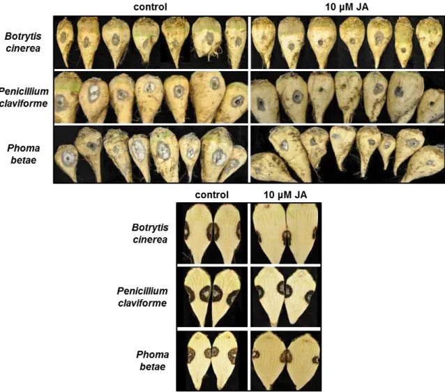
17 Visual evaluation of roots treated with JA and inoculated with B. cinerea, P. claviforme, or P. betae indicated that JA reduced the progression of disease symptoms in the storage tissue of sugarbeet roots, but did not prevent infection (Fig. 2). Signs of infection were evident at all inoculation sites regardless of the treatment applied (Fig. 2A). However, the progression of disease symptoms, as evidenced by the depth and breadth of necrotized tissue extending from the infection site, was reduced in JA-treated roots relative to water-treated controls (Fig. 2B). Zhang et al. (2009) similarly found that jasmonate treatment reduced infection area but not incidence of infection for P. expansum on pears. This contrasts with other studies that have found a reduction in both incidence and size of infection after jasmonate treatment (Yao and Tian, 2005b; Cao et al., 2008; Wang et al., 2010).
Fig. 2. Surface symptoms (A) and internal symptoms (B) of storage rot on water- (control) and
4 CONCLUSIONS
Postharvest JA treatment reduced rot due to B. cinerea, P. claviforme, and P. betae, three common storage pathogens of sugarbeet root. JA concentrations of 0.01-100 µM reduced rot due to B. cinerea and P. betae by an average of 51 and 71%, respectively. JA concentrations of 0.01-10 µM reduced rot due to P. claviforme by an average of 44%, while 100 µM JA reduced rot due to P. claviforme by 65%. JA treatment reduced rot by reducing the progression of disease symptoms in root storage tissue, but it had no effect on the incidence of infection. Relative to other postharvest products, sugarbeet roots were unusually sensitive to jasmonate treatment and were protected against fungal pathogens over a wide range of JA concentrations.
Acknowledgements
19
5 REFERENCES
Akeson, W.R., Yun, Y.M., Sullivan, E.F., 1979. Effect of chemicals on sucrose loss in sugarbeets during storage. J. Am. Soc. Sugar Beet Technol. 20, 255-268.
Bolton, M.D., 2009. Primary metabolism and plant defense-fuel for the fire. Mol. Plant-Microbe Interact. 22, 487-497.
Buchholz, K., Bliesener, H.-M., Buczys, R., Thielecke, K., 1998. Reactions of beet cellwall constituents. In: van der Poel, P.W., Schiweck, H., Schwartz, T. (Eds.), Sugar Technology: Beet and Cane Sugar Manufacture. Verlag Dr. Albert Bartens KG, Berlin, pp. 157-162.
Bugbee, W.M., 1975. Penicillium claviforme and Penicillium variabile: pathogens of stored sugar beets. Phytopathology 65, 926-927.
Bugbee, W.M., 1982. Storage rot of sugar beet. Plant Dis. 66, 871-873.
Bugbee, W.M., 1986. Storage rot of sugar beet. In: Whitney, E.D., Duffus, J.E. (Eds.), Compendium of Beet Diseases and Insects. APS Press, St. Paul, MN, USA, pp. 37-39.
Bugbee, W.M., Campbell, L.G., 1990. Combined resistance in sugar beet to Rhizoctonia solani, Phoma betae, and Botrytis cinerea. Plant Dis. 74, 353-355.
Bugbee, W.M., Cole, D.F., 1976. Sugarbeet storage rot in the Red River Valley, 1974-1975. J. Am. Soc. Sugar Beet Technol. 19, 19-24.
Bugbee, W.M., Cole, D.F., 1979. Comparison of thiabendazole and genetic resistance for control of sugar beet storage rot. Phytopathology 69, 1230-1232.
Cao, S., Zheng, Y., Yang, Z., Tang, S., Jin, P., Wang, K., Wang, X., 2008. Effect of methyl jasmonate on the inhibition of Colletotrichum acutatum infection in loquat fruit and the possible mechanisms. Postharvest Biol. Technol. 49, 301-307.
Creelman, R.A., Mullet, J.E., 1997. Biosynthesis and action of jasmonates in plants. Annu. Rev. Plant Physiol. Plant Mol. Biol. 48, 355-381.
Dutton, J., Huijbregts, T., 2006. Root quality and processing. In: Draycott, A.P. (Ed.), Sugar Beet. Blackwell Publishing Ltd., Oxford, UK, pp. 409-442.
Fugate, K., Campbell, L., 2009. Postharvest deterioration of sugar beet. In: Harveson, R.M., Hanson, L.E., Hein, G.L. (Eds.), Compendium of Beet Diseases and Pests. , 2nd ed. APS Press, St. Paul, MN, USA, pp. 92-94.
Gaskill, J.O., Seliskar, C.E., 1952. Effect of temperature on rate of rotting of sugarbeet tissue by two storage pathogens. Proc. Am. Soc. Sugar Beet Technol. 7, 571-574.
González-Aguilar, G.A., Buta, J.G., Wang, C.Y., 2003. Methyl jasmonate and modified atmosphere packaging (MAP) reduce decay and maintain postharvest quality of papaya ‘Sunrise’. Postharvest Biol. Technol. 28, 361 -370.
Kays, S.J., Paull, R.E., 2004. Postharvest Biology. Exon Press, Athens, GA.
Miles, W.G., Shaker, F.M., Nielson, A.K., Ames, R.R., 1977. A laboratory study of the ability of fungicides to control beet rotting fungi. J. Am. Soc. Sugar Beet Technol. 19, 288-293.
Moline, H.E., Buta, J.G., Saftner, R.A., Maas, J.L., 1997. Comparison of three volatile natural products for the reduction of postharvest decay in strawberries. Adv. Strawberry Res. 16, 43-48.
21 Rohwer, C.L., Erwin, J.E., 2008. Horticultural applications of jasmonates: a
review. J. Hortic. Sci. Biotechnol. 83, 283-304.
Thomma, B.P.H.J., Eggermont, K., Penninckx, I.A.M.A., Mauch-Mani, B., Vogelsang, R., Cammue, B.P.A., Broekaert, W.F., 1998. Separate jasmonate-dependent and salicylate-dependent defense-response pathways in Arabidopsis are essential for resistance to distinct microbial pathogens. Proc. Natl. Acad. Sci. U.S.A. 95, 15107-15111.
Tripathi, P., Dubey, N.K., 2004. Exploitation of natural products as an alternative strategy to control postharvest fungal rotting of fruits and vegetables. Postharvest Biol. Technol. 32, 235-245.
Tungland, B.C., Watkins, R.E., Schmidt, P.-V., 1998. Sugarbeet storage. In: van der Poel, P.W., Schiweck, H., Schwartz, T. (Eds.), Sugar Technology: Beet and Cane Sugar Manufacture. Verlag Dr. Albert Bartens KG, Berlin, pp. 267-289.
Wang, K., Jin, P., Cao, S., Shang, H., Yang, Z., Zheng, Y., 2009. Methyl jasmonate reduces decay and enhances antioxidant capacity in Chinese bayberries. J. Agric. Food Chem. 57, 5809-5815.
Wang, K., Jin, P., Shang, H., Zheng, Y., 2010. Effect of methyl jasmonate in combination with ethanol treatment on postharvest decay and antioxidant capacity in Chinese bayberries. J. Agric. Food Chem. 58, 9597-9604.
Yao, H.J., Tian, S.P., 2005a. Effects of a biocontrol agent and methyl jasmonate on postharvest diseases of peach fruit and the possible mechanisms involved. J. Appl. Microbiol. 98, 941-950.
Yao, H., Tian, S., 2005b. Effects of pre- and post-harvest application of salicylic acid or methyl jasmonate on inducing disease resistance of sweet cherry fruit in storage. Postharvest Biol. Technol. 35, 253-262.
23
4 ARTIGO 2
Jasmonic acid does not increase oxidative defense mechanisms or common defense-related enzymes in postharvest sugarbeet roots
JOCLEITA PERUZZO FERRAREZE(a)
, KAREN KLOTZ FUGATE(b),*, MELVIN D. BOLTON(c)
, EDWARD L. DECKARD (c), LARRY G. CAMPBELL(c) and FERNANDO L. FINGER (d),
*Corresponding author: Karen Klotz Fugate USDA-ARS, Northern Crop Science Laboratory 1605 Albrecht Blvd., N.
Fargo, ND 58102-2765, USA telephone: (701) 239-1356 fax: (701) 239-1349
email: karen.fugate@ars.usda.gov
(a) Departamento de Biologia Vegetal, Universidade Federal de Viçosa, 36570-000 Viçosa, MG,
Brazil.
(b) USDA-ARS, Northern Crop Science Laboratory, 1605 Albrecht Blvd. N., Fargo, ND
58102-2765, USA.
(c) Department of Plant Sciences, North Dakota State University, P.O. Box 6050, Fargo, ND
58108-6050.
ABSTRACT
Jasmonic acid (JA) treatment significantly reduces rot due to several sugarbeet (Beta vulgaris L.) storage pathogens. The mechanisms by which JA protects postharvest sugarbeet roots from disease, however, are unknown. In other plant species and organs, alterations in antioxidant defense mechanisms and elevations in common pathogen-related defense enzymes have been implicated in jasmonate-induced disease resistance. To investigate whether these mechanisms are involved in JA-induced disease resistance in stored sugarbeet roots, the activities of several reactive oxygen species (ROS)-scavenging and pathogen-related defense enzymes and the total concentration of antioxidant compounds were determined in harvested sugarbeet roots in the 60 d following treatment with JA. ROS-scavenging and pathogen-related defense enzymes and the concentration of antioxidant compounds were largely unaffected by JA as JA-treated roots exhibited small declines in superoxide dismutase (SOD) and chitinase activities, and were generally unaltered in ascorbate peroxidase (APX), catalase (CAT), peroxidase (POD), β
-1,3-glucanase (β-Gluc), polyphenol oxidase (PPO) and phenylalanine
ammonia-lyase (PAL) activities or antioxidant compounds concentration. The lack of increase in enzyme activities or metabolites related to defense against oxidative stress or pathogens suggests that JA-induced disease resistance in postharvest sugarbeet roots does not arise from a direct increase in any of the ROS-scavenging enzymes, defense-related enzymes, or the concentration of total antioxidant compounds that were examined in this study. However, ROS-scavenging enzymes and pathogen-related defense enzymes were affected by
storage duration with POD, SOD, β-Gluc, chitinase, and PPO activities elevated
25
1 INTRODUCTION
Jasmonates are endogenous plant hormones that activate native plant defense mechanisms (Ballaré, 2011). When applied exogenously, jasmonates reduce disease caused by a variety of fungal, bacterial and viral pathogens (Thaler et al., 2004; Rohwer and Erwin, 2008; Haggag et al., 2010) by reducing disease severity in most cases and disease incidence in others (Yao and Tian, 2005b; Cao et al., 2008; Zhang et al., 2009). Although some studies have suggested that jasmonate treatment reduces disease directly through antimicrobial activities that affect spore germination, germ tube elongation or mycelial growth (Yao and Tian, 2005a; Cao et al., 2008), exogenous jasmonate treatment is widely believed to protect plants by systemically inducing the same plant defense responses that are activated by endogenous jasmonates (Tripathi and Dubey, 2004; Pozo et al., 2005).
The defense mechanisms responsible for jasmonate-induced disease resistance are not well established. However, alterations in the activities of enzymes that scavenge reactive oxygen species (ROS), such as ascorbate peroxidase (APX), catalase (CAT), peroxidase (POD) and superoxide dismutase (SOD), and increases in the concentration of metabolites with antioxidant properties have been correlated with jasmonate-induced disease resistance in multiple plant species (Yao and Tian, 2005a; Cao et al., 2008; Wang et al., 2009). Modifications in antioxidative enzymes and metabolites are thought to promote disease resistance by altering the internal concentrations of ROS such as superoxide anion (O2-) and hydrogen peroxide. These
compounds have direct antimicrobial activity and act as signals that activate plant defense pathways (Lamb and Dixon, 1997; Mittler, 2002). Although beneficial for plant defense, ROS also damage membranes and other cellular components, necessitating control of their accumulation (Mittler, 2002). Because of both beneficial and detrimental effects of ROS, both increases and decreases in ROS-scavenging enzymes have been observed after jasmonate treatment as plants presumably balance protection against pathogens with protection from oxidative stress. Increases in the activities of common
oxidase (PPO) and phenylalanine ammonia-lyase (PAL) also have been associated with jasmonate-induced disease resistance in many plant species (Yao and Tian, 2005a; Haggag et al., 2010). These enzymes presumably increase disease resistance by their direct antimicrobial activity or their involvement in the synthesis of antimicrobial compounds. Although ROS-scavenging and pathogen-related defense enzymes are commonly altered after jasmonate treatment, the combination of enzymes affected differ between plant species (Rohwer and Erwin, 2008) and even plant organs (Parra-Lobato et al., 2009).
In sugarbeet (Beta vulgaris L.), exogenous jasmonate treatment reduced foliar symptoms due to Beet Mosaic Virus and root rot symptoms due to postharvest infections of Botrytis cinerea Pers. ex Fr., Penicillium claviforme Bainier, and Phoma betae Frank (Haggag et al., 2010; Fugate et al., 2012). In sugarbeet leaves, jasmonate treatment caused elevations in POD, chitinase, and PPO activities and the concentration of phenolic compounds that are thought to act as antioxidants (Haggag et al., 2010). In sugarbeet roots, no information is available on the effect of jasmonates on these or other ROS-scavenging or defense-related enzymes.
27
2 MATERIAL AND METHODS
2.1 Plant material and JA treatment
Sugarbeet hybrid VDH66156 (SESVanderHave, Tienen, Belgium) was greenhouse grown in Sunshine Mix #1 (Sun Gro Horticulture, Vancouver, BC, Canada) in 15-L pots with supplemental light under a 16 h light/8 h dark regime. Taproots were harvested 16 to 18 weeks after planting, all leaf and petiole material was removed, and roots were gently washed to remove potting media. Roots were submerged in 0 or 10 µM JA (Cayman Chemical Co., Ann Arbor, MI, USA) for 1 h at room temperature, then incubated at 20 °C and 90% relative humidity for up to 60 d in a controlled environment chamber (Model MTR30, Conviron, Winnipeg, MB, Canada). Peripheral and internal tissues were collected at 0, 1, 2, 3, 10, 30, and 60 d post-treatment. Peripheral tissue contained the epidermis and approximately 2 mm of underlying tissue; internal tissue contained all remaining tissue. Tissue was collected from the main portion of the root, free of tissue from the above-ground root crown or lower tail region where root girth was less than 2 cm. Samples were flash frozen in liquid N2, lyophilized, ground to a powder, and stored at -80 °C until analysis.
Individual roots were the experimental unit with 8 replicates per treatment per time point.
2.2 Protein extraction and enzyme activity assays
Protein extracts were prepared by adding 8 to 10 volumes (w/v) of an assay-specific extraction buffer to tissue. The resulting suspension was vortexed for 30 s, sonicated for 10 min (Model 4.6, Mettler Electronics, Anaheim, CA, USA), and centrifuged at 21,000 × g for 20 min, with all operations conducted at 4 °C. Extraction buffers contained 0.1 M sodium
acetate, pH 6.4 for chitinase assays, 2 mM EDTA, 5 mM β-mercaptoethanol, 10 mM sodium sulfite and 0.1 M sodium borate, pH 8.8 for PAL assays, and 0.1 M potassium phosphate, pH 7.5 for APX, CAT, POD, PPO, and SOD assays.
Enzyme activities were determined using established, end-point spectrophotometric assays modified for use with a microplate reader
(SpectraMAX Plus, Molecular Devices Corp., Sunnyvale, CA, USA). β-Gluc
activity was determined by measuring glucose formation at 37 °C in solutions containing 5 g L-1 laminarin and 0.1 M sodium acetate, pH 5.2 (Kauffmann et
al., 1987). Controls assayed for glucose concentration in the absence of laminarin. Chitinase activity was determined by absorbance changes at 550 nm and 37 °C in solutions containing 0.5 g L-1 carboxymethyl-chitin-remazol brilliant
29
2.3 Total phenolic compounds concentration and antioxidant capacity
Freeze-dried tissue was extracted using the protocol of Policegoudra and Aradhya (2007) with modification. Tissue was suspended in 5 volumes (w/v) of 80% methanol by rapid vortexing. The suspension was sonicated for 30 min, centrifuged at 21,000 × g for 20 min, and the supernatant was removed, with all operations performed at 4 °C. Total phenolic compounds (TPC) concentration was determined with Folin-Ciocalteu reagent (Sigma-Aldrich Corp., St. Louis, MO, USA) by the method of Magalhães et al. (2010) and expressed in gallic acid equivalents per dry weight. Antioxidant capacity (AOC) was measured as DPPH (2,2-diphenyl-1-picrylhydrazyl) radical scavenging activity by the method of Brand-Williams et al. (1995) and expressed as Trolox (EMD Chemicals, Inc., San Diego, CA, USA) equivalents per dry weight.
2.4 Statistical analysis
3 RESULTS
3.1 ROS-scavenging enzyme activities
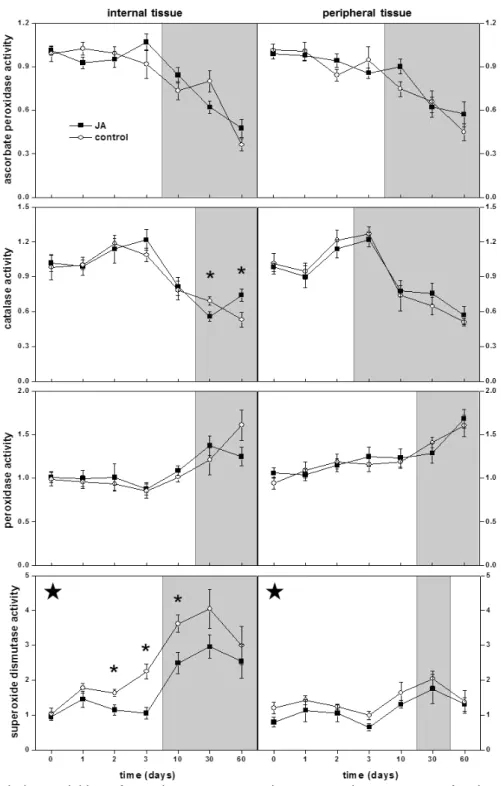
Enzymes that scavenge for ROS were generally unchanged or reduced in activity by postharvest JA treatment of sugarbeet roots throughout the 60 d following treatment (Fig. 1). APX and POD activities were unaltered by JA treatment in root internal and peripheral tissues. CAT activity was unaltered by JA treatment in root peripheral tissue and in root internal tissue for the first 10 d after treatment, but exhibited a small decline and a small increase in activity in internal tissues 30 and 60 d after treatment, respectively. JA treatment reduced SOD activity in both internal and peripheral tissues over the 60 d of the experiment. Overall, SOD activity was reduced in internal and peripheral tissue by 27% and 19%, respectively. No differences between localized and systemic effects of JA on ROS-scavenging enzymes were apparent in sugarbeet roots in this study, since internal and peripheral tissues responded similarly to JA treatment for all enzymes examined.
31 increased 250 and 90% for internal and peripheral tissues, respectively, relative to activity at time of harvest.
Fig. 1. Relative activities of reactive oxygen species-scavenging enzymes after jasmonic acid
3.2 Pathogen-related defense enzyme activities
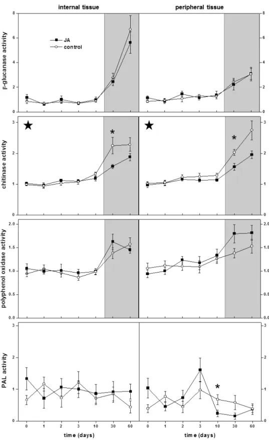
Postharvest JA treatment did not increase the activity of common pathogen-related defense enzymes during the 60 d following treatment (Fig. 2). β-Gluc and PPO activities were unaltered by JA treatment in root internal and peripheral tissues. PAL activity, which was barely detectable in sugarbeet roots (Supplementary Anexo A ), was also unaltered by JA treatment except for a transient decrease in activity occurring 10 d after harvest in peripheral tissue. JA treatment reduced chitinase activity in both internal and peripheral tissues, with the greatest suppression of activity occurring 30 to 60 d after treatment. During this period, chitinase activity was reduced in internal and peripheral tissues by 24 and 27%, respectively. Localized and systemic effects of JA were similar for the defense-related enzymes examined in this study, since enzymes in internal and peripheral tissues responded similarly to JA treatment.
33
Fig. 2. Relative activities of pathogen-related defense enzymes after jasmonic acid (JA)
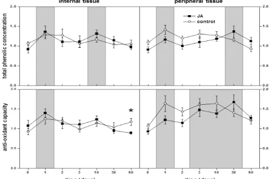
3.3 Total phenolic compounds (TPC) concentration and antioxidant capacity (AOC)
The concentration of antioxidants in sugarbeet roots, as measured by total phenolic compounds concentration and antioxidant capacity, was generally unaltered by postharvest JA treatment of sugarbeet roots during the 60 d following treatment (Fig. 3). TPC concentration and AOC in root internal and peripheral tissues were unaltered by JA treatment, except for a 24% decrease in AOC occurring 60 d after treatment in internal tissue. However, antioxidant concentrations were affected by storage duration in both internal and peripheral tissues. One day after harvest, there was a transient increase in TPC concentration by 33 and 28% and in AOC by 33 and 43%, in internal and peripheral tissues, respectively, relative to their values at harvest. Additional elevations in TPC concentration occurred 10 d after harvest in internal tissue and 30 d after harvest in peripheral tissue, while AOC was additionally elevated in peripheral tissue 3 to 30 d after harvest.
Fig. 3. Relative concentration of antioxidants, as measured by total phenolic compounds (TPC)
35
4 DISCUSSION
Enzymes that scavenge for reactive oxygen species such as APX, CAT, POD and SOD, and metabolites that act as antioxidants have been implicated in jasmonate-induced disease resistance in many plant species and organs (Seema et al., 2003; Yao and Tian, 2005b; Ali et al., 2006; Haggag et al., 2010; Chen et al., 2011). In harvested sugarbeet roots, however, JA treatment generally had no effect on APX, CAT, and POD activities or the concentration of antioxidants in the 60 d following treatment, indicating that these enzymes and metabolites are not directly involved in JA-induced resistance in this crop. SOD activity, in contrast, was reduced by JA treatment. In root internal and peripheral tissues, JA treatment decreased SOD activity an average of 27 and 19%, respectively. Jasmonate-induced reductions in SOD activity have been reported in other plant species and organs (Seema et al., 2003; Ali et al., 2006), and it has been suggested that these reductions contribute to disease resistance by allowing accumulation of O2-, a ROS implicated in defense
against pathogens (Lamb and Dixon, 1997). Whether the relatively minor reduction in SOD activity that was observed in sugarbeet roots could effectively alter O2- accumulation upon pathogen attack and contribute to plant defense is
unknown. Certainly, however, the lack of increase in any ROS-scavenging enzyme or total antioxidants indicates that JA-induced resistance in sugarbeet roots is not due to enhanced protection against oxidative stresses.
commonly induced by SA (Ohme-Takagi and Shinshi, 1990). Nevertheless, the lack of increase in β-Gluc, chitinase, PPO, and PAL activities indicates that these pathogen-related defense enzymes do not contribute to JA-induced disease resistance.
Storage duration significantly altered the activities of all ROS-scavenging enzymes examined in this study, with all changes occurring after 10 d storage. With prolonged storage, activities of APX and CAT declined and activities of POD and SOD increased. Similar storage-related changes in ROS-scavenging enzymes have been observed in other postharvest products. For example, APX activity declined in peach and loquat fruits, CAT activity or gene expression declined in peach, loquat, tomato and kiwi fruits, POD activity increased in loquat, sweet cherry, and kiwi fruits, and SOD activity increased in peach and kiwi fruits during storage (Ding et al., 2002; Yao and Tian, 2005b; Cao et al., 2008; Zhu et al., 2008; Jin et al., 2009a; Cai et al., 2011). The significance of these storage-related changes in ROS-scavenging enzymes is unknown. However, it is evident that the ability to detoxify O2- and H2O2 and
mount ROS-mediated defense mechanisms and signals is altered in sugarbeet root by prolonged storage.
Storage duration also affected the activity of several defense-related enzymes. After 30 and 60 d in storage, β-Gluc, chitinase, and PPO activities
increased in both peripheral and internal root tissues. Similarly, β-Gluc and
chitinase gene expression increased in tomato fruit, chitinase activity increased in banana fruit, and PPO activity increased in loquat and peach fruits during storage (Ding et al., 2002; Cao et al., 2008; Jin et al., 2009b; Tang et al., 2010). The induction of defense-related enzyme activities in stored plant products, however, is unrelated to improved disease resistance since disease resistance declines with storage duration in sugarbeet root and other postharvest products (Bugbee, 1979; Tang et al., 2010).
37 Jin et al., 2009a) and a study that reported increased activities of POD, chitinase, and PPO and total phenolic compounds concentration in jasmonate-treated sugarbeet leaves (Haggag et al., 2010). It is unknown whether the disparities between this and other studies arise from differences between plant species, plant organs, tissue age, or other unidentified factor. However, differential responses to jasmonates among plant species are common (Rohwer and Erwin, 2008) and can occur between plant organs (Parra-Lobato et al., 2009). In sugarbeet root, alterations in the activities of ROS-scavenging enzymes and several pathogen-related defense enzymes were related to prolonged storage, when disease resistance is reduced. The inverse association between these activities and disease resistance further suggests that these enzymes are unlikely to promote disease resistance in postharvest sugarbeet root.
Acknowledgements
5 REFERENCES
Ali, M.B., Yu, K.-W., Hahn, E.-J., Paek, K.-Y., 2006. Methyl jasmonate and salicylic acid elicitation induces ginsenosides accumulation, enzymatic and non-enzymatic antioxidant in suspension culture Panax ginseng roots in bioreactors. Plant Cell Rep. 25, 613-620.
Ballaré, C.L., 2011. Jasmonate-induced defenses: a tale of intelligence, collaborators and rascals. Trends Plant Sci. 16, 249-257.
Bradford, M., 1976. A rapid and sensitive method for the quantification of microgram quantities of protein utilizing the principle of protein-dye binding. Anal. Biochem. 72, 248-254.
Brand-Williams, W., Cuvelier, M.E., Berset, C., 1995. Use of a free radical method to evaluate antioxidant activity. LWT-Food Sci. Technol. 28, 25–30.
Bugbee, W.M., 1979. The effect of plant age, storage, moisture, and genotype on storage rot evaluation of sugarbeet. Phytopathology 69, 414-416.
Cai, Y., Cao, S., Yang, Z., Zheng, Y., 2011. MeJA regulates enzymes involved in ascorbic acid and glutathione metabolism and improves chilling tolerance in loquat fruit. Postharvest Biol. Technol. 59, 324-326.
Cao, S., Zheng, Y., Yang, Z., Tang, S., Jin, P., Wang, K., Wang, X., 2008. Effect of methyl jasmonate on the inhibition of Colletotrichum acutatum infection in loquat fruit and the possible mechanisms. Postharvest Biol. Technol. 49, 301-307.
Chen, Y., Pang, Q., Dai, S., Wang, Y., Chen, S., Yan, X., 2011. Proteomic identification of differentially expressed proteins in Arabidopsis in response to methyl jasmonate. J. Plant Physiol. 168, 995-1008.
39 El-Khallal, S.M., 2007. Induction and modulation of resistance in tomato plants against Fusarium wilt disease by bioagent fungi (arbuscular mycorrhiza) and/or hormonal elicitors (jasmonic acid & salicylic acid): 2-Changes in the antioxidant enzymes, phenolic compounds and pathogen related-proteins. Aust. J. Basic Appl. Sci. 1, 717-732.
Fugate, K.K., Ferrareze, J.P., Bolton, M.D., Deckard, E.L., Campbell, L.G., 2012. Postharvest jasmonic acid treatment of sugarbeet roots reduces rot due to Botrytis cinerea, Penicillium claviforme, and Phoma betae. Postharvest Biol. Technol. 65, 1-4.
Giannopolitis, C.N., Ries, S.K., 1977. Superoxide dismutases: Part I. Occurrence in higher plants. Plant Physiol. 59, 309–314.
Haggag, W.M., Mahmoud, Y.S., Farag, E.M., 2010. Signaling necessities and function of polyamines/jasmonate-dependent induced resistance in sugar beet against beet mosaic virus (BtMV) infection. New York Sci. J. 3, 95-103.
Hung, J.T., Kao, C.H., 2004. Nitric oxide acts as an antioxidant and delays methyl jasmonate-induced senescence of rice leaves. J. Plant Physiol. 161, 43-52.
Jiang, Y.M., Fu, J.R., 1999. Biochemical and physiological changes involved in browning of litchi fruit caused by water loss. J. Hortic. Sci. Biotechnol. 74, 43-46.
Jin, P., Wang, K., Shang, H., Tong, J., Zheng, Y., 2009a. Low-temperature conditioning combined with methyl jasmonate treatment reduces chilling injury of peach fruit. J. Sci. Food Agric. 89, 1690-1696.
Jin, P., Zheng, Y., Tang, S., Rui, H., Wang, C.Y., 2009b. A combination of hot air and methyl jasmonate vapor treatment alleviates chilling injury of peach fruit. Postharvest Biol. Technol. 52, 24-29.
Lamb, C., Dixon, R.A., 1997. The oxidative burst in plant disease resistance. Annu. Rev. Plant Physiol. Plant Mol. Biol. 48, 251-275.
Magalhães, L.M., Santos, F., Segundo, M.A., Reis, S., Lima, J.L.F.C., 2010. Rapid microplate high-throughput methodology for assessment of Folin-Ciocalteu reducing capacity. Talanta 83, 441–447.
Mittler, R., 2002. Oxidative stress, antioxidants and stress tolerance. Trends Plant Sci., 7, 405-410.
Niki, T., Mitsuhara, I, Seo, S., Ohtsubo, N., Ohashi, Y., 1998. Antagonistic effect of salicylic acid and jasmonic acid on the expression of pathogenesis-related (PR) protein genes in wounded mature tobacco leaves. Plant Cell Physiol. 39, 500-507.
Ohme-Takagi, M., Shinshi, H., 1990. Structure and expression of a tobacco beta-1,3-glucanase gene. Plant Mol. Biol. 15, 941-946.
Parra-Lobato, M.C., Fernandez-Garcia, N., Olmos, E., Alvarez-Tinaut, M.C., Gómez-Jiménez, M.C., 2009. Methyl jasmonate-induced antioxidant defence in root apoplast from sunflower seedlings. Environ. Exp. Bot. 66, 9-17.
Policegoudra, R.S., Aradhya S.M., 2007. Biochemical changes and antioxidant activity of mango ginger (Curcuma amada Roxb.) rhizomes during postharvest storage at different temperatures. Postharvest Biol. Technol. 46, 189–194.
Pozo, M.J., Van Loon, L.C., Pieterse, C.J., 2005. Jasmonates-signals in plant-microbe interactions. J. Plant Growth Regul. 23, 211-222.
Rao, M.V., Paliyath, G., Ormrod, D.P., 1996. Ultraviolet-9- and ozone-induced biochemical changes in antioxidant enzymes of Arabidopsis thaliana. Plant Physiol. 110, 125-136.
41 Schneider-Müller, S., Kurosaki, F., Nishi, A., 1994. Role of salicylic acid and intracellular Ca2+ in the induction of chitinase activity in carrot suspension culture. Physiol. Mol. Plant Pathol. 45, 101-109.
Seema, G., Srivastava, M.K., Srivastava, S., Shrivastava, A.K., 2003. Effect of methyl jasmonate on sugarcane seedlings. Sugar Tech. 5, 189-191.
Tang, W., Zhu, S., Li, L., Liu, D., Irving, D.E., 2010. Differential expressions of PR1 and chitinase genes in harvested bananas during ripening, and in response to ethephon, benzothiadizole and methyl jasmonate. Postharvest Biol. Technol. 57, 86-91.
Tarrad, A.M., El-Hyatemy, Y.Y., Omar, S.A., 1993. Wyerone derivatives and activities of peroxidase and polyphenol oxidase in faba bean leaves as induced by chocolate spot disease. Plant Sci. 89, 161-165.
Thaler, J.S., Owen, B., Higgins, V.J., 2004. The role of the jasmonate response in plant susceptibility to diverse pathogens with a range of lifestyles. Plant Physiol. 135, 530-538.
Tripathi, P., Dubey, N.K., 2004. Exploitation of natural products as an alternative strategy to control postharvest fungal rotting of fruit and vegetables. Postharvest Biol. Technol. 32, 235-245.
Wang, K., Jin, P., Cao, S., Shang, H., Yang, Z., Zheng, Y., 2009. Methyl jasmonate reduces decay and enhances antioxidant capacity in chinese bayberries. J. Agric. Food Chem. 57, 5809-5815.
Yao, H.J., Tian, S.P., 2005a. Effects of a biocontrol agent and methyl jasmonate on postharvest diseases of peach fruit and the possible mechanisms involved. J. Appl. Microbiol. 98, 941-950.
Zhang, H., Ma, L., Turner, M., Xu, H., Dong, Y., Jiang, S., 2009. Methyl jasmonate enhances biocontrol efficacy of Rhodotorula glutinis to postharvest blue mold decay of pears. Food Chem. 117, 621-626
43
5 ARTIGO 3
Characterization of the sugarbeet (Beta vulgaris L.) transcriptome,
RNA-sequencing of jasmonic acid differentially expressed genes and JA effect on expression of defense-related genes.
JOCLEITA PERUZZO FERRAREZE(a)
, KAREN KLOTZ FUGATE(b), MELVIN D. BOLTON(c), EDWARD L. DECKARD (c)
, LARRY G. CAMPBELL(c) and FERNANDO L. FINGER (d),
*Corresponding author: Karen Klotz Fugate USDA-ARS, Northern Crop Science Laboratory 1605 Albrecht Blvd., N.
Fargo, ND 58102-2765, USA telephone: (701) 239-1356 fax: (701) 239-1349
email: karen.fugate@ars.usda.gov
(a) Departamento de Biologia Vegetal, Universidade Federal de Viçosa, 36570-000 Viçosa, MG,
Brazil.
(b) USDA-ARS, Northern Crop Science Laboratory, 1605 Albrecht Blvd. N., Fargo, ND
58102-2765, USA.
(c) Department of Plant Sciences, North Dakota State University, P.O. Box 6050, Fargo, ND
58108-6050.
ABSTRACT
45 disease resistance. Identification of genes altered by JA treatment in these studies provides targets for future research into the mechanisms by which JA protects postharvest sugarbeet roots against pathogens.
1 INTRODUCTION
Jasmonates are endogenous plant hormones that activate native plant defense mechanisms (Ballaré, 2011). When applied exogenously, jasmonates have been found to reduce disease caused by a variety of fungal, bacterial and viral pathogens (Thaler et al., 2004; Rohwer and Erwin, 2008; Haggag et al., 2010) by reducing disease severity in most cases and reducing disease incidence in others (Yao and Tian, 2005b; Cao et al., 2008; Zhang et al., 2009). Although some studies have suggested that jasmonate treatment reduces disease by directly limiting antimicrobial activity, and the ability of jasmonates to reduce spore germination, germ tube elongation and mycelial growth has been demonstrated (Yao and Tian, 2005a; Cao et al., 2008), jasmonate treatment is widely believed to protect plants by systemically inducing the same plant defense responses that are activated by endogenous jasmonates (Tripathi and Dubey, 2004; Pozo et al., 2005).
Changes in expression of defense-related genes is a key element in inducible defense mechanisms in plants (Ryder et al., 1984; Edreva, 2005). Changes in expression of phenylalanine ammonia-lyase (PAL), peroxidase (POD), pathogenesis related 1 (PR 1), and chitinases genes are commonly related to jasmonates-induced defense in several plants (Sharan et al., 1998; Curtis et al., 1997; Reymond and Farmer 1998; Ding et al., 2002).
Understanding the transcriptome is essential for interpreting the functional elements of the genome and revealing the molecular constituents of cells and tissues, and also for understanding development and disease (Wang et al., 2009). High-throughput RNA sequencing (RNA-seq) is an efficient and reliable platform for transcriptomic analysis in non-model organisms. The large number of sequences generated provides valuable sequence information at the transcriptomic level for novel gene discovery, or for the investigation molecular mechanisms (Wang et al., 2009; Wei et al., 2011).
47 Fugate et al., 2012). In sugarbeet roots, there is no information available on the effect of jasmonic acid (JA) on the expression of defense-related genes.
2 MATERIAL AND METHODS
2.1 Plant material and JA treatment
Sugarbeet hybrid VDH66156 (SESVanderHave, Tienen, Belgium) was greenhouse grown in Sunshine Mix #1 (Sun Gro Horticulture, Vancouver, BC, Canada) in 15-L pots with supplemental light under a 16 h light/8 h dark regime. Taproots were harvested 16 - 18 weeks after planting, all leaf and petiole material was removed, and roots were gently washed to remove potting media. Roots were submerged in 0 or 10 µM JA (Cayman Chemical, Ann Arbor, MI) for 1 hr at room temperature, then incubated at 20 °C and 90 % relative humidity for up to 60 d in a controlled environment chamber (Conviron, model MTR30, Winnipeg, MB, Canada). Root samples were collected at 0, 1, 2, 3, 10, 30 and 60 d post-treatment by collecting tissue from the main portion of the root, free of crown or tail tissue, with the epidermis and approximately 2 mm of subepidermal tissue excluded. Samples were flash frozen in liquid N2,
lyophilized, ground to a powder, and stored at -80 °C until analysis. Individual roots were the experimental unit with 4 replicates per treatment per time point. Experiment was repeated three times.
2.2 RNA extraction and qRT-PCR
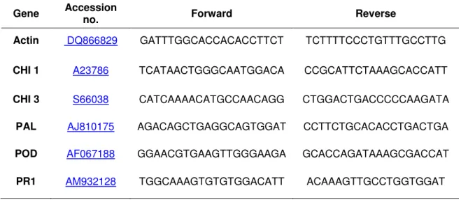
49 transcriptases. Quantitative real-time PCR (qPCR) was used to determine JA-related changes in transcript levels of genes for a chitinase 1 (CHI 1), a chitinase 3 (CHI 3), a phenylalanine ammonia-lyase (PAL), a cell wall peroxidase (POD), a pathogenesis-related protein 1 (PR1), using an actin (ACT) gene as a reference (Table 1). qPCR was performed on a MJ Research (Watertown, MA, USA) PTC-200 thermal cycler, equipped with a Chromo 4 real-time detector (Bio-Rad Life Science Hercules, CA, USA) using Power SYBR Green PCR Master Mix (Applied Biosystems, Foster, CA, USA) and the primer pairs listed in Table 1. For qPCR, samples were denatured for 2 min at 95°C and amplified during 39 cycles of 15 s at 95°C and 45 s at 60°C. Melting curves were used to confirm that single products were amplified. Primer efficiency (E) of each primer pair was determined by analyzing standard curves of Ct values generated by PCR amplification of 5-fold serial dilutions of the cDNA synthesized from each treatment. Primer efficiency was calculated according to the equation E = (10-1/slope-1). The relative quantification of real-time PCR results was performed using the mathematical model of Pfaffl (Pfaffl, 2001).
Table 1. Sugarbeet genes quantified by qPCR analysis, their accession numbers and primer
pairs used to effect their amplification. Genes analyzed include those encoding a chitinase 1 (CHI 1), a chitinase 3 (CHI 3), a phenylalanine ammonia-lyase (PAL), a cell wall peroxidase (POD), a pathogenesis-related protein 1 (PR1) and an actin (ACT). Actin served as a reference gene in these studies.
Gene Accession no. Forward Reverse
Actin DQ866829 GATTTGGCACCACACCTTCT TCTTTTCCCTGTTTGCCTTG
CHI 1 A23786 TCATAACTGGGCAATGGACA CCGCATTCTAAAGCACCATT
CHI 3 S66038 CATCAAAACATGCCAACAGG CTGGACTGACCCCCAAGATA
PAL AJ810175 AGACAGCTGAGGCAGTGGAT CCTTCTGCACACCTGACTGA
POD AF067188 GGAACGTGAAGTTGGGAAGA GCACCAGATAAAGCGACCAT
2.3 Reference transcriptome generation
A reference transcriptome for sugarbeet was generated by high-throughput, paired-end RNA sequencing using a pooled RNA sample. The pooled sample contained equivalent weights of RNA extracted from young newly developed leaf tissue, fully expanded leaf tissue, root tissue 5 weeks after planting, root tissue 16 weeks after planting, root tissue after 60 d postharvest storage, root tissue 2 d after a 10 μM JA postharvest treatment, and root tissue 2d after a postharvest 1 mM salicylic acid treatment. Roots subjected to storage, JA treatment or SA treatment were harvested 16 weeks after planting and maintained at 20 oC and 90% relative humidity after harvest. Sequencing
was performed on an Illumina, Inc. (San Diego, CA, USA ) HiSeq 2000 system by BGI Americas (Cambridge, MA, USA). Transcriptome assembly was carried out using SOAPdenovo assembly software (Li et al., 2009). BLASTx alignment
(e-value < 0.00001) of unigenes with protein sequences available in NCBI’s nr,
Swiss-Prot, KEGG and COG databases was used for function, biological pathway, cellular location, and biological process annotation.
When a Unigene happened to be unaligned to none of the above databases, a software named ESTScan (Iseli et al., 1999) was introduced to decide its sequence direction.
2.4 Unigene Annotation
51
2.5 RNA-sequencing of JA treated samples
RNA sequencing was performed on roots treated with 0 and 10 µM JA and collected at 2 and 60 days after treatment. Roots were harvested 16 weeks after planting and maintained at 20 oC and 90% relative humidity after harvest. Sequencing was performed on an Illumina, Inc. (San Diego, CA, USA) HiSeq 2000 system by BGI Americas (Cambridge, MA, USA).
Clean reads were mapped to reference sequences using SOAPaligner/soap2 (Li et al., 2009). Mismatches no more than 2 bases were allowed in the alignment. Statistical analysis was conducted to summarize the number of clean reads that align to the reference genome/genes, which provides the general information of the project. During the RNA-Seq experiment, mRNA are firstly broken into short segments by chemical methods and then sequenced. If the randomness is poor, reads preference from specific gene region will directly affect subsequent bioinformatics analysis. We use the distribution of reads locating on the genes to evaluate the randomness. Since reference genes have different lengths, the reads location on gene is standardized to a relative position (which is calculated as the ratio between reads location on the gene and gene length), and then the number of reads in each position is counted. If the randomness is good, the reads in every position would be evenly distributed. The gene expression is calculated by the numbers of reads mapped to the reference sequence and every gene. The gene expression level is calculated by using RPKM (Mortazavi et al., 2008) method (Reads Per kb per Million reads). The RPKM method is able to eliminate the influence of different gene length and sequencing discrepancy on the calculation of gene expression. Therefore, the calculated gene expression can be directly used for comparing the difference of gene expression among samples. If there is more than one transcript for a gene, the longest one is used to calculate its expression level and coverage.
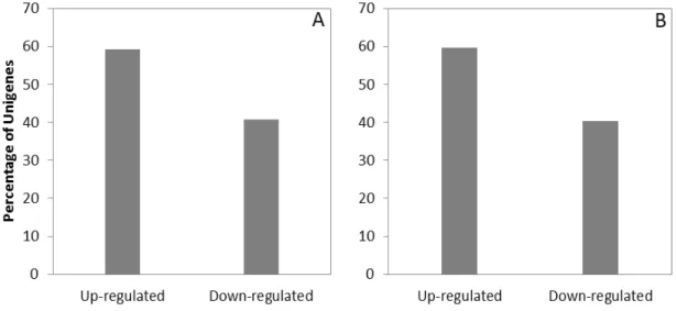
3 RESULTS
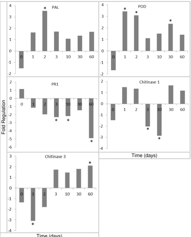
3.1 Defense-related genes expression
53
Fo
ld Re
gu
latio
n
Time (days)
Time (days)
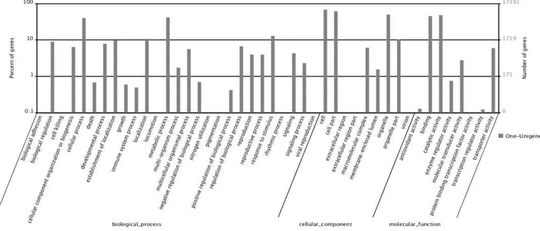
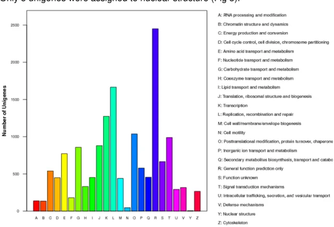
3.2 Gene ontology and clusters of orthologous groups of the RNA library
55
Fig. 2. Gene ontology classification of assembled unigenes of sugarbeet transcriptome. The unigenes were annotated to three categories: biological processes,