http://www.bipublication.com
Research Article
Study of carcinogenic activity of carnosic acid and rosmarinic acid in
cancer cells line of Hep- G2
Narges Njarpour1* and Masoud Mashhadi Akbar Boojar2
1
MSc studentof Biochemistry-Molecular Cell Biology, Department of Cell and Molecular Biology, Faculty of Biological Sciences, Kharazmi University, Tehran, Iran.
*narges_najarpour@yahoo.com, Code: 15719-14911Tel/Fax: +98-21-88848940
2
Associate Professor, PhD. of Biochemistry, Department of Cell and Molecular Biology, Faculty of Biological Sciences, Kharazmi University, Tehran, Iran . Mboojar@yahoo.com
ABSTRACT
Background: As anti-apoptotic properties of polyphenols have been proved in past years. In this study, carcinogenic activity of carnosic acid and rosmarinic acid inhuman hepatocellular carcinomaHep- G2cellswere studied.
Method:In this experimental study, Hep-G2 cells were cultured in DMEM supplemented containing bovine
fetal serum and antibiotics. Cells with double dilution were then cultured from 0 to 70 µM for 24 h and viability
of cells was determined by MTT method. In order to evaluate activity of caspase3 and caspase9 enzymes after 24 hours of incubation of the cells with treated materials, cells were centrifuged and cell lysis solution was added. This mixture was centrifuged and then an appropriate substrate was added into each enzyme and after incubation, absorbance of the resulted solution was read at 405 nm using a spectrophotometer device. To measure the level of ceramide, a recombinant acid ceramidase enzyme and naphthalene-2, 3-dialdehyde, which is fluorescent and is connected to sphingosine resulted from acid ceramidase, were added to the cell extract and was ultimately determined by HPLC.
Results: Carnosic acid increased cell viability and caused noinduction of apoptosis to a dose-dependent species in Hep-G2 cells by reducing ceramide levels and decreasing activity of caspase 3 and caspase 9 enzymes.
Rosmarinic acid in concentrations of up to 50 µM decreased cell viability by increasing ceramide levels and
activity of caspase 9 enzyme.also this substance in concentrations of up to 40 µM caused increasing activity of caspase 3 enzyme.
Conclusion: Although in most cases, polyphenols have caused induction of apoptosis and have decreased cell viability percentage. However in some cases, they have affected inversely and have caused cell growth.
Keywords:Carnosic acid, Rosmarinic acid, Ceramide, Cell line Hep-G2, Caspase3 and 9.
INTRODUCTION
Hepatocellular carcinoma (Hcc) is a type of liver cancer, which often occurs after infection with hepatitis B and C viruses and cirrhosis that is due to alcohol consumption in most cases. Hepatocellular carcinoma like other cancers occurs when a mutation occurs in the cellular machinery and leads to uncontrolled cell proliferation or it occurs as a result of cell avoiding apoptosis phenomenon (V. Kumar et
carnosic acid, which are in rosemary plant (Rosmarinusofficinalis.L), have special properties such as anti-cancer, anti-inflammatory and antioxidant properties (J.M.Kyoung et al., 2014; Md. ShahadatHoosa et al., 2014).
In recent research, it has been found that there is a relation between increasing levels of total ceramide in cancerous and malignant tumor tissue (M.Levy., 2010). Accordingly, this study is aimed to study whether rosmarinic acid and carnosic acid have an effect on Hep-G2 cell viability and if they are effective, dose the change in the metabolism of ceramides by these two substances explain effect of these substances on metabolism of these cells.
MATERIALS &METHODS
In this study, two substances of rosmarinic acid and carnosic acid were purchased from Sigma-Aldrich and concentrations of 0, 10, 20,
30, 40, 50, 60 and 70 µM of these two
substances were prepared and were cultured in 96-well culture plates with 5000 cells for 24 h and the effects of extracts on cell viability was determined by MTT test.
Cell Culture: Human hepatoma cell line(Hep-G2) were prepared from Razi vaccine and serum research institute (Hesarak, Iran). Then, they were cultured in DMEM mediumwith 5% fetal bovine serum (FBS), penicillin and streptomycin, the cells were incubated at 37 °C, humidity 90% and 5% of CO2 After 24
hours, the cells were exposed to different concentrations of two compounds, at the same time, triple control samples without treatment was considered.
Study of cells viability:Cells viability was tested by MTT [3(4,5dimethylthiazol2yl) -2, 5-diphenyl-2H-tetrazolium bromide] (Q.Xiang et al., 2010). Briefly, cells were plated at 5000 cells per 96-well culture plates and were treated with different concentrations of two compounds of rosmarinicacid and carnosic acid.The supernatant medium was discarded. The cells were placed in a solution of MTT (5 ml in PBS) for 4 h and after
preparing a solution offormazan by 100 µl of
DMSO, absorbance was read at 570 nm using a spectrophotometer.
Evaluation of caspse-3 enzyme activity:To measure the activity of caspase 3 enzyme, after 24 h of incubation of the cells with the treated material and centrifugation, cell lysis buffer was added to samples. Then the lysate mixture was centrifuged. A solution containing AC-DEVD-PNA substrate was added to the supernatant solution and incubated. Absorbance of supernatant solution resulting from separation of p-nitroanilide was measured at 405 nm.
Evaluation of caspse-9 enzyme activity:To measure the activity of caspase-9 enzyme, after 24 hours of incubation of the cells with the treated material and centrifugation, cell lysis buffer was added to samples. Then the lysate mixture was centrifuged.
A solution containing AC-DEVD-PNA substrate was added to the supernatant solution and incubated. Absorbance of resulted supernatant solution was measured at 405 nm. Evaluation of cellular ceramide level:To measure the level of ceramide, the recombinantacidceramidase enzyme was first added to cell extract that as a result, ceramide in the sample was fully hydrolyzed and converted to sphingosine. A special substance called naphthalene-2,3-dialdehyde (NDA), which is fluorescent, was connected to sphingosine. Sphingosine-NDA was then separated from cell extracts by reverse phase HPLC and was determined in fluorescence detector. Ceramide level is exactly as same as sphingosine obtained by HPLC in the same sample.
Statistical analysis: Each test was repeated three times and comparison of average data with the control and treatments with each other was obtained using SPSS software (ANOVA test).
RESULTS
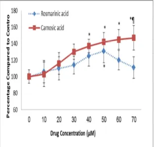
Under treatment with carnosic acid, cell viability was enhanced with increasing concentration of this treatment. In rosmarinic acid treatment, a reduction in percentage of cell viability was observed by increasing the
concentration from 50 to 70 µM.
Results of MTT color measurement by measuring optical density (OD) based on the concentration of used extracts compared with the molecular amplification rate were obtained as a plot (Figure 1).
Figure 1. The effect of various concentrations of two studied substance on Hep-G2 cells viability after 24 h of incubation.
* Significant difference compared to control (P <0.05); ¶ significant difference of a treatment compared to other treatment (P <0.05).
The effect of two treatments of rosmarinic acid and carnosic acid on caspase3 enzyme activity:carnosicacid treatment of Hep-G2 cell line reduced caspase3 enzyme activity in a dose-dependent trend.In rosmarinic acid treatment, it was observed that the enzyme activity of caspase3 following a downward trend was increased with increasing
concentration of treatment from 40 to 70 µM.
The results of evaluation of caspase3 enzyme activity under two treatments of rosmarinic acid and carnosic acid are seen in Figure
2.Figure 2.Caspase3 enzyme activity in Hep-G2 cells line treated with various concentrations of two substances of rosmarinic acid and carnosic acid.
* Significant difference compared to control (P <0.05); ¶significant difference of a treatment compared to other treatment (P <0.05).
Evaluation of caspase9 enzyme activity:Carnosic acid treatment of Hep-G2 cells reduced caspase9 enzyme activity in a dose-dependent trend.
In rosmarinic acid treatment, it was observed that the enzyme activity of caspase9 following a downward trend was increased with
increasing concentration from 50 to 70 µM.
Figure 3 demonstrates the results of evaluation of caspase9 enzyme activity.
Figure 3.Caspase9 enzyme activity under treatment with two substances of rosmarinic acid and carnosic acid in Hep-G2 cells line.
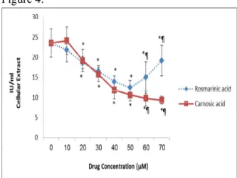
Evaluation of ceramide level in Hep-G2 cells:Carnosic acid treatment of Hep-G2 cells reduced ceramide level in a dose-dependent trend. In addition treatment of these cells with rosmarinic acid following a downward trend showed an increasing trend from concentration
of 50 µM. The results of evaluation of ceramide level in Hep-G2 cells are observed in Figure 4.
Figure 4.Ceramide level in Hep-G2 cells line treated with various concentrations of rosmarinic and carnosic acid.
* Significant difference compared to control (P <0.05); ¶ significant difference of a treatment compared to other treatment (P <0.05).
DISCUSSION
The sphingomyelin/ceramide signaling pathway is an evolutionary system conserved from yeast to humans ( M. Nardini et al., 2001). Function of ceramide has been reported as a secondary messenger in this pathway and inducing different responses depending on the type of cells including proliferation, distinction, growth arrest or mostly apoptosis (M .Nardini et al., 2001). In order to better understand the effective role of polyphenols in many pathological conditions involved in ceramide pathway (M.Nardini et al., 2001), the effects of rosmarinic acid and carnosic acid on Hep-G2 cell line have been studied in this study. Carnosic acid has increased Hep-G2 cells growth by increasing the dose. Rosmarinic acid afterincreasingtrend has increased Hep-G2 cells growth from concentration of 50 µM. Hep-G2 cells growth
can be attributed to the reduction of ceramidelevels (M.Nrddini et al., 2001). Caspase are a family of cysteine proteases that are central regulators of apoptosis. Among caspase family members, caspase3 is one of important performer caspases in running apoptosis. Activation of caspse3 is considered as a conventional marker of apoptotic cells. Activation of performer caspases such as caspase3 and caspase7 can cause disintegration of nuclear cytoplasmic substrates and fragmentation of DNA (Q.Xiang et al., 2015). In this study, it has also found that carnosic acid in a dose-dependent trend has reduced activity of caspase3 enzyme and as a result, cell viability of Hep-G2 cells has been increased.
Rosmarinic acid has increased enzyme activity after a decreasing trend from concentration of 40µM that seems is due to decreasingcell viability because of an increase in ceramide levels and cell’s tendency toward the apoptotic process. In this study, the activity of the caspse9 enzyme, which is an initiator or signaling caspase and its main duty is activating performer caspases(C.Kohler et al., 2003), has been decreased with increasing concentration of carnosicacid that reduction of activity of caspase9 is also because of increasing the percentage of cell viability. As a result of treatment by rosmarinic acid for active caspase9 as caspase3 is obtained but 50
µM concentration.
CONCLUSION
Although in most cases, polyphenols have resulted in induction of apoptosis and decreased cell viability, but in some cases they have inversely affected and caused cell growth.
REFERENCES
1. Abdullaev.F.(2001).Cancer
chemopreventive and tumorical properties of saffron (Crocus sativus L.).Mexicocity,
Mexico:Laboratory of Experimented Oncology.National Institute of Pediatncs. 2. Abdullaev.F.V, V.Rotenburd-Belacortu.(
2000).Saffron as chemopreventive agent. In: Abdullaev FI. Riveron-Negretts L. Rotenburd-Belacortu V, Kasumov FJ, eds. Food of 21st century: Food and resource technology environment. China: Ligh Industry Press, 185-95.
3. Goni, F. M.,Alonso,
A.(2002).Sphingomyelinases:
enzymology and membrane activityFEBS Letters. 531, 38-46.
4. KyoungJ.M.,Kyong, j. j.,Taeg,
K.K.(2014).Carnosic acid induces
apoptosis through reactive oxygen
species-mediated endoplasmic reticulum stress induction in human renal carcinoma Caki cells. Journal of Cancer Prevention. 19, 170–178.
5. Kohler, C., Orrenius, T.Zhivotovsky, B.(2002).Evaluation of caspase activity in apoptoticcells. J. Immun Methods. 265, 97-110.
6. Kumar, V., Fausto, N., Abul, A.(2003).Robbins &Cotran pathologic basis of disease. Philadelphia: W.B. Saunders.: 914–7.
7. Levy Michal &Futerman Anthony H. Mammalian ceramide synthases. IUBMB Life.2010; 62: 347-56.
8. Nardini, M., Leonardi, F., Scaccini, C., Virgili, F. (2011). Modulation of ceramide-induced NF-KB binding activity and apoptotic respons by ceffeic acid in U937 cells: comparison with other
antioxidants. Free radical biology. 30(7), 722-33.
9. Park, J.H., Schuchman, E. H. (2006). Acid ceramidase and human disease,BiochimicaetBiophysicaActa. 1758, 2133–2138.
10. Pavoine, C.,Pecker,F. o. (2009).Sphingomyelinases: their regulation and roles in cardiovascular pathophysiology. Cardiovascular Research.82, 175–18.
11. Shahadat, H. Md., Rahman Sh., Anwarul, B. A.B.M., Jahan R., Al-Nahain A., Rahmatullah M., Al-Nahain A., Rahmatullah, M,.(2014).Rosmaric acid: a review of its anticancer action.World journal of pharmacy and pharmaceutical sciences. 3(9), 57-70.