Jasmonic acid elicits oxidative defense and
detoxification systems in
Cucumis melo
L. cells
eetezaz nafie*, tahany hathout, al shyma al Mokadem
Botany Department, University College of Women’s for Art, Science and Education, Ain Shams University, Cairo, Egypt.
* Corresponding author: e-mail: [email protected]; Tel: +202-22736361 Received: 20 November 2010; Accepted: May 09 2011
abstract
This study investigated whether a jasmonic acid (JA) elicitation strategy developed in a conventional cell suspension culture could evoke melon resistance mechanisms, including secondary metabolite production. Twenty one day cultured melon cell suspensions grown in MS1 medium were supplemented with JA at the concentrations of 0.5, 5.0 and 10 µmol. Melon cultures were sampled 24, 48 and 72 h post elicitation to evaluate different defense related factors such as antioxidant enzymes, ascorbate metabolism and phenolic compounds. Results suggest that melon cells respond to JA reprogramming the primary and secondary metabolism which will result in melon plantlets with enhanced resistance against diverse stress conditions through the production of specific bioactive molecules. Jasmonic acid elicited melon cells exhibited enhanced oxidative enzymes activities and ascorbic acid, coumarin and p-coumaric amounts were found without growth retardation. Induced intracellular JA functions as a signal transducer acting upstream
to H2O2, which is a secondary messengertriggering jasmonic signaling cascades by activating certain late genes that regulate the
activity of catalase, peroxidase and de novo synthesis of five isozymes, ascorbic peroxidase detoxifying enzymes concomitant with
ascorbate compound. Secondary metabolite production in melon cells seems to be activated upon JA exposure suggesting that this cell culture could be used as a source for rapid and increased production of coumarin, p-coumaric, ascorbic acid and likely other specific phenylpropanoids. These data provides further evidences for a role of jasmonic acid in the intracellular signal cascade that results in the accumulation of secondary compounds and ultimately induced melon resistance. This approach could assist further in understanding the metabolic mechanisms operating in melon cells under stress, and thus how to control them.
Key words: ascorbic metabolism, detoxification enzymes, peroxidase isozymes, Phenolics profile.
introduction
Melon (Cucumis melo L.) is an important vegetable
crop that is widely cultivated in South East Asia, China, East Africa and throughout the tropical and subtropical regions
(Yadav et al., 1996). The flesh fruit is a significant source
of carbohydrates and water and the seeds are rich in oil and protein (Martyn and Miller, 1996). Melons are also considered an important source of ascorbic acid, folic acid, and potassium (Richter, 2000).
There are many potential sources of reactive oxygen species (ROS) formation in plants, some are reactions involved in normal metabolism, such as photosynthesis and respiration, other sources belong to pathways enhanced during biotic and abiotic stresses. In contrast to O2, ROS are
that disrupt the cellular homeostasis of cells enhance the production of ROS and induce specific oxidative responses. This enhanced production of ROS can pose a threat to cells but it is also thought that ROS act as signals for the activation of several stress-responses and defense pathways (Knight and Knight, 2001). Thus, ROS can be viewed as cellular indicators of stress as well as elicitors of stress-response signal transduction pathways.
Low molecular weight antioxidants, such as ascorbate, glutathione, and tocopherol, are information-rich redox buffers that interact with numerous cellular components. In addition to crucial roles in defense and as enzyme cofactors, these cellular antioxidants can also influence plant growth and development (Potters et al., 2004; Tokunaga et al., 2005). Notably, antioxidants provide essential information on cellular redox state, and they influence gene expression associated with biotic and abiotic stress responses to maximize the plant defense. Increasing evidence suggests a model for redox homeostasis in which the ROS–antioxidant interaction acts as a metabolic interface for signals derived from metabolism and from the environment. This interface modulates the appropriate induction of acclimation processes or, alternatively, execution of cell death programs. further evidences point to a pivotal role for the ascorbic-glutathione (AA-GSH) cycle that scavenges ROS, and
particularly H2O2. Its activity relies on the sequential
oxidation and re-reduction of AA and GSH in reactions catalyzed by the enzymes constituting the cycle, namely ascorbate peroxidase (APX), monodehydroascorbate reductase (MDHAR), dehydroascorbate reductase (DHAR), and glutathione reductase (GR) (Noctor and foyer, 1998). In plant cells this cycle has been shown to operate in all organelles in which ROS detoxification is needed (Jimenez et al., 1997; Asada, 1999).
Apart from its function directly to detoxify ROS, the AA-GSH cycle is also involved in redox sensing and signaling (Pastori and foyer, 2002). Hence, the specific interplay between ROS and the AA-GSH cycle constituents could generate compartment-specific changes in both the absolute concentrations of ROS and the antioxidant compounds, and in ascorbate and glutathione redox ratios. Under stress conditions these redox signals could interfere with signaling networks complementary to the antioxidant system and regulate defense gene expression (Vranova
et al., 2002; Kiddle et al., 2003), thus coordinating the necessary readjustments in the redox-regulated plant defense to overcome the oxidative stress.
Although most of the AA is localized in the cytoplasm, certain amount is exported and localized in the apoplast (Noctor and foyer, 1998). Apoplastic AA is thought to represent the first line of defense against potentially damaging external oxidants, and may play an important role in mediating response to stresses generating an enhanced oxidative burden (Barnes et al., 2002; Pignocchi and foyer, 2003). In the apoplast, ascorbate oxidase oxidizes AA to the unstable radical monodehydroascorbate, which rapidly disproportionate to yield dehydroascorbate and AA (Smirnoff, 2000).
Jasmonic acid, and its derivatives are a family of important signal transducers, that can efficiently stimulate secondary metabolism in plant cells (Gundlach et al., 1992;
Naill and Roberts, 2005; Wang et al., 2005). Since the first
report regarding the effect of methyl jasmonate (MJ) on the accumulation of secondary metabolites in plant cell cultures (Gundlach et al., 1992), more than 100 plant species have been demonstrated to respond to the addition of MJ to the culture medium by accumulating secondary metabolites
(Haider et al., 2000). Regarding the inducible mechanisms of
Material and Methods
This investigation was carried out from 2005 to 2009 at Gene Transfer Lab (GTL), and University College of Women’s for Arts, Science and Education, Botany Department.
Material: Melon seeds (Cucumis melo L.) Egyptian cultivar Shahd El-Dokki was obtained from the Horticulture Research Institute, ARC,Giza, Egypt. Tissue culture chemicals: Hormones, jasmonic acid (JA) and other chemicals were purchased from Sigma and fisher group.
biochemical and Molecular
responses of Jasmonic acid
Melon regeneration protocol: As previously described in (Nafie et al., 2009) three week-old-MS1 (MS medium
supplemented with (2 mg-1L 2, 4-D; 0.1 mg-1 L BA and 30
g-1L sucrose) melon cell suspension culture was elicited by
adding JA at different concentrations (0.5, 5 and 10 µmol), melon cells were harvested after 24, 48, and 72 hours post elicitation. In the case of control cultures, 10 µL of 70% ethanol was added to the three week-old-MS1 cell culture and cells were harvested at the same time intervals. Melon biochemical and molecular responses relevant to plant cell defense were assessed in JA-treated and control cells. The medium in which cells had been grown was also investigated by gradient elution high performance liquid chromatography (GC- HPLC).
preparation of extracts and assay of antioxidant enzymes: for determination of antioxidant enzyme activities, lyophilized melon tissues (0.5 g) were homogenized in 10 mL of respective extraction buffer in a prechilled mortar and pestle under liquid nitrogen. The extract was immediately centrifuged at 19,000 x g for 20 min at 20ºC. The supernatant was then used immediately for measuring the following enzymes activities.
catalase enzyme extraction and enzyme activity assay: Catalase (EC 1.11.1.6) was extracted following Jiang and Zhang (2002) method in which 10 mL of 50 mmol potassium phosphate buffer (pH 7.0) containing 1 mmol ethylene diamine tetra acetic acid (EDTA) and 1% polyvinylpyrrolidone (PVP) was the extraction media. After filtration and centrifugation, the supernatant was used as crude
enzyme extract. Catalase activity was estimated following Aebi (1984) protocol in which the reaction mixture was: 50
mmol potassium phosphate buffer (pH 7.0), 10 mmol H2O2
and 200 µL of enzyme extract in a final 3 mL volume. Reaction was left for 10 min then stopped by adding 1 mL of 1 mol HCl
as reported by Yingsanga et al. (2008). Catalase activity was
estimated spectrophotometrically (JENWAY 6305 UV/VIS) at
240 nm, expressed as mmol H2O2 decomposed min-1 g-1 fW.
soluble and cell wall-bound peroxidase enzymes extraction and activity assay: Soluble and cell wall-bound peroxidase (POD) (EC 1.11.1.7) was extracted following Jiang et al. (1984). Lyophilized melon tissues (0.5 g) were homogenized in cold extraction buffer containing 10 mL of 100 mmol sodium phosphate buffer (pH 7.0) and 0.5 g polyvinyl polypyrrolidone (PVPP). After centrifugation, supernatant was considered crude enzyme. Cell–wall bound was extracted by washing pellet twice with same previous phosphate buffer, twice with water and, then shacked in 2% Triton X-100 for 1 h at 4 °C, then again rinsed five times with
water. Pellet after that was treated with 1 mol CaCl2 with
gentle shaking for 12 h. After centrifugation, the supernatant
was used for cell wall-bound POD activity assay.Peroxidase
activity was measured following Ranieri et al. (1995) method .Reaction mixture consisting of 50 µL enzyme extract, 20 mmol guaiacol and 100 mmol sodium phosphate buffer, pH 7.0 in 3 mL, total volume. Reaction was initiated by 20 µL
H2O2 addition and terminated after 10 min by adding 1 mL of
1 mol HCl as reported by Yingsanga et al. (2008). POD activity was determined by measuring the increase in absorbance at
470 nm and expressed as Δ min−1gm−1.
peroxidase isozyme analysis
isozymes extraction: Peroxidase isozymes were extracted from lyophilized samples by homogenizing tissues sampled from different treatments in extraction buffer contains: 150 mmol Tris HCl (pH 6.8) and 30% glycerol. The extract was then transferred into clean Eppendorf tubes and centrifuged at 10000 rpm for 5 min. Then supernatant was transferred to new clean Eppendorf tubes and kept at
–20oC. Peroxidase isozyme assay native polyacrylamide gel
electrophoresis (PAGE) was done similar to Yamunarani et al.
gel preparation and sample application: Separating
gel 12% was prepared by mixing 3.3 mL H2O, 4 mL of
30% acrylamide, 2.5 mL of 1.5 mol Tris HCI pH (8.8), 100 µL (10%) ammonium persulfate and 4 µL N,N,N,N -Tetramethylethylene diamine (TEMED). Stacking gel 5%
was prepared by mixing 3.4 mL H2O, 0.83 mL of 30%
acrylamide, 0.63 mL of 0.5 mol Tris HCI pH (6.8), 50 µL ammonium persulfate (10%) and 10 µL TEMED and then poured over the separating gel. A volume of 40 µL extract from each sample was mixed with 10 µL bromophenol blue, and then 40 µL from this mixture was applied to each well. The run electrode buffer (0.1 mol potassium phosphate buffer, pH 5.0) was added to both lower and upper tanks. The apparatus was connected to the power supply. Electrophoresis apparatus was attached to circulating cool system during running which last 3-4 h.
isozyme assay:Peroxidase (POD) isoforms were then visualized by incubating the gel under dark condition in freshly prepared diaminobenzidine solution (10 mg in 20 mL 0.05 mol phosphate buffer, pH 5.0) for 5-10 min, then the gel washed twice with water and incubated carefully with 10 µL of 30% H2O2 in 20 mL of the 0.05 mol phosphate buffer, pH 5.0 until
the POD activity-containing brown bands visualized.
polyphenol oxidase enzyme extraction and activity assay: Lyophilized cells were homogenized in cold extraction buffer of 100 mmol sodium phosphate buffer pH (7.0) and 0.5 g PVPP for polyphenol oxidase (PPO)(EC 1.10.3.1) for extraction as described by Luh and Phithakpol (1972). The homogenate was filtered then centrifuged and the supernatant was considered the crude enzyme extract. Enzyme activity
in the supernatant fraction was determined at 25°C within
6 h after preparation. for the PPO enzyme assay, 1ml of supernatant was mixed with 1 mL of sodium phosphate buffer (100 mmol, pH 7.0) and 1 mL catechol 50 mmol. Reaction was terminated after 10 min as reported by Yingsanga et al. (2008). Activity was measured and expressed as Δ A410
min−1g−1 fW.
phenylalanine ammonia-lyase extraction and activity assay: Melon cells were homogenized in extraction buffer contains 0.1 mol borate buffer, pH 7.0 and 0.1 g PVP. The homogenate after filtration and centrifugation was collected and used for enzyme assay. Phenylalanine ammonia-lyase (PAL) (EC 4.3.1.24) activity was determined spectrophotometrically by assaying the rate of conversion of L-phenylalanine to
trans-cinnamic acid at 290 nm, as described by Dickerson et al. (1984). Reaction mixtures consisted of 0.4 mL of enzyme extract, 0.5 mL of 0.1 mol borate buffer pH 8.8 and 0.5 mL of 12 mmol phenylalanine in the same buffer. The reaction mixtures were incubated for 15 min at 30°C in a water bath and reaction was terminated and stopped as described by
Yingsanga et al. (2008). In reference cell, 0.4 mL of enzyme
extract was taken along with 1.0 mL borate buffer. The amount of trans-cinnamic acid synthesized was calculated using a calibrated sample. Enzyme activity was expressed µmol trans-cinnamic min-1g-1 fW.
ascorbate oxidase extraction and activity assay: Ascorbate oxidase (AO) (EC 1.10.3.3) enzyme extraction was done following Oberbacher and Vines (1963) method. Lyophilized melon cells were ground in extraction buffer (0.1 mol potassium phosphate, pH 7.0) on ice, then filtered to discard any remaining tissues, homogenate was centrifuged and the supernatant was used as crude enzyme extract.
Enzyme activity assay was determined according to
Esaka etal. (1988). The reaction mixture contained 20 mmol
K-phosphate buffer (pH 7.5) and 2.5 mmol ascorbic acid. The reaction was initiated with the addition of enzyme extract and left for 10 min then terminated as described by Yingsanga et
al. (2008). Enzyme activity was measured by the decrease
in absorbance at 265 nm due to ascorbate oxidation and
expressed as mmol oxidized ascorbate min-1 g-1 DW.
ascorbate peroxidase extraction and activity assay: Soluble proteins were extracted by homogenizing the lyophilized cell tissues in 10 mL of 50 mmol potassium phosphate buffer (pH 7.0) containing 1 mmol EDTA and 1% polyvinylpyrrolidone (PVP) with the addition of 1 mmol ascorbic acid (ASC). The homogenate was filtered and centrifuged, and then the supernatant was used for enzyme assay (Jiang and Zhang, 2002). Ascorbate peroxidase (EC 1.11.1.11) activity was measured by monitoring the decrease in absorbance at 290 nm. The assay reaction mixture contained 50 mmol potassium phosphate buffer (pH 7.0), 0.5 mmol ascorbic acid, 0.1 mmol hydrogen peroxide, and 200 µL of enzyme extract. The reaction was initiated by adding enzyme extract (Nakano and Asada, 1981) and left for 10 min then terminated as described by Yingsanga et al. (2008). Enzyme activity was
glutathione reductase extraction and enzyme activity assay: Glutathione reductase (GR) (EC 1.8.1.10) was extracted according to (Hodges et al., 1997) method with some modification. Lyophilized tissues were ground in extraction buffer contains 50 mmol phosphate buffer (pH 7 containing 0.1mmol EDTA and 1% PVP) in pre chilled mortar and pestle, after filtration and centrifugation the supernatant was used as crude enzyme extract. Glutathione reductase activity was monitored at 340 nm in 1 mL reaction mixture containing 50 mmol potassium phosphate buffer (pH 7.8), 15 mmol ethylenediaminetetraacetic acid, 5 mmol oxidized glutathione (GSSG), 1.5 mmol NADPH and enzyme extract. The reaction was initiated by adding NADPH lifted for 5 min and then reaction was terminated as described by Yingsanga et al. (2008). Enzyme activity was expressed as mmol oxidized NADPH min-1g-1 DW.
extraction and Quantificational
of secondary Metabolites
phenolics profile analysis in melon cells and its culture media: Melon cell suspensions were comminuted
with a mortar and pestlein the presence of liquid nitrogen.
freezed 1 g tissue sample was suspendedin 5 mL of ethanol
95% (v/v), homogenized for 3 min, and then incubated at
48°C for1 h. Samples, from cell suspensions were then
prepared for HPLC analysis as detailedby Buschmann et al.
(2000). While cell suspension media was directly injected to the HPLC, chlorophyll was removed from 200 µL samples in
methanol by addition of 200 µL of waterat 0°C; chlorophyll
precipitated after about 4 h,samples were centrifuged (2 min,
4°C, 10 000 g),filtered through micro filter 0.45 µmol and
injected. Phenolic compound analysis was performed on HPLC model (HP1050) equipped with UV detector.
Phenolics were identifiedby their retention times, as detailed
by Buschmann etal. (2000). Quantification was based on
reference compoundsas external standards; standards were
obtained from Sigma.
total flavonoids: Total flavonoids was extracted from lyophilized melon tissue (0.1 g) by homogenizing in 4 mL of ethanol: acetone 7: 3, (v/v) for 1 h at 37ºC according to Lee and Wicker (1991). Residue was re extracted using same conditions. The two filtrates were combined and then stored at -20ºC until needed for assay. Total flavonoids content was
determined following Yong et al. (2008). In a 10 mL Eppendorf tube, 0.3 mL melon extract, 3.4 mL 30% ethanol, and 0.15
mL of 0.3 mol L-1 AlCl
3.6H2O were added and mixed. After
5 min, 1 mL of 1 mol L-1 NaOH was added, and the mixture
was measured at 506 nm. Total flavonoids concentration was calculated from a calibrated sample using querctein and
expressed as µg querctein g-1 DW.
ascorbic acid content in melon cells and culture media: Intracellular ascorbic acid (AA) was measured as described by Jagota and Dani (1982) in which 0.1 g lyophilized tissue sample was homogenized in 1.5 mL 10% (w/v) trichloroacetic acid (TCA) at 4ºC and centrifuged at 3000 g for 5 min, 0.3 mL of the supernatant was made up to 2 mL volume with distilled water. Extracellular AA was estimated in culture media. 0.2 mL 10% (v/v) folin phenol reagent (Sigma Chemical, Egypt) was then added to the mixture, and vigorously shaken. After 10 min reaction time, maximum absorbance was measured at 760 nm, a result of the reaction between ascorbic acid and folin phenol reagent. L (+)-ascorbic acid (Sigma, Egypt) was used as a standard. The intra and extracellular ascorbate content was calculated from the prepared standard curve and
expressed as mg g-1 DW and µg mL-1. for the preparation of
standard curve , ascorbic acid solutions, 1000-50 µg mL-1
serial solutions were prepared. for the colorimetric assay folin phenol reagent was used, maximum absorbance was measured at 760 nm.
statistical analysis: Experiments were set up with three replicates. The data were subjected to one-way analysis of variance (ANOVA) and means were compared using the least significant difference (LSD) at the 5% probability level, standard deviation (SD) was calculated and represented in the figures.
results
80
70
60
50
40
catalase (mmol h
2
o2
decomposed min
-1 g -1 fW)
24 48 72
harvesting time (h)
0 0.5 5 10 (µM JA)
figure 1. Changes in the catalase activity in suspension-cultured melon cells 24, 48 and 72 h harvested post JA-elicitation. The data are mean values ± SD (n=3).
Soluble peroxidase enzyme activity increased in response to exogenous JA application, compared to control as illustrated in figure2a. Elicited melon cells using 0.5–10 µmol JA and harvested 24 and 72 h post elicitation, induced a statistically significant (P < 0.05) average (3.2, 21 and 43%) and (3.5, 52.6 and 83.5%) increase, respectively.
Melon cells under the influence of external jasmonic application, respond by increment in the cell wall–bound peroxidase activity over the control. Data was represented in (figure2b).
Results demonstrate that melon cells activated this enzyme after 24 h in the presence of 0.5–10 µmol JA to approximately 17.6, 29.2 and 137% respectively. The enzyme reached its maximum activity after 48 h after exposure to JA and then dropped after 72 h but to levels still higher than that estimated after the 24 h levels. The highest estimated activity was induced by 10 µmol JA reaching 139% over the control after two days exposure. In melon cells, peroxidase enzyme exhibit as multiple isoforms and the spectrophotometer analysis indicate only the combined activity of different isoforms. Therefore, we analyzed the isoform composition of peroxidase enzyme by native polyacrylamide gel electrophoresis (native-PAGE). Alterations in isoform profile of melon cell culture peroxidases resulted from applying JA were examined by discontinuous native-PAGE results were illustrated in (figure2c).
Scanning the native stained gel for the enzyme activity revealed the presence of five peroxidase isoforms namely (A, B, C, D and E) detected in melon cells extract. The yielded isoform named (A) was a single sharp protein band, intensively observed in elicited cells and was JA dependent, compared to low intensive band in non elicited cells. Band (B) was induced by10 µmol JA
as a de novo synthesized isoform. The other two isoforms (C,
D) were sharper in melon cells elicited by 5 µmol JA after 48 h than all other treatments including the control. finally, the isoform
named (E) was de novo synthesized in cells treated with (5, 10
µmol JA) throughout the experiment period and disappeared from the lower jasmonic dose and the non elicited control as if it needs certain JA concentration to be expressed.
75
55 65
45
35
25
soluble peroxidase activity
(
∆
min
-1 g -1)
a
0 0.5 5 10 (µM JA)
cell wall-bound peroxidase
(
∆
min
-1 g -1)
b
40 50
30
20
10
– A
– B – C – D – E
C1 1 2 3 C2 4 5 6 C3 7 8 9
c
figure 2. Effect of jasmonic acid (JA) on the soluble (A) and cell wall-bound (B) activities during different harvesting times for the melon cell suspension culture cultivated in MS1 medium. The data are mean values ± SD (n=3). Peroxidase isoforms revealed in a 12% native PAGE on the day 21st upon JA treatment. Lane C1: from calli untreated harvested 24h, lane 1: from calli treated with 0.5 µmol JA harvested 24h, Lane 2: 5 µmol JA harvested 24h, Lane 3: 10 µmol JA harvested 24h, Lane C2: from calli untreated harvested 48h, Lane 4: from calli treated with 0.5 µmol JA harvested 48h, Lane 5 : 5 µmol JA harvested 48h, Lane 6: 10 µmol JA harvested 48h, Lane C3: from calli untreated harvested 72h,Lane 7: from calli treated with 0.5 µmol JA harvested 72h, Lane 8: 5 µmol JA harvested 72h, Lane 9: 10 µmol JA harvested 72h. Symbols (A, B, C, D, E)
alterations in ascorbic acid Metabolism in the Melon cell suspension: To investigate the ascorbic acid (AA) metabolism under the influence of 0.5,5 and 10 µmol JA, alterations in AA in the medium and in the cells, the activity
of three related enzymes ascorbic peroxidase (APO), ascorbic oxidase (AO) and glutathione reductase (GR) compared to the control after experiment different time points. Data were represented analyzed statistically and recorded in Tables 1-4.
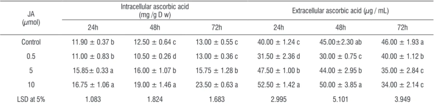
table 1. Effect of jasmonic acid (JA) application on the intra - and extracellular ascorbic acid content in melon cell suspension culture cultivated in MS1 medium.
JA (µmol)
Intracellular ascorbic acid
(mg /g D w) Extracellular ascorbic acid (µg / mL)
24h 48h 72h 24h 48h 72h
Control 11.90 ± 0.37 b 12.50 ± 0.64 c 13.00 ± 0.55 c 40.00 ± 1.24 c 45.00±2.30 ab 46.00 ± 1.93 a
0.5 11.00 ± 0.83 b 10.50 ± 0.26 d 13.00 ± 0.36 c 31.50 ± 2.36 d 30.00 ± 0.75 c 40.00 ± 1.12 b
5 15.85± 0.33 a 16.00 ± 1.07 b 15.75 ± 1.28 b 47.50 ± 1.00 b 44.00 ± 2.95 b 35.00 ± 2.84 c
10 16.75 ± 1.06 a 19.00 ± 1.46 a 23.50 ± 0.63 a 52.50 ± 1.42 a 50.00 ± 3.85 a 34.00 ± 2.14 c
LSD at 5% 1.083 1.824 1.683 2.995 5.101 3.949
Values (means of three replicates ± SD) in the same column followed by the same letter are not significantly different at p < 0.05.
table 2. Effect of jasmonic acid (JA) on ascorbate peroxidase enzyme activity during different harvesting times in the melon cell suspension culture cultivated in MS1 medium.
JA (µmol)
Ascorbate peroxidase activity
(m mol oxidized ascorbate min-1 gm-1 fw)
24h 48h 72h
Control 0.87 ± 0.03 d 0.88 ± 0.04 c 0.91 ± 0.04 c
0.5 1.00 ± 0.08 c 1.25 ± 0.02 b 1.5 ± 0.03 b
5.0 1.20 ± 0.03 b 1.27 ± 0.09 b 1. 6 ± 0.09 b
10.0 1.30 ± 0.04 a 1.5 ± 0.10 a 1.8 ± 0.07 a
LSD at 5% 0.090 0.134 0.117
Values (means of three replicates ± SD) in the same column followed by the same letter are not significantly different at p < 0.05.
table 3. Effect of jasmonic acid (JA) on ascorbate oxidase enzyme activity during different harvesting times in the melon cell suspension culture cultivated in MS1 medium.
JA
(µmol)
Ascorbate oxidase activity
(m mol oxidized ascorbate min-1 g-1 DW)
24h 48h 72h
Control 0.88 ± 0.03 b 0.39 ± 0.02 c 0.82 ± 0.03 b
0.5 0.71 ± 0.05 c 0.39 ± 0.01 c 0.41 ± 0.01 c
5.0 0.91 ± 0.02 b 0.82 ± 0.06 b 0.76 ± 0.06 b
10.0 2.71 + 0.07 a 2.70 ± 0.21 a 1.47 ± 0.09 s
LSD at 5% 0.088 0.205 0.107
Values (means of three replicates ± SD) in the same column followed by the same letter are not significantly different at p < 0.05.
table 4. Effect of jasmonic acid (JA) on glutathione reductase enzyme activity during different harvesting times in the melon cell suspension culture cultivated in MS1 medium.
JA (µmol)
Glutathione reductase activity
(m mol oxidized NADPH min-1 g-1 DW)
24h 48h 72h
Control 15.80 ± 0.49 c 16.0 ± 0.85 a 16.00 ± 0.67 a
0.5 19.10 ± 1.43 b 20.5 ± 0.34 b 22.5± 0.34 b
5.0 20.66 ± 0.43 ab 22.8 ± 1.13 a 24.5 ± 1.37 a
10.0 20.90 ± 0.56 a 25.9 ± 1.33 a 28.8 ± 0.87 b
LSD at 5% 1.571 1.854 1.684
Values (means of three replicates ± SD) in the same column followed by the same letter are not significantly different at p < 0.05.
During time–course experiments the AA content in intracellular and extracellular control cells showed slow increment in parallel to the cells growth. Melon callus exposed
to 0.5 µmolJA triggered significant negative changes in the
the other hand, estimated AA at elicited growth medium with 5 or 10 µmol JA was significantly higher than its control after 24 h. In contrast after 72 h estimated AA was lower in all elicited growth media, the highest decrease was in the 10 µmol JA growth medium (26% lower than the control).
Ascorbate peroxidase (APX) share crucially in the detoxification of H2O2, according to the following reaction:
2 ascorbate + H2O2→ 2 monodehydroascorbate + 2H2O
Effect of exogenous application of JA as chemical elicitor on the activity of ascorbate peroxidase enzyme in melon cell suspension culture was studied (Table2). The enzyme activity enhancement was JA dose and harvesting time depended with respect to the control. Jasmonic acid at 0.5 µmol concentration induced a statistically significant average 14.9, 42 and 64.8% increase at interval harvesting times, respectively. Moreover, JA at 10 µmol induced statistically highly significant average 49.4, 70.5 and 97.8% increase respective to interval harvesting times.
The effect of extracellular addition of JA as chemical elicitor in melon cell suspension culture on the activity of ascorbate oxidase AO enzyme was also studied. The data was tabulated in Table3. As shown in Table 3, JA at 10 µmol concentrations induced highly significant average 207.9, 592.3 and 79.2% increase respective to interval harvesting time. In contrast JA at 0.5 and 5 µmol concentrations induced significant average 50, 7.3% lower than the control respectively at 72 h harvesting time.
We further examined the effect of JA on glutathione reductase enzyme activity in melon cell suspension. Under the influence of 0.5, 5, and 10 µmol JA, changes in the antioxidant enzyme GR compared to the control non elicited was recorded. The data was statistically analyzed and illustrated in Table 4.
The GR enzyme activity in melon cells was activated in dose and time dependent manner with respect to the control. Jasmonic acid at 0.5 µmol concentration induced a statistically significant average 20.8, 28.1 and 40.6% increase respective to interval harvesting time. Moreover the 10 µmol JA concentration induced the highest average 32.2, 61 and 80% increases during 24-48 and 72 h post elicitation compared to the control.
alterations in phenolic compound Metabolism in Melon cell suspension culture: The effect of JA on the secondary compound induction and two related enzymes phenylalanine ammonia–lyase (PAL) and polyphenol oxidase (PPO) were examined in melon cell suspension to evaluate its response after exogenous JA application.
The effect of exogenous JA application on PAL activity was analyzes in melon cell suspension to assess its response. Under the influence of 0.5, 5 and 10 µmol JA an increase in enzyme activity over the control was found. Data was analyzed statistically in Table5. The enzyme activity increased with the increase in exogenous JA concentrations at all exposure periods in comparison with the control. Jasmonic acid at 0.5, 10 µmol concentration range induced significant average (23, 33.3 and 43.3%) and (80, 86 and 86%) increases respectively, in PAL activity during elicitation time.
Table 5. Effect of jasmonic acid (JA) on the phenylalanine ammonia-lyase enzyme activity during different harvesting times in the melon cell suspension culture cultivated in MS1 medium.
JA (µmol)
Phenylalanine ammonia-lyase activity
(µmol cinnamic min-1 gm-1 fw)
24 h 48 h 72 h
Control 0.30 ± 0.01 d 0.30 ± 0.02 c 0.3 ± 0.01 c
0.5 0.37 ± 0.03 c 0.40 ± 0.01 b 0.43 ± 0.01 b
5.0 0.41 ± 0.01 b 0.43 ± 0.03 b 0.47 ± 0.04 b
10.0 0.54 ± 0.01 a 0.56 ± 0.04 a 0.56 ± 0.04 a
LSD at 5% 0.033 0.052 0.055
Values (means of three replicates ± SD) in the same column followed by the same letter are not significantly different at p < 0.05.
Table 6. Effect of jasmonic acid (JA) on polyphenol oxidase enzyme activity during different harvesting times in the melon cell suspension culture cultivated in MS1 medium.
JA (µmol)
Polyphenol oxidase activity
(Δ min−1gm−1)
24h 48h 72h
Control 1.44 ± 0.04 c 1.78 ± 0.09 c 1.81 ± 0.08 c
0.5 0.43 ± 0.03 b 1.00 ± 0.03 d 1.25 ± 0.04 d
5.0 2.18 ± 0.05 a 2.24 ± 0.15 b 2.25 ± 0.18 b
10.0 2.22 ± 0.06 a 2.50 ± 0.19 a 3.03 ± 0.19 a
LSD at 5% 0.087 0.244 0.260
Values (means of three replicates ± SD) in the same column followed by the same letter are not significantly different at p < 0.05.
The induction of POX, PAL and PPO in elicited cells suggested that phenolics might be enhanced during elicitation. So changes in the profile of phenolic compounds extracted from melon cells and the medium in which they had been grown was investigated by gradient elution
high performance liquid chromatography (GC-HPLC). After visual examination of the complete set of HPLC chromatograms, it was apparent that melon cell suspension was secreting several common and distinctive secondary metabolites under our experimental condition .Peaks were selected according to several criteria: the compounds displayed characteristic production within an 5-10 µmol elicitor treatment as well as the control; the compounds appeared on a specific harvest day(s), and concentration of the compounds which was enhanced compared to the other compounds secreted. We characterized 5 compounds on the basis of their potentials as elicitors of defense compounds previously described as endowed with this biological activity. The phenolics coumarin, caffeic, P-coumaric, salicylic and benzoic were identified in the cells and culture medium. Coumarin was the highest phenolic to increase concomitantly with JA elicitation in melon cells and the culture medium (Tables 7 and 8).
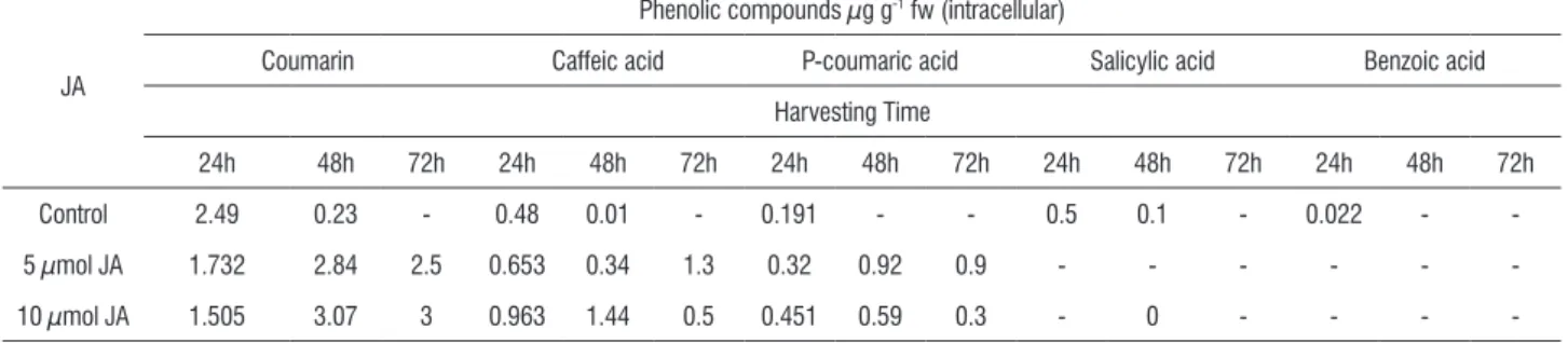
Table 7. Effect of jasmonic acid (JA) on the intracellular phenolic compounds production during different harvesting times in the melon cell suspension culture cultivated in MS1 medium. Values are expressed as µg /g fW.
JA
Phenolic compounds µg g-1 fw (intracellular)
Coumarin Caffeic acid P-coumaric acid Salicylic acid Benzoic acid
Harvesting Time
24h 48h 72h 24h 48h 72h 24h 48h 72h 24h 48h 72h 24h 48h 72h
Control 2.49 0.23 - 0.48 0.01 - 0.191 - - 0.5 0.1 - 0.022 -
-5 µmol JA 1.732 2.84 2.5 0.653 0.34 1.3 0.32 0.92 0.9 - - -
-10 µmol JA 1.505 3.07 3 0.963 1.44 0.5 0.451 0.59 0.3 - 0 - - -
-In melon cells the changes were recorded in abundance of three of these phenolics (coumarin, caffeic and P-coumaric), and other compounds (salicylic and benzoic) generally disappeared under JA elicitation from the melon cells compared to the control (Table7). On the other hand while still there is great increase under elicitation between
table 8. Effect of jasmonic acid (JA) on the extracellular phenolic compounds production during different harvesting times in the melon cell suspension culture cultivated in MS1 medium. Values are expressed as µg /g fW.
JA
Phenolic compounds µg g-1 fw (intracellular)
Coumarin Caffeic acid P-coumaric acid Salicylic acid Benzoic acid
Harvesting Time
24h 48h 72h 24h 48h 72h 24h 48h 72h 24h 48h 72h 24h 48h 72h
Control - 2.9 - 0.248 0.12 - 0.451 0.17 - - -
-5 µmol JA - 25.4 30 - - 1 0.224 3.64 3.4 - 0.1 0.1 - -
-10 µmol JA 19.64 3.7 26 0.424 0.72 - 2.77 0.77 3.8 0.056 - 0.1 - - 0
changes in total flavonoids content: The effect of extracellular application of JA on the total flavonoids content in melon cell suspension culture is demonstrated in figure 3. The data revealed that application of JA to melon culture medium enhanced melon total flavonoids. At low dose, melon treated cells significantly accumulate fairly low amount (5.5%) after 24 h, meanwhile the other two doses accumulate higher amounts in dose depend manner (49 and 58%) compared to the control. Two days post elicitation exhibited different responses as compared to the first day, while JA at 0.5 µmol significantly increased the flavonoids (30%), at 5 and 10 µmol JA induced lower amounts (33.3 ,36.2%) compared to the control. Going to the third day the highest JA concentration induced the highest amount 64% in melon culture compared to the control.
500
400
300
200 600
total flavonoids
µ
g g
-1 dW)
24 48 72
harvesting time (h)
0 0.5 5 10 (µM JA)
figure 3. Effect of jasmonic acid (JA) on the total flavonoids production during different harvesting times in the melon cell suspension culture cultivated in MS1 medium.
discussion
Cell-suspension cultures are handled relatively easily, which make them valuable and attractive for standardized experiments to study elicitor-induced defense responses. for different cell-suspension cultures, a variety of elicitor molecules have been shown to induce defense-related responses that are similar to the responses in elicited plants. In the present study we found that elicitation could be a promising strategy to drive melon cells toward modulating specific secondary metabolites production concomitant with specific oxidative defense mechanisms. Exogenous JA application which lead to endogenous JA enhancement probably result from and activates JA signaling pathways and the related defense gene expression, without damaging
the melon nucleic acids (Nafie et al., 2009), and also may be
responsible for other cascades of biochemical and molecular events. Our results together with some previous studies on JA as influential integral elicitor, suggest that JA can potentiate
melon cells defense responses via modulating ascorbic acid
and phenolic metabolism, total flavonoids, coumarins and ROS -detoxification enzyme systems.
Antioxidant enzyme enhancement, a response related to ROS detoxification, was increased under diverse environmental stress situations (Mittler, 2002). Catalase, a
detoxifying system member, converts H2O2 to form oxygen
and water. Ali et al. (2005) found that CAT activity declined
severely in Panax ginseng and Panax quinquefolium plants,
al. (2005) shows that CAT activity was lower in JA treated
Morinda elliptica cell cultures after 6 days of elicitation. The suppression in CAT activity have similarly been reported in fungal-elicited Catharanthus roseus cell cultures (Zhao et al., 2001) and water-stressed in strawberry plants (Wang, 1999). In contrast, MJ treatment of strawberry plant increases CAT activity (Wang, 1999). In the present study, CAT in melon cells exposed to the highest JA concentrations cause significant increase in enzyme activity, probably to overcome the transient
increase in H2O2 levels which require a prompt generation of
ROS-scavenging systems to neutralize its adverse effects in cell membranes stability.
Peroxidases participate in a variety of defense mechanisms (Liu et al., 2008), to ameliorate oxidative burst a common event in defense response (Lamb and Dixon, 1997). In our work, induction of peroxidases (POD) in melon cells during the time course of elicitation, may result from enzyme
activation and/or the de novo differential expression in POD
isoforms, implicating them in stress alleviation, as POD expression is controlled by multiple genes (Gomez-vasquez et al., 2004). On the other hand, Bestwick et al. (1998); Martinez
et al. (1998) and Zhao et al. (2001) found that apoplastic
peroxidases have been responsible for the oxidative burst in an increasing list of plant species. Cell wall-bound POD enzyme activity in melon tissues treated with JA collaborates in H2O2 production, but it is not the major factor, but implicates
it in cells membrane stability disturbance resulted in transient
leakage of important compounds into growth media. Liuet
al. (2008) found that cell wall POD activity in JA treated pea leaves were not parallel to the H2O2 accumulation which is in
line with our data. In the present work, assessing exogenous JA application on ascorbic acid metabolism revealed that JA
modulates its content. Plants synthesize ascorbic acid via
de novo synthesis and salvage pathways, but the regulation of its biosynthesis and the mechanisms behind ascorbate homeostasis are largely unknown (Valpuesta and Botella, 2004). Jasmonic acid and its methyl ester (jasmonates) mediate plant responses to many biotic and abiotic stresses by triggering a transcriptional reprogramming that allows cells to cope with these stress. In this regard, Wolucka et al. (2005) proposed that, in tobacco BY-2 cells, MJ stimulated the de novo biosynthesis of L-ascorbic acid, at least in part, by enhanced transcription of MJ-responsive genes encoding key enzymes of vitamin C synthesis. Also they found that, in Arabidopsis cell suspension phytohormones (auxin and/
or cytokinin) not only are necessary to support growth but also affect jasmonates-elicited L-AA synthesis, but such relationship is not understood. In our results, MS1 (MS
medium supplemented with 2 mg-1L 2, 4-D; 0.1 mg-1L BA and
30 g-1L sucrose) medium the hormonal composition seems
crosstalk with JA signaling pathways, including AA genes enhancement.
It was found an AA imbalance between melon cells and its growth medium, where the highest level in the elicited growth medium was recorded after only one day compared to control, resulting in membrane efficient permeability under transient hydrogen peroxide production. Estimated AA levels undergoing the JA control as stated previously to affect directly genes for AA biosynthesis.
Ascorbate peroxidase is a key hydrogen peroxide scavenging enzyme and its isoforms are present in different
cell compartments (Jimenez et al., 1997). In plant cells, APX
expression is induced in response to many environmental stresses that result in the accumulation of ROS (Karpinski et al., 1999; Mittler, 2002). In particular, methyl jasmonate stimulated APX expression at the levels of mRNA (Orvar et
al., 1997), protein (Rakwal et al., 1999) and enzyme activity
(Maksymiec and Krupa, 2002). In contrast, Chong et al.
(2005) showed that ascorbic peroxidase activity was lower in JA elicited Morinda elliptica cells than in control. In our work
and during 24h, transient increase in H2O2 concomitant with
AA availability stimulating APX activity to counter balance hydrogen peroxide impact and thus preventing the cell membrane permeability.
Significant increase in glutathione reductase (GR) enzyme activity in JA treated melon cells should play a crucial role in protecting the tissues against oxidative stress by maintaining the GSH level so antagonizing the consumption of AA and recycling it for APX and ascorbic oxidase (AO) as well as to maintain AA level for its crucial role as antioxidant metabolite.
fotopoulos et al. (2006) reported that lowering the
apoplast AA redox state, throughincreased AO expression in
transgenic tobacco (Nicotiana tabacumL. cv. Xanthi), exerts
no effects on the expression levels ofgenes involved in AA
recycling under normal growth conditions,but plants display
enhanced sensitivity to various oxidativestress-promoting
complex transcriptionaland translational controls (Esaka et al., 1992). Kato and Esaka (2000), found that the activity and expression of AO are closely correlated with cell expansion
and Pignocchi et al. (2003) stated that the enzyme transcript
levels increased upon growth promoters exposure, such as auxin and jasmonates (Sanmartin, 2002).
Perturbation in ascorbic acid oxidase (AO) activity in treated melon cells was evaluated and the resulted data imply that 5 and 10 µmol JA enhanced the enzyme activity, while JA at 0.5 µmol inhibited this enzyme. Increase in enzyme activity may compromise the tissue detoxification potential ability and JA may be implicated in activating AA-recycling genes to protect cells from membrane damage.
Elicitation of in vitro cultures is a useful approach to
enhance and extend production of desirable products (Oksman-Caldentey and Inze, 2004). Elicitors are now considered as signal molecules that activate the signal-transduction cascade which lead to the activation and expression of genes related to the biosynthesis of secondary metabolites (Zhao et al., 2005). Wang and Wu (2005) stated that MJ, is an effective inducer of the secondary metabolite diterpenoid taxol (paclitaxel) in Taxus
spp. and that exogenously supplied MJ at 100 µmol induced rapid production of NO concomitantly with the phenylalanine ammonia-lyase (PAL) activation. Liu et al. (2008) found that
10 µmol JA treatment of pea leaves (Pisum sativum), led
to a significant increase in activities of plasma membrane NADPH oxidase and PAL. Jasmonates elicitation was also found to increase the production of phenylpropanoids and
naphtodianthrones in Hypericum perforatum cell suspensions
(Gadzovska et al., 2007).
Secondary metabolite production is usually associated with rapid, transient increases in activities of key enzymes of the phenylpropanoid / flavonoid pathway such as PAL
and chalcone isomerase (Gundlach et al., 1992; Dixon et al.,
2002). In addition, many studies have shown that polyphenol oxidase (PPO) is induced in response to mechanical wounding, fungal and bacterial infection, and by treatment with signaling molecules, such as JA /MJ and salicylic acid (Constabel et al., 2000; Stewart et al., 2001). Defense proteins whose expression is dependent on the JA/COI1 pathway include polyphenol oxidase (Browse and Howe, 2008).
In JA-treated melon cells PAL and PPO enzymes and total flavonoid were significantly enhanced. As PAL controls
the initial step in the phenolic compounds biosynthesis pathway, factors induced enzyme activity will encourage phenolic compound enrichment. Also the present results indicate the presence of coumarin and four phenolic acids: caffeic, p-coumaric, salicylic and benzoic to different levels in both JA-elicited melon cells and in the MS1 growth medium with respect to the control.
Increase in the caffeic and p-coumaric acids was recorded in melon cells and growth medium, which was still detectable three days after JA-elicitation. These results agree with those reported by Kauss et al. (1992a) who found in the dark-grown parsley culture that MJ-treatment at 1 to 5 µmol results in a synergistic increase in coumarin secretion and incorporation of phenolics into the cell walls. Zulak et al.
(2008) studying opium poppy (Papaver somniferum) have
found that coumarate levels initially increased in elicitor-treated cells more rapidly than in controls, but subsequently decreased from 50–80 h in both cases. Benzoic acid (BA) and its derivatives also play important roles in biotic and abiotic stress responses and are incorporated into several secondary defense-related metabolites (Wildermuth, 2006). Present data analysis revealed that BA and salicylic acid (SA) were not involved in the melon responses to the JA-elicitation since they were undetectable in both the elicited melon cells and growth medium. But worthy note is that SA was recorded but only in the non elicited melon cells during the 48 h and then disappeared. In contrast, in Arabidopsis, leaves challenged with various biotic and abiotic stresses, methylbenzoate was found to be induced (Chen et al., 2003) and benzoic carboxymethyl-transferases were also induced under similar conditions (Effmert et al., 2005). On the other hand, Zulak et al. (2008) stated that BA have not yet been identified in
opium poppy (Papaver somniferum ) as a key component of
functional genomics platform during investigating the interplay between primary and secondary metabolism in cultured opium poppy cells treated with a fungal elicitor.
modulated and controlled by the AA availability, which had been induced and its synthesis was shown previously to be reprogrammed by JA in melon cells.
acknowledgements: Authors greatly appreciate technical cooperation with Gene Transfer Lab (GTL), Agricultural Genetic Engineering Research Institute (AGERI), Agricultural Research Centre (ARC), Cairo, Egypt.
references
Aebi H (1984) Catalase in vitro. Methods in Enzymology. 105:121- 126.
Ali MB,Yu KW, Hahn EJ, Paek KY (2005) Differential responses of anti-oxidants enzymes, lipoxygenase activity, ascorbate content and the production of
saponins in tissue cultured root of mountain panax ginseng C.A. Mayer and
Panax quinquefolium L. in bioreactor subjected to methyl jasmonate stress. Plant Sci. 169: 83–92.
Asada K (1999) The water–water cycle in chloroplasts: scavenging of active oxygen and dissipation of excess photons. Annul. Rev. Plant Physiol. Plant Mol. Biol. 50: 601–639.
Barnes JD, Zheng Y, Lyons TM (2002) Plant resistance to ozone: the role of ascorbate. In: Omasa K, Saji H, YousseWan S, Kondo N (eds) Air pollution and plant biotechnology, Springer, Berlin Heidelberg New York, pp 235–252. Bestwick SC, Brown IR, Mansfield JW (1998) Localized changes in peroxidase activity accompanies hydrogen peroxide generation during the development of a nonhost hypersensitive reaction in lettuce. Plant Physiol. 118: 1067–1078.
Browse J, Howe GA (2008) New weapons and a rapid response against insect attack. Plant Physiol. 146: 832-838.
Buschmann H, Rodriguez, MX, Tohme J, Beeching JR (2000) Accumulation of hydroxycoumarins during post-harvest deterioration of tuberous roots of
cassava (Manihot esculenta Crantz).Ann Bot 86: 1153-1160.
Chen f, D’Auria JC, Tholl D, Ross JR, Gershenzon J, Noel JP, Pichersky E
( 2003) An Arabidopsis thaliana gene for methylsalicylate biosynthesis,
identified by a biochemical genomics approach, has a role in defense. Plant J. 36:577-588.
Chong TM , Abdullahd MA, fadzillahb NM, Laia OM, Lajisc NH (2005) Jasmonic acid elicitation of anthraquinones with some associated enzymic
and non-enzymic antioxidant responses in Morinda elliptica. Enzyme
MicrobTechnol. 36: 469–477.
Constabel CP,Yip L, Patton JJ, Christopher ME (2000) Polyphenol oxidase from hybrid poplar, cloning and expression in response to wounding and herbivore. Plant Physiol. 124: 285–95.
Dickerson DP, Pascholati Sf, Hagerman AE, Butler LG, Nichol son RL (1984) Phenylalanine ammonia-lyase and hydroxycinnamate: CoA ligase in maize
mesocotyls inoculated with Helminthosporium maydis or Helminthosporium
carbonum. Physiol. Plant Pathol. 25: 111-123.
Dixon RA, ferreira D, Genistein S (2002) The phenylpropanoid pathway and plant defense - a genomics perspective. Mol. Plant Pathol. 3: 371–390. Effmert U, Saschenbrecker S, Ross J, Negre f, fraser CM, Noel JP, Dudareva
N, Piechulla B (2005) floral benzenoid carboxyl methyltransferases: from in
vitro to in planta function. Phitochem. 66:1211-1230.
Esaka M, fujisawa K, Goto M, Kisu Y (1992) Regulation of ascorbate oxidase expression in pumpkin by auxin and copper. Plant Physiol. 100: 231–237. Esaka M, Uchida M, fukui H, Kubota K, Suzuki K (1988) Marked increase in ascorbate oxidase protein in pumpkin callus by adding copper. Plant Physiol. 88: 656-660.
fotopoulos v, Sanmartin M, Kanellis AK (2006) Effect of ascorbate oxidase over-expression on ascorbate recycling gene expression in response to agents imposing oxidative stress. J Exp Bot. 1-11
Gadzovska S, Maury S, Delaunay A, Spasenoski M, Joseph C, Hagege D
(2007) Jasmonic acid elicitation of Hypericum perforatum L. cell suspensions
and effects on the production of phenylpropanoids and naphtodianthrones. Plant Cell Tiss. Organ Cult. 89:1–13.
Gomez-Vasquez R, Day R, Buschmann H, Randles S, Beeching JR, Cooper RM (2004) Phenylpropanoids, phenylalanine ammonia-lyase and peroxidases
in elicitor-challenged Cassava (Manihot esculenta) suspension cells and
leaves. Ann. Bot. 94: 87 97.
Gundlach H, Muller M J, Kutchan M, Zenk M H (1992) JA is a signal transducer elicitor-induced plant cell cultures. Proc Nat Acad Sci USA. 89:2389-2393. Haider NB, Jacobson SG, Cidecyan AV, Swiderski R, Streb LM, Searby C, Beck G, Hockey R, Hanna DB, Gorman S (2000) Mutation of a nuclear receptor gene, nr2e3, causes enhanced cone syndrome, a disorder of retinal cell fate. Nat. Genet. 24: 127-31.
Hodges D, Andrews C, Johnson DA, Hamilton RI (1997) Antioxidant enzyme responses to chilling stress in differentially sensitive inbred maize lines. J. Exp. Bot. 48: 1105-13.
Jagota SK, Dani HM (1982) A new colorimetric technique for the estimation of vitamin C using folin phenol reagent. Anal. Biochem. 127: 178–182.
Jiang AL, Tian SP, Xu Y (1984) Effect of controlled atmospheres with high
O2 or high-CO2 concentrations on postharvest physiology and storability of
“Napoleon” sweet cherry. J. Integrative Plant Biol. 44: 925–930.
Jiang M, Zhang J (2002) Water stress-induced abscisic acid accumulation triggers the increased generation of reactive oxygen species and up-regulates the activities of antioxidant enzymes in maize leaves. J Exp Bot 53(379): 2401-2410.
Jimenez A, Hernandez JA, Del Rio LA, Sevilla f (1997) Evidence for the presence of the ascorbate–glutathione cycle in mitochondria and peroxisomes of pea leaves. Plant Physiol. 114: 275–284.
Karpinski S, Reynolds H, Karpinska B, Wingsle G, Creissen G, Mullineaux P (1999) Systemic signalling and acclimation in response to excess excitation
energy in Arabidopsis. Science. 284: 654–657.
Kato N, Esaka M (2000) Expansion of transgenic tobacco protoplasts expressing pumpkin ascorbate oxidase is more rapid than that of wild-type protoplasts. Planta. 210: 1018–1022.
Kauss, H, Krause, K, Jeblick, W (1992a) Methyl jasmonate conditions parsley suspension cells for increased elicitation of phenyl propanoid defense responses. Biochem. Biophys. Res. Commun. 189: 304-308.
Kiddle G, Pastori GM, Bernard S, Pignocchi C, Antoniw J, Verrier PJ, foyer CH (2003) Effects of leaf ascorbate content on defense and photosynthesis gene
expression in Arabidopsis thaliana. Antiox Redox Signal. 5: 3–32.
Knight H , Knight MR (2001) Abiotic stress signaling pathways: specificity and cross-talk. Trends Plant Sci. 6: 262–267.
Lamb C, Dixon RA (1997) The oxidative burst in plant disease resistance. Annul. Rev. Plant Physiol Plant Mol. Biol. 48:251–75.
Lee HS, Wicker L (1991) Anthocyanin pigments in the skin of lychee fruit. J food Sci. 6(2): 466–468, 483.
Liu Y,Pan QH, Yang HR, Liu YY, Huang WD (2008) Relationship between H2O2
and Jasmonic acid in Pea leaf wounding response. Russ J Plant Physiol. 55(6): 851–862.
Luh BS, Phithakpol B (1972) Characteristics of polyphenol oxidase related to browning in cling peaches. J. food Sci. 37: 264–268.
Maksymiec W, Krupa Z (2002) The in vivo and in vitro influence of methyl
jasmonate on oxidative processes in Arabidopsis thaliana leaves. Acta
Martens S, Knott J, Seitz CA, Janvari L (2003) Impact of biochemical pre studies on specific metabolic engineering strategies of flavonoid biosynthesis in plant tissues. Biochem. Eng. J. 14: 227–235.
Martinez C, Montillet JL, Bresson E, Agnel JP, Dai GH, Daniel Jf, Geiger JP, Nicole M (1998) Apoplastic peroxidase generates superoxide anions in cells
of cotton cotyledons undergoing the hypersensitive reaction to Xanthomonas
campestris pv malvacearum. Race 18, Mol. Plant-Microbe Int. 11: 1038– 1047.
Martyn RD, Miller ME (1996) Monosporascus root rot/vine decline: An emerging disease of Melons worldwide. Plant Dis. 80: 716-725.
Nafie E, Hussein G, Al Mokadem AlS (2009) Jasmonic acid elicitation modulates anthraquinones production and evokes defense responses in
Cucumis melo L. cell suspension culture. African J. Biol. Sci. 5(2): 63-96. Naill MC, Roberts SC (2005) Cell cycle analysis of Taxus suspension cultures at the single cell level as an indicator of culture heterogeneity. Biotechnol. Bioeng. 90: 491–500.
Nakano Y, Asada K (1981) Hydrogen peroxide is scavenged by ascorbate-specific peroxidase in spinach chloroplasts. Plant Cell Physiol .22: 867-880. Noctor G, foyer C (1998) Ascorbate and glutathione: keeping active oxygen under control. Annul. Rev. Plant Physiol. Plant Mol. Biol. 49: 249–279 Oberbacher Mf, Vines HM (1963) Spectrophotometric assay of ascorbic acid oxidase. Nature. 197:1203–1204.
Oksman-Caldentey KM, Inze´ D (2004) Plant cell factories in the post-genomic era: new ways to produce designer secondary metabolites. Trends Plant Sci. 9: 433–440.
Orvar BL, McPherson J, Ellis BE (1997) Pre-activating wounding response in tobacco prior to high-level ozone exposure prevents necrotic injury. Plant J. 11: 203–212.
Pastori GM, foyer CH (2002) Common components, networks, and pathways of cross-tolerance to stress. The central role of ‘redox’ and abscisic acid-mediated controls. Plant Physiol. 129: 460–468.
Pignocchi C, fletcher J, Barnes J, foyer CH (2003) The function of ascorbate
oxidase (AO) in tobacco (Nicotiana tabacum L.). Plant Physiol. 132: 1631–
1641.
Pignocchi C, foyer CH (2003) Apoplastic ascorbate metabolism and its role in the regulation of cell signaling. Curr Opin Plant Biol. 6: 379–389. Potters G, Horemans N, Bellone S, Caubergs J, Trost P, Guisez Y, Asard H (2004) Dehydroascorbate influences the plant cell cycle through a glutathione independent reduction mechanism. Plant Physiol. 134:1479–1487. Rakwal R, Agrawal GK, Yonekura M (1999) Separation of proteins from
stressed rice (Oryza sativa L.) leaf tissues by two dimensional polyacrylamide
gel electrophoresis: induction of pathogenesis-related and cellular protecting proteins by jasmonic acid, UV irradiation and copper chloride. Electrophoresis. 20: 3472–3478.
Ranieri A, Nali G, D’Urso G (1995) Peroxidase activity in Cucurbita pepo L.
leaves exposed to ozone. Agricoltura Mediterranea. (Special volume) 47-54. Richter H (2000) fresh produce guide: Nutrition, selection, preparation, storage, handling, cooking. Try-foods Intl. Inc. Apopka, fL.
Mittler R (2002) Oxidative stress, antioxidants and stress tolerance. Trends Plant Sci .7 : 405-410.
Sanmartin M (2002) Regulation of melon ascorbate oxidase gene expression and effect of its modification in transgenic tobacco and melon plants .Spain ,University of Valencia .Ph.D. thesis.
Smirnoff N (2000) Ascorbic acid: metabolism and functions of a multi-facetted molecule. Curr. Opin. Plant Biol. 3:229–235.
Stewart RJ, Sawyeer BJB, Bucheli CS, Robinson SP(2001) Polyphenol oxidase is induced by chilling and wounding in pineapple. Aust. J. Plant Physiol. 28:181–191.
Tokunaga T, Miyahara K, Tabata K, Esaka M (2005) Generation and properties of ascorbic acid-overproducing transgenic tobacco cells expressing sense RNA for L-galactono-1,4-lactone dehydrogenase. Planta. 220: 854–863. Valpuesta V, Botella MA (2004) Biosynthesis of L-ascorbic acid in plants: new pathways for an old antioxidant. Trends Plant Sci. 9: 573–577.
Vranova E, Inze D, Breusegem fV (2002) Signal transduction during oxidative stress. J. Exp. Bot. 53: 1227-1236.
Wang JW, Wu JY (2005) Nitric oxide is involved in methyl jasmonate-induced defense responses and secondary metabolism activities of Taxus cells. Plant Cell Physiol. 46(6): 923-30.
Wang SY (1999) Methyl jasmonate reduces water stress in strawberry. Plant Growth Regul. 18: 127–34.
Wang W, Zhao ZJ, Xu Y f, Qian XH, Zhong JJ (2005) Efficient induction of ginsenoside biosynthesis and alteration of ginsenoside heterogeneity
in cell cultures of Panax notoginseng by using chemically synthesized
2-hydroxyethyl jasmonate. Appl. Microbiol. Biotechnol. 70: 298–307. Wildermuth MC (2006) Variations on a theme: synthesis and modification of plant benzoic acids. Curr. Opin. Plant Biol. 9: 288-296
Wolucka BA, Goossens A, Inze´D (2005) Methyl jasmonate stimulates the de
novo biosynthesis of vitamin C in plant cell suspensions. J Exp Bot. 56(419):
2527–2538.
Yadav RC, Salah MT, Grume R (1996) High frequency shoots regeneration from leaf explant of muskmelon. Plant Cell Tiss Organ Cult. 45: 207-214. Yamunarani K, Jaganathan R, Bhaskaran R, Govindaraju P, Velazhahan R
(2004) Induction of early blight resistance in tomato by Quercus infectoria
gall extract in association with accumulation of phenolics and defense-related enzymes. Acta Physiol Plantarum. 26(3): 281-290
Yingsanga P, Srilaong V, Kanlayanarat S, Noichinda S, McGlasson WB (2008) Relationship between browning and related enzymes (PAL, PPO and POD)
in rambutan fruit (Nephelium lappaceum Linn.) cvs. Rongrien and
See-Chompoo. Postharvest Biol Technol. 50: 164–168.
Yong SP, Soon TJ, Seong GK, Buk GH, Patricia AA, fernando T (2008) Antioxidants and proteins in ethylene-treated kiwi fruits. food Chem.107 (2): 640–648.
Zhao J, Davis LC, Verpoorte R (2005) Elicitor signal transduction leading to production of plant secondary metabolites. Biotechnol. Adv. 23: 283–333. Zhao J, Hu Q, Guo YQ, Zhu WH (2001) Elicitor-induced indole alkaloid
biosynthesis in Catharanthus roseus cell cultures is related to Ca2+ influx and
the oxidative burst. Plant Sci. 161:423–31.