ANDRÉ RICARDO E SILVA
CONTROLE BIOLÓGICO DE NEMATÓIDES GASTRINTESTINAIS DE OVINOS, OVOS DE Trichuris trichiura, Trichuris vulpis E DE
Anoplocephala perfoliata POR FUNGOS NEMATÓFAGOS
Tese apresentada à Universidade Federal de Viçosa, como parte das exigências do programa de Pós-Graduação em Medicina Veterinária, para obtenção do título de Doctor Scientiae
VIÇOSA
CONTROLE BIOLÓGICO DE NEMATÓIDES GASTRINTESTINAIS DE OVINOS, OVOS DE Trichuris trichiura, Trichuris vulpis E DE
Anoplocephala perfoliata POR FUNGOS NEMATÓFAGOS
Tese apresentada à Universidade Federal de Viçosa, como parte das exigências do programa de
Pós-Graduação em Medicina
Veterinária, para obtenção do título de Doctor Scientiae
APROVADA: 25 de outubro de 2011.
Profª. Vânia Rita Elias P. Bittencourt Prof. José Cola Zanuncio
Prof. Maria de Lurdes de A. Rodrigues Prof. José Eduardo Serrão
À Deus pelo dom da vida, sem ele nada existiria!
Aos meus pais Luiz de Almeida e Silva e Geny da Silva in memorian. À minha filha Carolina Vicentini de Azevedo Silva.
AGRADECIMENTOS
À Deus.
Aos meus pais e minha família.
A Universidade Federal de Viçosa e ao Departamento de Veterinária da UFV.
Aos órgãos de fomento FAPEMIG pelo financiamento do projeto e a CAPES pela bolsa de doutorado.
Ao Professor Jackson Victor de Araújo pela confiança, oportunidade, orientação e pelos ensinamentos indispensáveis a minha formação. Ao professor Laércio dos Anjos Benjamim pela inestimável ajuda, dedicação e trabalho árduo no Núcleo de Microscopia Eletrônica e Microanálise (NMM)
e no Departamento de Veterinária (DVT) /UFV.
Aos funcionários José Geraldo (Tuim) e Sr. Ademir pela amizade e contribuição valiosa em minha formação, nos experimentos a campo e no Laboratório de Parasitologia do DVT/UFV, enfim durante todos os momentos
dessa trajetória.
A Lucinda, Aécio e Luiz Márcio pela contribuição no Laboratório de Patologia Clínica do DVT/UFV.
Ao Professor Dantas pela amizade e companheirismo, além dos ensinamentos e contribuição inequívoca a minha formação profissional. Ao professor Joaquim Hérnan Patarroyo Salcedo pelas cobranças e pelos
ensinamentos.
Aos professores Pacífico Antônio Diniz Belém, Ernane Paulino do Rego, Eduardo Paulino da Costa, Luís Augusto Nero e Abelardo da Silva Jr. pelos
ensinamentos, pela amizade e orientações durante toda esta jornada, aos quais agradeço em nome de todos os professores da Universidade Federal de Viçosa que contribuíram direta e indiretamente por minha formação e por
esta conquista.
Ao professor Adauto José Gonçalves de Araújo e a colega Daniela Leles de Souza da Fundação Oswaldo Cruz (Fiocruz), Rio de Janeiro, Brasil, pelo
fornecimento de material e pelas contribuições.
A todos os funcionários do DVT e do NMM da UFV, que contribuíram para a realização deste trabalho.
Aos Co-orientadores e membros da banca examinadora, pelas correções e sugestões na elaboração final deste trabalho.
A todos que direta ou indiretamente me ajudaram e motivaram na conquista desse objetivo.
Enfim agradeço a Deus por ter me dado esta oportunidade. Meu,
BIOGRAFIA
ANDRÉ RICARDO E SILVA, filho de Luiz de Almeida e Silva e Geny da Silva, in memorian, nasceu em 30 de novembro de 1969, em Viçosa, Minas Gerais. Em março de 1993, graduou-se em Medicina Veterinária pelo Departamento de Veterinária da Universidade Federal de Viçosa (UFV). Nessa data ingressou na indústria química e farmacêutica veterinária, Smithkline Beecham Laboratórios Ltda. Posteriormente, atuou na Pfizer Saúde Animal e em seguida na Indústria Química e Farmacêutica Schering-Plough S/A, exercendo atividades nas áreas técnicas e comercial dessas empresas. Após este período, atuou como autônomo, prestando consultorias a propriedades rurais de gado leiteiro nas regiões de Viçosa-MG e Colatina-ES, sendo que, nessa última, atuou como técnico do Projeto Educampo, em parceria com o SEBRAE daquele Estado. Realizou pesquisa junto à equipe do Prof. Dr. Sebastião Teixeira Gomes, do Departamento de Economia Rural, da UFV, na região central do estado do Tocantins-TO. O objetivo dessa pesquisa foi diagnosticar a situação da bacia leiteira da região central desse estado, para posteriormente possibilitar ações de revitalização e desenvolvimento dessa bacia leiteira. Após esta pesquisa foi convidado pela Nestlé, Dairy Partners Americas (DPA) para desenvolver e implementar o projeto Núcleo de Assistência Técnica Autorizada (NATA), na região do Centro-Oeste, mais especificamente, aos produtores fornecedores da indústria da região de Goiânia e Rialma no estado de Goiás. Em outubro de 2006, ingressou no Programa de Mestrado em Medicina Veterinária, no Departamento de Veterinária/UFV, submetendo-se à defesa de dissertação, em julho de 2008. Em agosto de 2008, ingressou no Programa de Doutorado em Medicina Veterinária, no Departamento de Veterinária/UFV, submetendo-se à defesa de tesubmetendo-se, em outubro de 2011. Atualmente é revisor das revistas Veterinary Parasitology da WAAVP (World Association for the Advancement of Veterinary Parasitology e da Ciência Rural (UFSM). Durante o doutoramento participou como: Membro da Comissão Científica do III
Simpósio de Pesquisa em Medicina Veterinária 2010 e III Semana da
Pós-Graduação em Medicina Veterinária/UFV na área de Doenças e
(APG/UFV) - Gestão 2010/2011. Conselheiro efetivo no Conselho
Universitário da UFV (CONSU/UFV). Membro da Comissão Própria de
Avaliação da UFV, junto a Pró-Reitoria de Planejamento e Orçamento.
Membro da Comissão Organizadora do Simpósio de Integração Acadêmica
(SIA) da UFV 2011 junto a Pró-Reitoria de Pós-Graduação. Membro da Comissão de Planejamento do Projeto UFV 2026, criada pelo ex-reitor da
UFV e atual Secretário da Educação Superior do MEC Luiz Cláudio Costa.
Membro da Comissão Organizadora da Solenidade de Entrega de diplomas
aos mestres e doutores da UFV de fevereiro e agosto de 2011.
Representante discente suplente no programa de pós-graduação em
Medicina Veterinária da UFV (2010-2011). Membro da Comissão Eleitoral da
UFV que coordenou a eleição dos representantes dos professores e
servidores no Conselho Universitário (CONSU) e no Conselho de Ensino,
Pesquisa e Extensão (CEPE), e a eleição dos representantes dos
professores na Comissão Permanente do Pessoal Docente (CPPD),
CONTEÚDO
LISTA DE FIGURAS... IX LISTA DE TABELAS... XIII RESUMO...XIV ABSTRACT...XVI
(I) INTRODUÇÃO GERAL...1
OBJETIVOS DO PRESENTE TRABALHO...6
(I) REFERÊNCIAS BIBLIOGRÁFICAS...7
(II) CAPÍTULO 1 - Biological control of sheep gastrointestinal nematodiosis in a tropical region of the southeast of brazil with the nematode predatory fungi Duddingtonia flagrans and Monacrosporium thaumasium...12
Abstract ...13
Introduction ...14
Material and Methods ...15
Results...18
Discussion ...19
Conclusion ...22
References ...22
(II) CAPÍTULO 2 - Activity in vitro of fungal conidia of Duddingtonia flagrans and Monacrosporium thaumasium on Haemonchus contortus infective larvae...32
Abstract ...33
Introduction ...34
Material and methods ...34
Results and discussion ...36
References ...38
(II) CAPÍTULO 3 - Comparative analysis of destruction of the infective forms of Trichuris trichiura and Haemonchus contortus by nematophagous fungi Pochonia chlamydosporia, Duddingtonia flagrans and Monacrosporium thaumasium by scanning electron microscopy...44
Abstract ...45
Material and methods ...47
Results...49
Discussion ...50
Conclusion ...51
References ...51
(II) CAPÍTULO 4 - In vitro ovicidal activity of the nematophagous fungi Duddingtonia flagrans, Monacrosporium thaumasium and Pochonia chlamydosporia on Trichuris vulpis eggs...60
Abstract ...61
Introduction ...62
Material and methods ...63
Results...64
Discussion ...64
Conclusion ...65
References ...66
(II) CAPÍTULO 5 - Destruction of Anoplocephala perfoliata eggs by the nematophagous fungus Pochonia chlamydosporia...71
Abstract...72
Introduction ...73
Material and methods ...73
Results ...75
Discussion ...75
Conclusion ...76
References ...76
LISTA DE FIGURAS
Página (ii) Capítulo 1
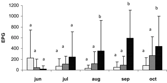
Figura 1 Monthly averages and standard deviations of the countings of eggs per gram of feces (EPG) of the animals in the groups treated with the nematophagous fungi Duddingtonia flagrans and Monacrosporium thaumasium (0,2g of fungus/10Kg of live weight) and the control group, collected from June to October 2007, Viçosa, Minas-Gerais, Brazil. The values followed by the same capital letters (A) are similar (p >0.05) Tukey Test...29
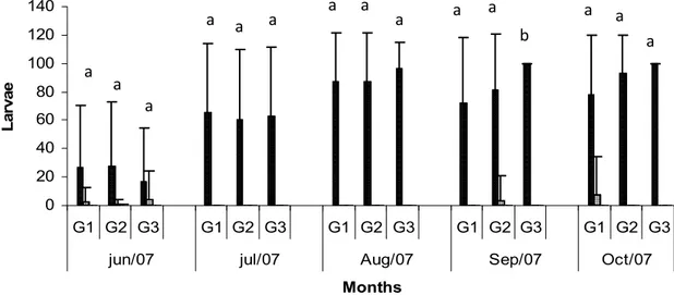
Figura 2 Monthly average values and standard deviations of the larvae of gastrointestinal nematodes (Cooperia sp., Haemonchus spp., Oesophagostomum sp., Trichostrongylus spp.) recovered from the coprocultures of the animals in the groups treated with the fungus Duddingtonia flagrans (G1) and the fungus Monacrosporium thaumasium (G2) (0.2g de fungus/10Kg of live weight) and the control group without fungus (G3), collected from June to October 2007, Viçosa, Minas-Gerais, Brazil. The values followed by the same capital letters (A) are similar (p >0.05) Tukey Test.. .. 30
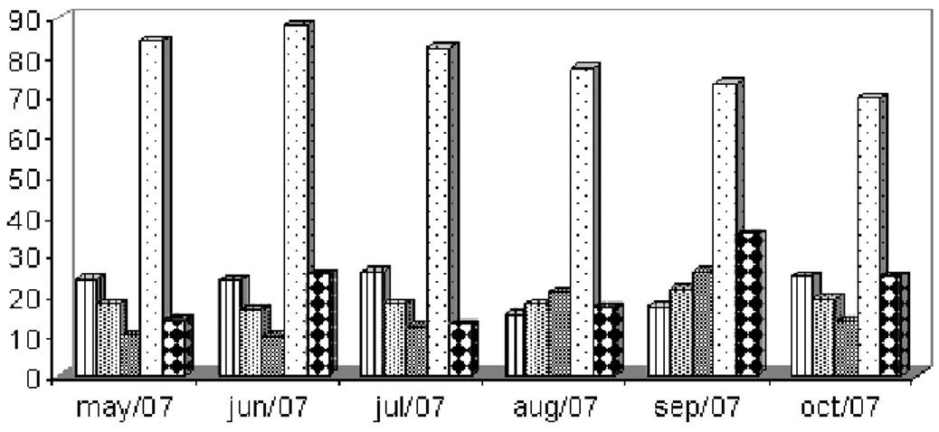
Figura 3 Monthly average temperature; minimum, average and maximum temperature (ºC) temperature, air relative humidity (%) and rainfall (mm3) recorded from May to October 2007, Viçosa, Minas Gerais, Brazil...31
(ii) Capítulo 2
fungus). Lines on bars represent standard deviation. Means followed by at least one common capital letter (A) in the row are not significantly different by Tukeys test at a 5% probability level ...42
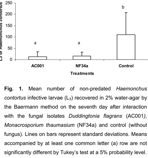
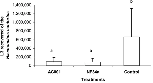
Figura 2 - Mean number of non-predated Haemonchus contortus infective larvae (L3) recovered in coprocultures by the Baermann method on the seventh treatment day after interaction with the fungal isolates Duddingtonia flagrans (AC001), Monacrosporium thaumasium (NF34a) and control (without fungus). Lines on bars represent standard deviation. Means followed by at least one common capital letter (A) in the row are not significantly different by Tukeys test at a 5% probability level . ..43
(ii) Capítulo 3
Figura 1 - (a and b)Destruction of Trichuris trichiura eggs (white arrow) after the 6-h observation period by isolates of Pochonia chlamydosporia (VC1 and VC4) (black arrow). SEM. Bar: (a) 10µm; (b) 10µm .. 55
Figura 2 - (a and b) Formation of appressoria of Pochonia chlamydosporia (white arrow) on Trichuris trichiura eggs (a and b), causing its destruction (black arrow) (b). SEM. Bar: (a) 10µm; (b) 10µm...55
Figura 3 - (a and b) Hyphae of fungi (a) Duddingtonia flagrans (AC001) and (b) Monacrosporium thaumasium (NF34a) (white arrow) attached to the surface of eggs (black arrow) during the entire observation period, without destruction of eggs. SEM. Bar: (a) 20µm; (b) 20µm . .56
Figura 4 - Trichuris trichiura eggs (black arrow) without fungus (control). Bar:
20µm ..56
arrow), causing its destruction, after 6h. Scanning electron microscopy (SEM). Bar: (a) 40 µm; (b) 100 µm; (c) 20 µm...57
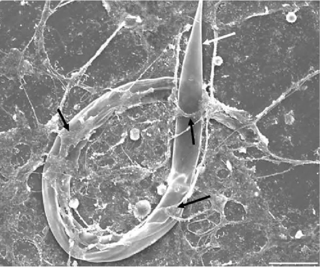
Figura 6 - SEM of infective larvae (L3) of preyed Haemonchus contortus (white arrow) by predator nematophagous fungi stimulating the production of traps (black arrow). Bar: 40µm...58
Figura 7 - (a and b) Observation of specific structures of the predator fungus Duddingtonia flagrans attached to infective larvae (L3) of Haemonchus contortus (white double arrow) and later causing its destruction. (a) Conidia (white arrow); (b) traps (black arrow). SEM. Bar: (a) 100µm; (b) 10µm...58
Figura 8 - (a and b) Production of chlamydospores (black arrow) of fungus Duddingtonia flagrans in Petri dishes. SEM. Bar: (a) 100µm; (b) 50µm...59
(ii) Capítulo 4
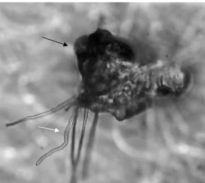
Figura 1 - Broken eggs of Trichuris vulpis (white arrow) and hyphae the Pochonia chlamydosporia (black arrow), after 21 days of interaction. Optical Microscopy - 40x objective lens .. ..70
Figura 2 Show Trichuris vulpis of the control group, without fungus ...70
(ii) Capítulo 5
Figura 1 Proglottids (black arrow) of an adult specimen (Anoplocephala perfoliata) obtained from the large intestines of parasitized horse, dead from gastrointestinal complications (intussusception) ....80
Anoplocephala perfoliata egg (black arrow) of the control group, without
fungus . ...80
Figura 3 (A and B) A; Anoplocephala perfoliata eggs (white arrow) and hyphae the Pochonia chlamydosporia (black arrow), effect type 1. Optical Microscopy - 40x objective lens. B; Anoplocephala perfoliata egg (white arrow), lytic effect with morphological alteration of embryo and eggshell
(effect type 2) 81
LISTA DE TABELAS
Página (ii) Capítulo 2
Tabela 1 - Daily means and standard deviations of non-predated infective larvae (L3) of Haemonchus contortus per 4mm diameter field in 2% water-agar during 7 days of treatment with the fungal isolates Duddingtonia flagrans (AC001), Monacrosporium thaumasium (NF34a) and control without
fungus . . ..41
(ii) Capítulo 4
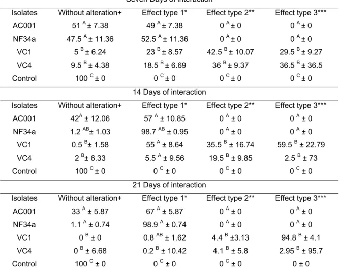
Tabela 1 - Percentages and standard desviation of ovicidal activity for the nematophagous fungus Duddingtonia flagrans (AC001), Monacrosporium thaumasium (NF34a), Pochonia chlamydosporia (VC1 and VC4) and the control without fungal treatment, against eggs of Trichuris vulpis after 7,14 and 21 days of interaction . . 69
(ii) Capítulo 5
RESUMO
SILVA, André Ricardo, D. Sc., Universidade Federal de Viçosa, Outubro de 2011. Controle biológico de nematóides gastrintestinais de ovinos, ovos de Trichuris trichiura, Trichuris vulpis e de Anoplocephala perfoliata por fungos nematófagos. Orientador: Jackson Victor de Araújo. Co-orientadores: Laércio dos Anjos Benjamim e Abelardo da Silva Jr.
As helmintoses gastrointestinais provocam danos e podem matar
ruminantes e outros animais, incluindo o homem. O uso indiscriminado de
anti-helmínticos provocou a seleção de populações de helmintos
resistentes aos diferentes grupos químicos utilizados. Fungos
nematófagos, como Duddingtonia flagrans, Monacrosporium thaumasium e Pochonia chlamydosporia são utilizados no controle biológico das helmintoses gastrintestinais. No primeiro experimento, ovelhas foram
tratadas com péletes contendo D. flagrans e M. thaumasium evidenciando
que esses fungos podem ser usados como uma alternativa para o
controle das nematodioses gastrintestinais de ovinos. No segundo
perfoliata de equinos. Os fungos D. flagrans (AC001) e M. thaumasium (NF34a) foram efetivos no controle das nematodioses gastrintestinais de
ABSTRACT
SILVA, André Ricardo, D. Sc., Universidade Federal de Viçosa, October, 2011. Biologic control of gastrointestinal nematodes of ovine, eggs of Trichuris trichiura, Trichuris vulpis and Anoplocephala perfoliata by nematophagous fungi. Adviser: Jackson Victor de Araújo. Co-Advisers: Laércio dos Anjos Benjamim and Abelardo da Silva Jr.
Gatrointetistinal helminthosis cause damage and can kill ruminants
and other animals, including man. The indiscriminate use of anthelminthic
caused the selection of populations of helminthes resistant to different
chemical groups used. Nematophagous fungi are used in the biological
control of gastrointestinal helminthosis, such as Duddingtonia flagrans, Monacrosporium thaumasium and Pochonia chlamydosporia. In the first experiment, sheep were treated with pellets with nematophagous fungi D. flagrans and M. thaumasium evidencing that the fungus can be used as an alternative for control of ovine gastrointestinal nematodiosis. In the second
experiment, D. flagrans (AC001 isolate) and M. thaumasium (NF34a isolate) were efficient in controlling in vitro larvae in the third stage (L3) of Haemonchus contortus of ovine and can be used in the biological control of this nematode. In the third experiment, isolates of P. chlamydosporia (VC1 and VC4) had an important role in the destruction of eggs of human
Trichuris trichiura. In addition, fungi D. flagrans (AC001) and M. thaumasium (NF34a) preyed L3 of H. contortus, but no predation by P. chlamydosporia was found. Thus, isolates of P. chlamydosporia (VC1 and VC4) can be potential biological controllers of Trichuris trichiura in humans and D. flagrans (AC001) and M. thaumasium (NF34a) of H. contortus for ovine. In the fourth experiment, isolates of P. chlamydosporia (VC1 and VC4) showed ovicidal activity (p < 0.05) in eggs of T. vulpis of dogs. Fungi of D. flagrans (AC001) and M. thaumasium (NF34a) did not show ovicidal
chlamydosporia isolates (VC1 e VC4) destroyed eggs of T. trichiura in humans and T. vulpis of dogs. The isolate (VC1) destroyed eggs of A. perfoliata in horses.
(I) INTRODUÇÃO GERAL
Grandes perdas econômicas em pequenos ruminantes estão associadas ao parasitismo por helmintos gastrintestinais, principalmente Haemonchus contortus, que se alimenta de sangue. Prejuízos por essas infecções estão relacionados, principalmente, com a queda na produção, retardo no crescimento do animal, custos com tratamento médico veterinário e, em algumas situações, com a morte desses animais (ARAÚJO, 2006; SILVA et al., 2008; 2009).
No Brasil, grande parte da criação de ovinos é feita em regime de pasto ou parcial, levando a constante infecção por parasitos nas pastagens. As perdas econômicas mundiais anuais causadas pela infecção por nematóides gastrintestinais são estimadas em milhões de dólares, devido ao impacto que causam na produção de carne e leite e aos altos custos das medidas de controle, além de perdas na produção de lã e couro (ANUALPEC, 2003; SILVA et al., 2009).
O controle de verminoses em ovinos é geralmente feito com anti-helmínticos, os quais não têm sido eficazes no controle destes nematóides, devido à sua ação restrita aos parasitos adultos, ao aparecimento de resistência aos benzimidazóis, levamisois e avermectinas além da baixa possibilidade da formulação de novos compostos químicos de maior eficiência (ROCHA et al., 2006; SILVA et al., 2009).
Problemas com a resistência e ecotoxicidade enfatizam a necessidade de programas integrados de controle parasitário que assegurem saúde e segurança dos organismos vivos, por meio de tratamentos estratégicos baseados na epidemiologia, eliminação de vermifugações desnecessárias, utilização de pastoreio alternado e higienização de pastagens. Além disso, o uso continuado de uma mesma classe de anti-helmíntico deve ser evitado, como a rápida rotação de compostos e a utilização de doses inferiores às recomendadas (MOTA et al., 2003; THAMSBORG et al., 1999; SILVA et al., 2008; 2009; 2010a; 2011).
de origem animal. Medidas eficazes para o controle desses organismos devem ser buscadas, pois o controle químico tem problemas econômicos e ecológicos.
Controle biológico é uma alternativa viável e com resultados promissores in vitro e in vivo (LARSEN, 2000; SILVA et al; 2009; 2010a,b,c; 2011). Fungos nematófagos, constituem uma opção ao controle dos nematóides gastrintestinais de animais domésticos com ação concentrada no ambiente fecal para o combate de larvas de vida livre dos parasitos (GRONVOLD, et al., 1996; ARAÚJO et al., 1998; SILVA et., al 2009). O emprego destes fungos apresenta oportunidade de controle dos estágios de vida livre de nematóides nas pastagens, reduz as reinfecções e contribui para o controle das parasitoses (GRONVOLD et al., 1996; FAEDO et al., 1997; 2002; LARSEN, 2000; ROCHA et al., 2007; SILVA et., al 2009). O controle biológico das helmintoses gastrintestinais com fungos nematófagos visa diminuir a contaminação ambiental por estádios de vida livre dos parasitos que, em muitas situações, podem causar doenças tanto nos animais quanto nos humanos. Algumas helmintoses se destacam, pois parte de seu ciclo vital ocorre no ambiente e o homem pode ser hospedeiro acidental (SILVA et., al 2009; 2011).
A ação dos fungos nematófagos não é bem elucidada no controle biológico de ovos de Trichuris sp. e de Anoplocephala perfoliata (SILVA et., al 2010b,c; 2011).
Nematóides
Helmintos de interesse médico veterinário são divididos em dois filos: O Nemathelminthes engloba os Nematoda, e o Platyhelmintes é formado pelos Cestoda e Trematoda (URQUHART, et al., 1998).
O ciclo biológico de H. contortus é direto, com uma fase pré-parasitária, tendo larvas (L1 a L3) no ambiente e uma fase parasitária (adultos machos e fêmeas) dentro do hospedeiro que se reproduzem e os ovos são eliminados juntamente com as fezes do hospedeiro. Os sintomas observados caracterizam-se, principalmente, pela anemia e hipoproteinemia, perda do apetite, emagrecimento, que pode resultar também na morte dos animais.
A parasitose cosmopolita trichurose causada pelo nematóide Trichuris sp., acomete o intestino grosso do trato gastrintestinal de animais domésticos, mamíferos silvestres e inclusive do homem. Possui um período de latência no solo antes de atingir o hospedeiro, não necessita de hospedeiro intermediário e apresenta ciclo biológico direto. Dípteros muscóides como Chrysomya megacephala, Musca domestica e outros, são vetores mecânicos de ovos de vários helmintos, inclusive Trichuris sp. presentes no corpo e no conteúdo intestinal das moscas (OLIVEIRA et al., 2002; SILVA et., al 2010b; 2011). Ovos de Trichuris sp. são eliminados com as fezes e dependendo da temperatura desenvolvem em um ou dois meses no ambiente para o estágio infectante L1 que quando ingerido pelos animais, torna-se adulto no intestino. Um dos aspectos mais importantes relacionados à epidemiologia deste parasito é que os ovos poderão sobreviver nas pastagens e no meio ambiente por três ou quatro anos. Os sinais clínicos dessa parasitose podem não ser aparentes, por isto seu diagnóstico deve ser feito com métodos laboratoriais como exame de fezes. O controle e o tratamento eficiente estão ligados respectivamente ao conhecimento de sua epidemiologia e à utilização de drogas anti-helmínticas (URQUHART et al., 1998). No entanto, os fungos nematófagos aparecem como uma possível ferramenta no controle desta parasitose (SILVA et al., 2010b, 2011).
por ácaros das forragens, nos quais se desenvolvem até o estágio de cisticercoide em 2-4 meses. Os cestóides adultos são encontrados no intestino dos equinos 1 ou 2 meses após a ingestão de foragens infectadas com ácaros (URQUHART et al., 1998).
Fungos nematófagos
Pesquisas com fungos que parasitam helmintos começaram com as observações de Lohde, em 1874, com o fungo endoparasita Harposporium anguilullae (KERRY, 1984). Fungos nematófagos como controladores biológicos vem sendo amplamente estudados no mundo para a redução de populações de nematóides no meio ambiente. Essas ações ocorrem através de organismos vivos que atuam como antagonistas naturais no ambiente (controle biológico). Fungos nematófagos são a alternativa mais promissora para o controle de helmintos. Eles são antagonistas naturais e capazes de promover a captura, a morte ou mesmo sua destruição, como endoparasitas, predadores, oportunistas (parasitas de ovos e cistos) e aqueles que produzem metabólitos tóxicos. A seleção de animais resistentes, vacinas e o controle biológico com fungos representam os últimos avanços contra as parasitoses gastrintestinais de animais domésticos (MANKAU, 1980; WALLER & LARSEN, 1993; SILVA et al., 2009; 2010a,b).
Os gêneros Duddingtonia e Monacrosporium, são os fungos predadores mais estudados (DIMANDER et al., 2003). Duddingtonia flagrans é a espécie mais promissora e estudada no controle das helmintoses de animais domésticos, com ação predatória por hifas adesivas simples e, além disso, possui dois tipos de estruturas (conídios e clamidósporos) intercalados em hifas maduras que permitem seu uso como controlador biológico, crescendo de forma lenta em temperaturas abaixo de 25° C, possuindo rápido crescimento em temperatura em torno de 30° C, (LARSEN, 1999).
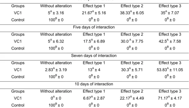
O fungo ovicida Pochonia chlamydosporia é uma das espécies mais promissoras cuja ação é medida por: sem alteração; efeito tipo 1, efeito lítico sem prejuízo morfológico à casca do ovo, onde hifas são observadas aderidas à casca; tipo 2, efeito lítico com alteração morfológica da casca e embrião do ovo, sem penetração de hifas através da casca; e tipo 3, efeito lítico com alteração morfológica do embrião e da casca, além de penetração de hifas e colonização interna do ovo (LYSEK, 1976; 1978).
Duddingtonia flagrans e Monacrosporium thaumasium foram eficientes no controle de nematóides em laboratório e campo (MELO et al., 2003; TERRIL et al., 2004; SILVA et al., 2009;2010a). Pochonia chlamydosporia é pouco estudada, mas com atividade ovicida comprovada in vitro (ARAÚJO et al., 2008; BRAGA et al., 2007; 2008; SILVA et al., 2010c,d).
OBJETIVOS DO PRESENTE TRABALHO:
1) Avaliar a efetividade dos fungos nematófagos Duddingtonia flagrans (AC001) e Monacrosporium thaumasium (NF34a) em formulação, pellets destes fungos em matriz de alginato de sódio, no controle biológico dos namatóides gastrintestinais de ovinos a campo.
2) Avaliar a atividade predatória dos fungos Duddingtonia flagrans (AC001) e Monacrosporium thaumasium (NF34a) sobre larvas infectantes (L3) de Haemonchus contortus de ovinos.
3) Demonstrar comparativamente por Microscopia Eletrônica de Varredura (MEV) a atividade in vitro dos fungos nematófagos ovicidas Pochonia chlamydosporia (isolados VC1 e VC4) e predadores D. flagrans (AC001) e M. thaumasium (NF34a) sobre ovos de Trichuris trichiura de humanos e larvas infectantes (L3) de Haemonchus
contortus de ovinos.
4) Avaliar o efeito in vitro de quatro isolados de fungos nematófagos, Duddingtonia flagrans (AC001), Monacrosporium thaumasium (NF34a) e Pochonia chlamydosporia (VC1 e VC4) sobre ovos de Trichuris vulpis de cães.
(I) REFERÊNCIAS BIBLIOGRÁFICAS
ANUALPEC. Anuário estatístico da produção animal. São Paulo: FNP Consultoria & Comércio. 2003. 380 p.
ARAÚJO, J.V. Diagnostico das helmintoses. Universidade Federal de Viçosa, 2006. p. 9-46 (Caderno didático, 113).
ARAÚJO, J.V.; BRAGA, F.R.; SILVA, A.R.; ARAÚJO, A.M.; TAVELA, A.O. In vitro evaluation of the effect of the nematophagous fungi Duddingtonia flagrans, Monacrosporium sinense and Pochonia chlamydosporia on Ascaris suum eggs. Parasitology Research, v.102, p.787790, 2008.
ARAUJO, J.V.; GOMES, A.P.S.; GUIMARÃES, M.P. Biological control of bovine gastrointestinal nematode parasites in Southern Brazil by nematode-trapping fungus Arthrobotrys robusta. Revista Brasileira de Parasitologia Veterinária, v. 7, n. 2, p. 117-122, 1998.
BARRON, G.L. The nematode-destroying fungi. Topics in Mycobiology. Canadian Biological Publications. p.140, 1997.
BRAGA, F.R.; ARAUJO, J.V.; CAMPOS, A.K.; CARVALHO, R.O.; SILVA, A.R.E.; TAVELA, A.; MACIEL, A.S. Observação in vitro da ação dos isolados fúngicos Duddingtonia flagrans, Monacrosporium thaumasium e Verticillium chlamydosporium sobre ovos de Ascaris lumbricoides (Lineu, 1758). Revista da Sociedade Brasileira de Medicina Tropical, v. 40, p. 356-358, 2007.
DIMANDER, S. O.; HOGLUND, J.; UGGLA.; SPÖRNDLY, E.; WALLER, P. J. Evaluation of gastro-intestinal nematode parasite control strategies for first season grazing cattle in Sweden. Veterinary Parasitology, v. 11, n. 2, p. 192-209, 2003.
FAEDO, M.; LARSEN, M.; DIMANDER, S.O.; YEATES, G.W.; HÖGLUND, J.; WALLER, P.J. Growth of the fungus Duddingtonia flagrans in soil surrounding feces deposited by cattle or sheep fed the fungus to control nematode parasites. Biological Control, v.23, p.64-70, 2002.
FAEDO, M.; LARSEN, M.; WALLER, J.P. The potential of nematophagous fungi to control free-living stages of nematodes parasites of sheep, comparison between Australian isolates of Arthrobotrys spp. and Duddingtonia flagrans. Veterinary Parasitology, v. 72, p. 149155, 1997.
GRÖNVOLD, J.; NANSEN, P.; HENRIKSEN, S.A.; LARSEN, M.; WOLSTRUP, J; BRESCIANI, J.; RAWAT, H.; FRIBERT, L. Induction of traps by Ostertagia ostertagi larvae, chlamisdospore production and growth rate in nematode-trapping fungus Duddingtonia flagrans. Journal of Helminthology, v.70, p.291-297, 1996.
KERRI, B.R. Nematophagous fungi and the regulation of nematode poppulations in soil. Helminthology, v.53, p.1-14, 1984.
LARSEN, M. Biological control of helminthes. Internacional Journal for Parasitology, v.29, p.139-146, 1999.
LARSEN, M. Prospects for controlling animal parasitic nematodes by predacious micro fungi. Parasitology, v.120, p.121-131, 2000.
LYSEK, H. Classification of ovicide fungi according to type of ovicidity. Acta University Palacki Olomue, v. 76, p. 9-13, 1976.
MANKAU, R. Biological control of nematode pest by natural enemies. Annual Review of the Phytopatholgy, v. 18, p. 415-440, 1980.
MELO L. M., BEVILAQUA C. M. L. ARAÚJO J. V., MELO A. C. F. L. Atividade predatória do fungo Monacrosporium thaumasium contra o nematóide Haemonchus contortus, após passagem pelo trato gastrintestinal de caprinos. Ciência Rural, v.33, n.1, p.169-171, 2003.
MOTA, M.A.; CAMPOS, A.K.; ARAÚJO, J.V. Controle biológico de helmintos parasitos de animais: estágio atual e perspectivas futuras. Pesquisa Veterinária Brasileira, v.23, n.3, p.93-100, 2003.
OLIVEIRA V.C.; MELLO R.P.; DALMEIDA J. M.; Dípteros muscóides como vetores mecânicos de ovos de helmintos em jardim zoológico, Brasil. Revista de Saúde Pública, v.36, p.614-620, 2002.
ROCHA, R.A. Efficacy of the nematode-trapping fungus Duddingtonia flagrans against infections by Haemonchus and Trichostrongylus species in lambs at pasture. Journal of Helminthology, v.16, p.1-6, 2007.
ROCHA, R.A.; PACHECO, R.D.L.; AMARANTE, A.F.T. Efficacy of homeopathic treatment against natural infection of sheep by gastrointestinal nematodes. Revista Brasileira de Parasitologia Veterinária, Rio de Janeiro, v.15, n.1, p.23-27, 2006.
SILVA, A.R.; ARAUJO, J.V.; Braga, F.R.; Alves, C.D.F.; Frassy, L.N. Comparative analysis of destruction of the infective forms of Trichuris trichiura and Haemonchus contortus by nematophagous fungi Pochonia chlamydosporia; Duddingtonia flagrans and Monacrosporium thaumasium by scanning electron microscopy. Veterinary Microbiology, v. 147, p. 214-219, 2011.
SILVA, A.R.; ARAUJO, J.V.; BRAGA, F.R.; Alves, C.D.F.; Frassy, L.N. In vitro ovicidal activity of the nematophagous fungi Duddingtonia flagrans, Monacrosporium thaumasium and Pochonia chlamydosporia on Trichuris vulpis eggs. Veterinary Parasitology, v. 172, p. 76-79, 2010b.
SILVA, A.R.; ARAÚJO, J.V.; BRAGA, F.R.; FRASSY, L.N.; TAVELA, A.O., CARVALHO, R.O.; CASTEJON, F.V. Biological control of sheep gastrointestinal nematodiasis in a tropical region of the southeast of Brazil with the nematode predatory fungi Duddingtonia flagrans and Monacrosporium thaumasium. Parasitology Research, v. 105, p. 1707-1713, 2009.
SILVA, A.R.; ARAÚJO, J.V.; BRAGA, F.R.; ALVES, C.D.F.; FILHO, J.D.R. Destruction of Anoplocephala perfoliata eggs by the nematophagous fungus Pochonia chlamydosporia, v. 30, p. 701-704, 2010c.
SILVA, A.R.; ARAUJO, J.V.; BRAGA, F.R.; OLIVEIRA, A.C.; CARVALHO, R.O.; ARAUJO, J.M.; CASTEJON, F.V. Avaliação da eficácia de compostos anti-helmínticos sobre nematóides parasitos gastrintestinais (Strongyloidea) de caprinos. Revista Brasileira de Parasitologia Veterinária, v.17, p. 123-128, 2008.
THAMSBORG, S.M.; POEPSTORFF, A.; LARSEN, M. Integrated and biological control of parasites in organic and conventional production systems. Veterinary Parasitology, v.84, p. 169-186, 1999.
TERRIL, T.H.; LARSEN, M.; SAMPLES, O.; HSTED, S.; MILLER, J.E.; KAPLAN, R.M.; GELAYE,S. Capability of the nematode-trapping fungus Duddingtonia flagrans to reduce infective larvae of gastrointestinal nematodes in goat feces in the southeastern United States: dose titration and dose time interval studies. Veterinary Parasitology, v.120, p.285-296, 2004.
URQUHART, G.M., ARMOUR, J., DUNCAN, J.L., DUNN, A.M., JENNINGS, FW. 1998. Parasitologia Veterinária. (Guanabara Koogan, Rio de Janeiro).
(ii) Capítulo 1
BIOLOGICAL CONTROL OF SHEEP GASTROINTESTINAL NEMATODIASIS IN A TROPICAL REGION OF THE SOUTHEAST OF BRAZIL WITH THE NEMATODE PREDATORY FUNGI Duddingtonia
flagrans AND Monacrosporium thaumasium
Abstract
Fungus can be used as an alternative for control of ovine gastrointestinal
nematodiosis. Formulations in matrix of sodium alginate (pellets) of the nematode predatory fungi Duddingtonia flagrans and Monacrosporium thaumasium were evaluated in the biological control of sheep gastrointestinal nematodiasis. Three groups (1, 2 and 3), each one with eight sheep of the Santa Inês breed, at the ages of 15-48 months, were placed in paddocks of Brachiaria decumbens for five months. In group 1, each animal received 1g/10 kg of live weight (l.w.) of pellets of D. flagrans (0.2g of fungus/10Kg l.w.). In group 2, each animal received 1g/10 kg of l.w. of pellets of the fungus M. thaumasium (0.2g of fungus/10Kg l.w.), twice a week, for five months. In group 3 (control), the animals received 1g/10 kg of live weight of pellets without fungus. The monthly average of the egg countings per gram of feces of the animals of groups 1 and 2 treated, respectively were 71.6% and 61.1% smaller, respectivelly, in comparison to the animals of group 3 (control). The treatment of sheep with pellets containing the nematophagous fungi D. flagrans and M. thaumasium may be used as an alternative for the control of sheep gastrointestinal nematodiasis.
Introduction
Sheep raising is a widely explored activity in tropical countries, seeking meat, milk and leather production (Vieira 2003). The management practices and technologies used are not always adequate, which leads to health problems, especially those related to helminthosis (Ahid et al. 2008).
The gastrointestinal parasitism by nematodes causes diseases and can kill ruminants throughout the world, but the greatest economic impact is in the growth decrease of young animals, resulting in low productivity (Araújo et al. 2007). Younger sheeps are more susceptible to helminthosis than adults and with increasing age acquires immunity against these parasites. All age groups are affected by helminths, but the most susceptible to this helminthosis are the lambs, and pregnant females (Urquhart et al. 1998).
The severity of the disease and the losses in production and productivity depend on the intensity of the infection, immunity and the host nutritional status (Stear et al. 2007). It is very important to understand the epidemiology of the ruminant helminthosis in order to provide an efficient strategic control in a region.
The indiscriminate use of antihelminthic provoked the selection of helminthes populations that are resistant to the different chemical groups used in sheep treatments (Mota et al. 2003). Besides, the chemical residues of such products have a negative impact on the environment and public health (Boray et al. 1990).
present in surviving the passage through the gastrointestinal tract of ruminants without losing their predatory activity is an important pre-requisite in the biological control of helminthosis (Araújo and Sampaio, 2000; Dias et al. 2007).
Sodium alginate-based formulations have been evaluated experimentally in the control of animal parasite nematodes by some research laboratories. Such formulations have provided good results under laboratory and field conditions (Campos et al. 2007).
The objective of this work was to evaluate the effectiveness of the nematophagous fungi D. flagrans and M. thaumasium, by testing pellets formulations of these fungi in a sodium alginate matrix, in the biological control of gastrointestinal nematodiasis in sheep raised in the field.
Material and Methods
Organisms
Panagrellus sp. (free-living nematodes) were kept in Petri plaques with a medium of dampened and mashed oat flakes. These nematodes were removed from the culture medium through the immersion of small amounts of oat in distilled water in the Baermann funnel and collected in tubes of hemolysis after 6 hours of decantation. The nematodes were counted, by means of light microscopy (40x), by taking six aliquots of 10 µL, taking the average and extrapolating for the total volume.
Two isolates of the predatory fungi D. flagrans (AC001) and M. thaumasium (NF34a) were kept at 4ºC, in the dark and in test tubes containing cornmeal-agar 2%. These isolates came from a Brazilian soil and belonged to the mycology collection of the Universidade Federal de Viçosa.
which were made in a sodium alginate matrix, according to Walker and Connick (1983) and modified by Lackey et al. (1993).
In vivo experimental essay
The experiment was developed in the experimental farm of the Universidade Federal de Viçosa, located in the city of Canaã, state of Minas Gerais, southeast of Brazil, latitude 20º4520 and longitude 42º5240, from May to October 2007.
In the beginning of the experiment, 24 Santa Inês sheep, at the ages from 15 to 48 months, with average l.w. of 32.5 kg were previously treated with a dose of Trimix®-Merial from Brazil, [closantel (10%), albendazole (5%), levamisole (6.4%), ivermectina B1a (0.2%), selenium (0.1g) and cobalt (0.44%)], with the oral dose of 1ml/10Kg of l.w.
After the 15th day of the antihelminthic treatment, the sheep were placed into three groups 1, 2 and 3, each of them with eight animals. The characteristics considered for this division were the weight and age similarity between the animals of the groups. Later, each group was placed in three pickets, each one with 2.0 ha and pasture of Brachiaria decumbens, naturally infested with gastrointestinal parasite helminths, due to the previous grazing of young and adult animals. In group 1, each animal received 1 gram of pellets (0.2 g of fungal mycelium) for each 10 kg of l.w. containing the fungus D. flagrans (AC 001). In group 2, each animal received 1 gram of pellets (0.2 g of fungal mycelium) for each 10 kg of l.w. containing the fungus M. thaumasium (NF34a). In group 3, the animals received 1 gram of fungus-free pellets for each 10 kg of l.w. All the animals received the pellets orally, twice a week, mixed in concentrated and balanced ration, provided daily for meat sheep (22% of total protein Universidade Federal de Viçosa), and water ad libitum during five months, starting from June 2007.
Samples of feces were collected from the animals to observe the growth of the fungi. The feces were placed in plaques containing water-agar 2% and 1.000 Panagrellus sp. and put into a greenhouse, at 25oC, for 10 days.
Simultaneously to the EPG exam, coprocultures were carried out, for each animal, according to the methodology described by Roberts and O´Sullivan (1950). The identification of the infectant larvae in the coprocultures was performed according to Keith (1953).
EPG count curves (EPG) and larvae recovered from coprocultures of animals of both treated and control groups were recorded, and percentage of larval reduction was determined according to Mendoza-De-Guives et al. (1999):
reduction (%)= Mean L3 recovered from control group - Mean L3 recovered from treated group x 100 Mean L3 recovered from control group
Every 15 days, in each picket of the several groups of animals, two samples of pasture were collected, in W of varied points, alternates of up to 20 cm from the fecal mass and 20-40 cm far from the fecal mass, according to Amarante et al. (1996). Then, 500g of pasture were weighed, and larvae of sheep parasite nematodes were recovered from there. The pasture samples were put into a drying greenhouse at 100oC, for 3 days, for the achievement of dry matter. The data obtained were transformed in number of larvae by kilogram of dry matter.
Every 15 days, the scores of color intensity of the ocular mucosa were achieved in the animals, by the Famacha method, according to Bath and Van Wick (2001).
The weather conditions, such as temperature, air relative humidity and rainfall are represented in Figure 3.
multiple comparison test with 1% probability. The analyses were performed using the BioEstat 3.0 Software (Ayres et al., 2003).
Results
The monthly average values of the EPG countings are represented in Figure 1. In the beginning of the experiment (June), the EPG of the animals in group 1 was higher than that of the animals in groups 2 and 3, but no statistical difference was observed (p>0.05). From the month of July, the animals in group 3 (control) presented higher averages for the EPG than the animals in groups 1 and 2, until the end of the experiment (October). On the other hand, in the last three months of the experiment (August, September and October), a statistical difference was observed (p<0.05) between the animals in groups 1 and 2 and the animals of the control group. There was no difference (p>0.05) between the animals in groups 1 and 2 treated with the fungi D. flagrans and M. thaumasium, respectively, during the 5 months of the experiment. Besides, the monthly average of the EPG of the animals in group 1 treated with pellets containing the fungus D. flagrans was 71.6% lower, compared to the animals in group 3 (control). For the animals in group 2 treated with pellets containing the fungus M. thaumasium, the reduction observed was of 61.1%, in comparison to the animals in group 3 (control). This decrease for the percentage values found for the animals in groups 1 and 2, was probably determined by the action of the fungi D. flagrans and M. thaumasium, also considering that this action was satisfactorily demonstrated in the last 3 months of the experiment.
gastrointestinal nematodes (Cooperia, Oesophagostomum. and Trichostrongylus), no difference (p>0.05) was observed during all the experiment between the animals of the three groups and they were presented in very low levels.
As to the number of L3 of Cooperia sp., Haemonchus sp., Oesophagostomum sp. and Trichostrongylus spp. recovered from the paddocks in the distances of up to 20 cm from the fecal mass and 20-40 cm far from the fecal mass, no difference (p>0.05) was observed between the groups during all the experiment (from June to October). For the group 1, in the distances of up to 20 cm and 20-40 cm from the fecal mass, the reduction percentual was 33.33% and 50%, respectively, in relation to group 3. Group 2 showed percentual reduction of 33.33% and 47.22%, at the same distances, in relation to group 3.
As to the weight of the animals, it was not observed any difference (p>0.05) between the animals from the three groups during all the experiment.
The Famacha method revealed a difference between the animals in groups 1 and 2 (p<0.05) and the animals in group 3 in August and September.
Discussion
For the animals of group 2 treated with M. thaumasium, this reduction was of 61.1% in comparison to the animals in group 3. Fungi of the genus Monacrosporium sp. (M. sinense, M. thaumasium, M. appendiculatum, M. ellypsosporum) were evaluated by several authors, demonstrating effectiveness in the control of gastrointestintinal nematodiasis of different animal species (Araújo et al. 1992; Gomes et al. 1999; Castro et al. 2003; Assis and Araújo 2003; Campos et al. 2006; Araújo et al. 2007). These results are also in accordance with the present work, since the EPG decrease was observed in the animals in the group treated with M. thaumasium.
In the coprocultures, Haemonchus sp. was the most prevailing gastrointestinal parasite nematode in all the groups, reaching percentages of 77.85%, 92.86% and 100% for the animals in groups 1, 2 and 3, respectively, in October. Such results agree with Amarante et al. (2004) and Rocha et al. (2008), who demonstrated that in the southeast region of Brazil, Haemonchus contortus and Trichostrongylus colubriformis were the most prevailing gastrointestinal nematodes in sheep. H. contortus was also the most prevailing gastrointestinal nematode in goats, followed by T. colubriformis, in the Brazilian semiarid region (Araújo et al. 2007). These results are also in accordance with the present work (Figure 2).
In Malaysia, Chandrawathani et al. (2002) observed the effectiveness of the treatment with the fungus D. flagrans, daily administered to ovine. On the other hand, Terrill et al. (2004), using this same fungus, found a reduction in the larvae found in feces of goats infected mainly with H. contortus. According to Casillas et al. (2008), D. flagrans can be considered a viable alternative for the control of haemonchosis in sheep, which were treated with chlamydospores of this fungus. Araújo et al. (2007) also recorded a larva decrease in the coprocultures of the animals treated with the fungus M. thaumasium in the Brazilian semi-arid. These results are in accordance with the present work, where it was observed the effectiveness of the fungi D. flagrans and M. thaumasium on sheep gastrointestinal parasite nematodes.
experiment took place. However, Larsen et al. (1998) evaluated the potential of the fungus D. flagrans in the control of the free living stages of the animal parasites and achieved a reduction of more than 80% of the number of L3 in paddocks.
The correlation coefficient between EPG and infective larvae recovered from paddocks of group 1 within 020 cm from fecal pats was 0.7262; and for the distance 2040 cm was 0.8076. For group 2, the correlation coefficient between EPG and infective larvae recovered within 020 cm from the fecal pats was -0.5760 and within 2040 cm was 0.2436. For group 3, the correlation coefficient recovered within 020 cm from the fecal pats was -0.7519 and within 2040 cm was -0.3864. In group 1, was observed strong positive correlation between EPG and infective larvae. In group 2, these results showed negative correlation within 020 cm and positive within 2040 cm. Although in the group 3 was observed negative correlations between EPG and infective larvae. Dias et al. (2007) pointed out, there might be dependence between EPG and infective larvae recovered from pastures even if the correlations are null. Besides, the availability of larvae on pasture may be determined by contamination from animals, as well as environmental factors, parasite and host (Lima et al., 1997).
Weather conditions, such as temperature, air relative humidity and rainfall were responsible for the development of the free living stages of the nematodes in paddocks, since they were observed in the pickets of all groups and in all the months. According to Honer and Bianchin (1987), during the rainy season, pasture contamination would reach maximum levels, while in the dry season, the helminth population would survive almost exclusively inside the hosts, due to the less favorable weather. These results agree with the present work, in which the highest recovery of larvae from paddocks was observed in the periods with higher rainfall indices (August and September; Fig. 3).
gain for the animals (Santa Inês x Corriedale) treated with the fungus D. flagrans in an experiment carried out in the field during approximately one year. A higher weight gain in the animals was also observed by Araújo et al. (2007) when testing the fungus M. thaumasium in goats in the Brazilian semi-arid.
In the present work, the application of the Famacha method in the animals revealed difference (p<0.05) between the animals in groups 1 and 2 and the animals in group 3 only for August and September. Molento (2004) did not observe a satisfactory result either, when using this method along with the chemical control in small ruminants.
The nematophagous fungi D. flagrans and M. thaumasium used in the present work were responsible for the EPG decrease of the animals in groups 1 and 2, in comparison to the animals in group 3.
Conclusion
The treatment of sheep with pellets containing the nematophagous fungi D. flagrans and M. thaumasium can be used in the control of sheep gastrointestinal nematodes.
Acknowledgements
This study was supported by the Research Project of the Universidade Federal de Viçosa, Brasil. Thanks are also due to FAPEMIG for the financial support and CAPES for the scholarships granted.
References
Ahid SMM, Suassuna DAC, Maia MB, Costa VMM, Soares HS (2008) Parasitos gastrintestinais em caprinos e ovinos da região oeste do Rio Grande do Norte, Brasil. Ciênci Anim Brasileira. 9:212-218.
Monacrosporium thaumasium (Drechsler, 1937) no controle de nematóides de bovinos. Arq. Bras. Med. Vet. Zootec. 55:568-573.
Amarante AFT, Padovani CR, Barbosa MA (1996) Contaminação de
pastagens por larvas de nematóides gastrintestinais parasitos de bovinos e
ovinos em Botucatu- SP. Rev. Bras. Parasitol. Vet. 5:25-73.
Amarante AFT, Bricarello PA, Rocha RA, Gennari SM (2004) Resistence of Santa Ines, Suffolk and Ile de France sheep to naturally acquired gastrointestinal nematodes infections. Vet. Parasitol. 120:91-106.
Araújo JV, Sampaio WM (2000a) Effects of temperature, mineral salt and passage through gastrointestinal tract of calves on alginate formulation of Arthrobotrys robusta. Rev. Bras. Parasitol. Vet. 9:55-59.
Araújo JV, Santos MA, Ferraz S, Maia AS, Magalhães ACM (1992) Controle de larvas infectantes de Haemonchus placei por fungos predadores da espécie Monacrosporium ellypsosporum em condições de laboratório. Arq. Bras. Med. Vet. Zootec. 44:521-526.
Araújo JV, Sampaio WM, Vasconcelos RS, Campos AK (2000). Effects of different temperatures and mineral salt on pellets of Monacrosporium thaumasium - a nematode-trapping fungus. Veterinarski Arhiv. 80:181-190.
Araújo JV, Guimarães MP, Campos AK, Sá NC, Sarti P, Assis CI (2004). Control of bovine gastrointestinal nematode parasites using pellets of the nematode-trapping fungus Monacrosporium thaumasium. Ciência Rural. 34:457-463.
Araújo JV, Rodrigues MLA, Silva WW, Vieira LS (2007) Controle biológico de nematóides gastrintestinais de caprinos em clima semi-árido pelo fungo Monacrosporium thaumasium. Pesq. Agropec. Bras. 42:1177-1181.
Assis RCL, Araújo JV (2003) Avaliação da viabilidade de fungos predadores do gênero Monacrosporium em predar ciatostomíneos após a passagem pelo trato gastrintestinalde eqüinos em formulação de alginato de sódio. Rev. Bras. Parasitol. Vet. 12:109-113.
Ayres M, Ayres JRM, Ayres DL, Santos AS (2003) Aplicações estatísticas nas áreas de ciências biológicas. Belém: Sociedade Civil Mamirauá: Brasília CNPq, 290p.
Bath GF, Van Wyk JA (2001) Using the Famacha system on commercial sheep farms in South Africa. In: INTERNATIONAL SHEEP VETERINARY CONGRESS, 1., 1992, Cidade do Cabo. Anais... Cidade do Cabo: University of Pretoria. 1, 3.
Boray JC, Martin PJ, Roush RT (1990) Resistance of parasites to antiparasitic drugs: The report of a Round Table Conference held at the VIIth International Congress of Parasitology. Int. J. Parasitol. 4:549-550.
Campos AK (2006) Fungos nematófagos no controle de nematóides gastrintestinais de ruminantes. 138f. Tese (Doutorado) Instituto de Ciências Biológicas, Universidade Federal de Minas Gerais, Belo Horizonte.
Campos AK, Araújo JV, Assis RCL, Gandra JR, Guimarães MP (2007) Viabilidade de formulação peletizada do fungo nematófago Monacrosporium sinense, no controle biológico de nematóides parasitos gastrintestinais de bezerros. Arq. Bras. Med. Vet. Zootec. 59:14-20.
Castro AA, Oliveira CRC, Anjos DHS, Ornelas EI, Bittencourt VREP, Araújo JV, Sampaio IBM, Rodrigues MLA (2003) Potencial dos fungos nematófagos Arthrobotrys sp. e Monacrosporium thaumasium para o controle de larvas de ciatostomíneos de eqüinos (Nematoda: Cyathostominae). Rev. Bras. Parasitol. Vet. 12:53-57.
Chandrawathani P, Omar J, Peter JW, Johan H, Larsen M, Wan MZ (2002) Nematophagous fungi as a biological control agent for nematode parasites of small ruminants in Malaysia: a special emphasis on Duddingtonia flagrans. Vet. Res. 33:685696.
Chandrawathani P, Jamnah O, Adnan M, Waller PJ, Larsen M, Gillespie AT (2004) Field studies on the biological control of nematode parasites of sheep in the tropics, using the microfungos Duddingtonia flagrans. Vet Parasitol 120:177-187.
Dias AS, Araújo JV, Campos A K, Braga FR, Fonseca TA (2007) Relação
entre larvas recuperadas da pastagem e contagem de ovos por gramas de
fezes (OPG) de nematóides gastrintestinais de bovinos na micro região de
Viçosa, Minas Gerais. Rev. Bras. Parasitol. Vet. 16:33-36.
Dimander SO, Höglund J, Uggla A, Spörndly E, Waller PJ (2003) Evaluation of gastro-intestinal nematode parasite control strategies for first-season grazing cattle in Sweden. Vet. Parasitol. 111:192-209.
Fontenot ME, Miller JE, Peña MT, Larsen M, Gillespie A (2003) Efficiency of feeding Duddingtonia flagrans chlamydospores to grazing ewes on reducing availability of parasitic nematode larvae on pasture. Vet. Parasitol. 118:203-213.
Gordon HM, Whitlock HV (1939) A new technique for counting nematode eggs in sheep faeces. J. Council Scientific Ind. Research 12:50-52.
Honer MR, Bianchin I (1987) Considerações básicas para um programa de controle estratégico da verminose em gado de corte no Brasil. Circular técnica, EMBRAPA-CNPGC. 20, 53p.
Keith RK (1953) The differentiation of infective larval of some common nematode parasites of cattle. Aust. J. Zool. 1:223-235.
Knox MR, Faedo M (2001) Biological control of field infections of nematode parasites of young sheep with Duddingtonia flagrans and effects of spore in take on efficacy. Vet. Parasitol. 101:155-160.
Lackey BA, Muldoon AE, Jaffe BA (1993) Alginate pellet formulation of Hirsutella rossiliensis for biological control of plant-parasitic nematodes. Biol. Cont. 3:155-160.
Larsen M, Faedo M, Waller PJ, Hennessy DR (1998) The potential of nematophagous fungi to control the free living stages of nematode parasites of sheep: Studies with Duddingtonia flagrans. Vet. Parasitol. 76:121-128.
Lima WS, Fakuri E, Guimarães MP (1997) Dinâmica de helmintoses de bovinos de leite na região metalúrgica de Minas Gerais. Rev. Bras. Parasitol. Vet. 6:97103.
Mendoza-De-Guives P, Davies KG, Clarck SJ, Behnke JM (1999) Predatory behaviour of trapping fungi against srf mutants od Caenorhabditis elegans and different plant and animal parasitic nematodes. Parasitol. 119:95104.
Mota MA, Campos AK, Araújo JV (2003) Controle biológico de helmintos parasitos de animais: estágio atual e perspectivas futuras. Pesq. Vet. Bras. 23:93-100.
Paraud C, Pors I, Chartier C (2007) Efficiency of feeding Duddingtonia flagrans chlamydospores to control nematode parasites of first-season grazing goats in France. Vet. Res. Commun. 31:305-315.
Roberts IH, O′Sulivan PJ (1950) Methods for egg counts and larval cultures for strongyles infesting the gastrointestinal tract of cattle. Aust. J. Agricult. Res. 1:99-102.
Rocha RA, Bresciani KDS, Barros TFM, Fernandes LH, Silva MB, Amarante AFT (2008) Sheep and cattle grazing alternately: Nematode parasitism and pasture decontamination. Small Rumin. Res. 75:135-143.
Sanyal PK, Sarkar AK, Patel NK, Mandal SC, Pal S (2008) Formulation of a strategy for the application Duddingtonia flagrans to control caprine parasitic gastroenteritis. J. Helminthol. 82:169-174.
Stear MJ, Doligalska M, Donskow-Schmelter K (2007) Alternatives to anthelmintics for the control of nematodes in livestock. Parasitol. 134:139 151.
Terril TH, Larsen M, Samples O, Husted S, Miller JH, Kaplan RM, Gelaye S (2004) Capability of the nematode-trapping fungus Duddingtonia flagrans to reduce infective larvae of gastrointestinal nematodesin goat faeces in the southeastern United States: dose titration and dose time interval studies. Vet. Parasitol. 120:285-296.
Vieira LS (2003) Alternativas de controle da verminose gastrintestinal dos pequenos ruminantes. Circular técnico. 29, 10 p.
Waghorn TS, Leathwick DM, Chen LY, Skipp RA (2003) Efficacy of the nematode-trapping fungus Duddingtonia flagrans against three species of gastro-intestinal nematodes in laboratory faecal cultures from sheep and goats. Vet. Parasitol. 118:227-234.
Figure 1 Monthly averages and standard deviations of the countings of eggs per gram of feces (EPG) of the animals in the groups treated with the nematophagous fungi Duddingtonia flagrans and Monacrosporium thaumasium (0,2g of fungus/10Kg of live weight) and the control group, collected from June to October 2007, Viçosa, Minas Gerais, Brazil. The values followed by the same letters (a) are similar (p>0.05) Tukey Test.
jun jul aug sep oct
B B 0 200 400 600 800 1000 1200
Junho Julho Agosto setembro outubro
E
P
G
D. flagrans M. thaumasium Control
a a a a a a a a b a a b a a b
0 20 40 60 80 100 120 140
G1 G2 G3 G1 G2 G3 G1 G2 G3 G1 G2 G3 G1 G2 G3
jun/07 jul/07 Aug/07 Sep/07 Oct/07
Months L a rv a e
Cooperia sp. Haemonchus spp. Oesophagostomum sp. Trichostrongylus spp.
Figure 2 Monthly average values and standard deviations of the larvae of gastrointestinal nematodes (Cooperia sp., Haemonchus spp., Oesophagostomum sp., Trichostrongylus spp.) recovered from the coprocultures of the animals in the groups treated with the fungus Duddingtonia flagrans (G1) and the fungus Monacrosporium thaumasium (G2) (0.2g de fungus/10Kg of live weight) and the control group without fungus (G3), collected from June to October 2007, Viçosa, Minas-Gerais, Brazil. The values followed by the same letters (a) are similar (p>0.05) Tukey Test.
a
a
a a
a a
a a
a a
a b
a a a
(ii) Capítulo 2
Activity in vitro of fungal conidia of Duddingtonia flagrans and Monacrosporium thaumasium on Haemonchus contortus infective
larvae
Journal of Helminthology, V.21, p. 1-4, 2010. DOI: 10.1017/S0022149X10000362
Abstract
Introduction
Significant economic losses in sheep production are associated with the parasitism by gastrointestinal helminths, mainly Haemonchus contortus. The annual world economic losses due to infections caused by gastrointestinal nematodes are estimated in millions of dollars (Amarante et al., 2009; Silva et al., 2009). In addition, the frequent use of anthelmintics for the prophylaxis of gastrointestinal nematode (GIN) infections has led to the dissemination of populations of resistant parasites (Getachew et al., 2007). Researches worldwide looks for alternative measures for control of domestic animals helminthiasis, aiming to reduce the usage of chemotherapeutics (Silva et al., 2009). The application of biological control using nematophagous fungi has become a viable alternative and has presented itself as promising. Some work done with the species Duddingtonia flagrans and Monacrosporium thaumasium demonstrate their effectiveness in the control of nematodes in the laboratory and field (Waller et al., 1994; Larsen, 1999; Terril et al., 2004). The objective of this work was to evaluate in vitro activity of fungal conidia of D. flagrans and M. thaumasium on H. contortus infective larvae.
Material and methods
Fungal culture and experimental assays
The isolates of the predator fungi species D. flagrans (AC001) and M. thaumasium (NF34a) were kept in test tubes containing 2% cornmealagar (CMA) in the dark, at 4°C for 10 days. The isolates were previously stored at the Laboratory of Parasitology in the Department of Veterinary Medicine, Federal University of Viçosa, Minas Gerais, Brasil. Petri dishes containing 2% water-agar (2% WA) were inoculated with one of the isolates and incubated at 26°C for ten days. After that, the conidia were collected according to Araújo et al. (1993).
fungal conidia of the isolates D. flagrans and M. thaumasium on sheep faeces naturally infected by H. contortus was evaluated.
Collection of L3 Haemonchus contortus
The H. contortus L3 were obtained by the Baermann method after coprocultures were carried out for 10 days, according to Gordon & Whitlock (1939), using naturally infected sheep faeces that were positive for superfamily Strongyloidea. Using light microscopy ( x10 objective lens) the samples with 100% of H. contortus were used in the experiment, having been washed with distilled water and centrifuged five times to remove the supernadant. The coprocultures were carried out, for each sheep, according to the methodology described by Roberts & Sulivan (1950). The identification of the infective larvae in the coprocultures was performed according to Keith (1953).
Collection of conidia
Culture discs (4mm in diameter) were removed from the fungal isolates kept in test tubes containing 2% CMA and transferred to 9.0-cm Petri dishes containing 20 ml of 2% potato dextrose agar and kept at 25°C in the dark for 10 days. After growth, new culture discs (4 mm in diameter) were transferred to 9.0-cm diameter Petri dishes containing 20 ml of 2% water-agar (2% WA) and 1ml of distilled water containing 1000 larvae of Panagrellus sp. was added daily for 21 days for induce conidia formation. When fungal development was complete, 5ml of distilled water were added to each Petri dish, and the conidial and mycelia fragments were removed as described by Araújo et al. (1993).
Assay A
After the last reading of the plates, the Baermann method was used for counting the non-predated larvae of all treated groups and control plates.
Assay B
In coprocultures made from 20g of faeces from sheep naturally infected by H. contortus, 1000 conidia of the fungal isolates D. flagrans (AC001) and M. thaumasium (NF34a) were homogeneized and plated. In the control group, only coprocultures made from 20g of faeces from sheep naturally infected by H. contortus were used. There were six replicates per group. At the end of 7 days, the H. contortus larvae were recovered from the treated and control groups using the Baermann method, according to the technique described by Araújo et al. (1993).
Data analysis
Means of recovered H. contortus L3 were calculated. Data were examined by analysis of variance at significance levels of 1 and 5% probability (Ayres et al., 2003). Predation efficiency of L3 relative to the control group was evaluated by the Tukeys test at 5% probability. The reduction percentage of L3 means was calculated according to the following equation: Reduction%= (Mean of L3 recovered from control Mean of L3 recovered from treatment)/Mean of L 3 recovered from control x 100.
Results and discussion
The mean of non-predated H. contortus L3 per 4mm diameter field during the experiment is shown in table 1. The D. flagrans (AC001) and M. thaumasium (NF34a) fungal isolates predated the larvae throughout the experiment assay. The predation was observed on the first reading of the two treated groups, 24 hours after the interaction between the larvae and the fungal isolates. However, the presence of fungus and formed traps predating the L3 were not observed in the control group Petri dishes.