ii
JULIANA MILANI ARAUJO
AVALIAÇÃO DE FUNGOS NEMATÓFAGOS SOBRE LARVAS INFECTANTES DE Strongyloides westeri E OVOS DE Toxocara canis.
Tese apresentada à Universidade Federal de Viçosa, como parte das exigências do Programa de Pós-graduação em Medicina Veterinária, para obtenção do título de Doctor Scientiae.
VIÇOSA
ii
JULIANA MILANI ARAUJO
AVALIAÇÃO DE FUNGOS NEMATÓFAGOS SOBRE LARVAS INFECTANTES DE Strongyloides westeri E OVOS DE Toxocara canis.
Tese apresentada à Universidade Federal de Viçosa, como parte das exigências do Programa de Pós-Graduação em Medicina Veterinária, para obtenção do título de Doctor Scientiae.
ii
A Deus por mais essa conquista, e aos meus pais, Otacílio e Ana,
iii
AGRADECIMENTOS
Agradeço a Deus, por mais uma jornada conquistada! Pela proteção, pela força de Sua presença, que me fez lembrar em diversos momentos o quanto Seu poder é maior que meus problemas. Por me fazer crer que o bem sempre reina e que nas dificuldades devemos ter fé.
Agradeço mais uma vez aos meus pais, meus heróis, meus amores. Pela força, pelo incentivo, pela vida! E mesmo na distância se faziam presente em cada conquista, a cada passo dado. Palavras sempre irão faltar para poder agradecer o quanto sou grata a vocês e quanto vocês são preciosos para mim!
Agradeço aos meus irmãos, Marcelo e Dayane, pelo carinho, pela amizade. Pela força dada em momentos tão precisos. Por me fazerem crer que sempre estarão presentes quando eu mais precisar.
Agradeço a minha cunhada Luciana, pela amizade, pelo carinho, e principalmente por dar a minha família um presente tão precioso enviado por Deus, nosso pequeno André.
Agradeço a esse Pequeno príncipe, nosso Pequeno polegar, Dedeco, Dedé...são tantos nomes para um pequeno ser tão importante em nossas vidas! Agradeço a você meu pequeno André, por trazer tantos momentos bons em nossas vidas. Pelas rizadas, pelas piadas, pelas histórias contadas de uma forma tão entusiasta que nos faz viajar e esquecer os momentos ruins, e acreditar que momentos bons existem!
Ao meu namorado Leomar, pelo amor dedicado, pela presença constante em minha vida. Por apoiar meus sonhos, por estar comigo na caminhada. Pela paciência quando eu mesma já não a tenho comigo. Por me amar assim, com tantos defeitos, e com tantas diferenças.
Aos meus amigos-irmãos Fabio e Cilene. Primeiro pela amizade de tantos anos! Agradeço ao Fabio, pois mais uma vez, nessa nova conquista sempre se fez presente, mais que um companheiro de laboratório. Tornou-se um “co-orientador” onde pude contar em todos os momentos de dificuldades. E à Cilene, pela amizade, confiança, carinho... A vocês, por confiarem a mim, seu bem mais precioso, o pequeno Davi!
iv
engraçados, mas principalmente os momentos bons. Ah... que saudade terei quando uma de nós ter que partir.
Aos meus amigos de longa data, que sempre torceram por mim e sempre acreditaram que eu chegaria ao final dessa jornada: Jamile, Leidynha, Dany, Cássia, Lucirene, Luiza, Vânia, José Fernando, Marinho, Bruno, Dedés, Sérgio, Nega, Nanado, Zé, Júnior... Enfim, vocês sempre me fazem tão bem!
Aos amigos dessa nova jornada, que tornaram minha estadia em Viçosa mais alegre: Thaís, Juliana, Carlos, Cris, Sebastião, Sanely, Carol, Luiza, Gláucia, Elizângela, Camilinha, Paulinho, Lucas, Filippe, Hugo, Fernanda, Rogério, Anderson, Luana, Laiane, Wendeo, Tavela, Lorendaine, Ingrid, Alessandra e Rosane. A todos vocês, muito obrigada pela ajuda, carinho e amizade.
Ao meu orientador, professor Jackson, pela orientação, pela confiança, e pela grande oportunidade de poder ter sido orientada por ele. Pela amizade, pelo apoio nas vezes que eu estava desanimada, e por acreditar que eu conseguiria chegar até aqui.
Ao meu co-orientador, Professor Laércio, pelo carinho, pela amizade, pelos 15 minutos de boa conversa sempre que há oportunidade. Por sua colaboração em meu trabalho, pela prontidão em me ajudar quando eu preciso. Por me fazer crer que pessoas boas existem!
Ao co-orientador Giovanni, por ceder gentilmente os animais e o espaço para a realização de parte da pesquisa.
Aos técnicos do laboratório de Parasitologia, meus queridos José Geraldo (Tuim) e Ademir. A vocês, meus agradecimentos, pela companhia, pela ajuda de grande importância, pela amizade, pelo carinho.
À nossa querida secretária Rose, pela amizade, pelo carinho que vejo em seu sorriso gostoso, pela paciência em todos os momentos em que eu a procurei para resolver qualquer tipo de situação.
Á secretária Bete, pela amizade, dedicação, carinho, e também por sua paciência e boa vontade quando eu precisei.
v
vi BIOGRAFIA
JULIANA MILANI ARAUJO, filha de Otacílio da Costa Araújo e Ana Maria Milani Araújo, nasceu em Muriaé – Minas Gerais, em 05 de Dezembro de 1983.
Em Dezembro de 2005 graduou-se em Ciências Biológicas pela Fic – Faculdades Integradas de Cataguases, em Cataguases – Minas Gerais.
Em Março de 2007, iniciou o curso de Mestrado em Medicina Veterinária pelo Departamento de Veterinária (DVT) da Universidade Federal de Viçosa (UFV) – Minas Gerais, submetendo-se à defesa de dissertação em Julho de 2008.
vii
SUMÁRIO
LISTA DE TABELAS...ix
LISTA DE FIGURAS...x
RESUMO...xii
ABSTRACT...xiv
1. INTRODUÇÃO GERAL...1
2. OBJETIVOS...6
CAPÍTULO 1 - IN VITRO PREDATORY ACTIVITY OF NEMATOPHAGOUS FUNGI AND AFTER PASSING THROUGH GASTROINTESTINAL TRACT OF EQUINE ON INFECTIVE LARVAE OF Strongyloides westeri………..……..………..……7
Abstract………..……….………..………..8
1. Introduction……...…………..………..…..………9
2. Material and Methods………..10
2.1. Experimental test A - In vitro efficacy of nematophagous fungi on infective larvae (L3) of Strongyloides westeri. ………...….10
2.2. Statistic analysis ……….……….…11
2.3. Experimental test B- Efficacy test about L3 of Strongyloides westeri after passage through equine gastrointestinal tract. ………..………11
2.4. Statistic analysis ………..12
3. Results ………...………..………12
3.1. Experimental test A………..………..………..…12
3.2. Experimental test B………..…13
4. Discussion………..……….……….………14
5. Conclusion………..……….……….…………...…16
References ………...……….…..…17
CAPÍTULO 2 - CONTROL OF Strongyloides westeri LARVAE BY NEMATOPHAGOUS FUNGI AFTER PASSAGE THROUGH THE GASTROINTESTINAL TRACT OF DONKEYS………..…….25
viii
Resumo……….……...………27
Texto………..………27
References………...…32
CAPÍTULO 3 - PREDATORY ACTIVITY OF CHLAMYDOSPORES OF THE FUNGUS Pochonia chlamydosporia ON Toxocara canis EGGS IN LABORATORY CONDITIONS………...……….………..………36
Abstract……….…….…...………....……37
Resumo……….…....………38
Texto………..………..…..………..…..38
References……….……….…..43
CAPÍTULO 4 - SURVIVAL OF Pochonia chlamydosporia IN THE GASTROINTESTINAL TRACT OF EXPERIMENTALLY TREATED DOGS………..……….……….…………48
Abstract………...……..………49
1.Introduction………..………..……..………..…50
2. Material and Methods………..……51
2.1.Fungus……….……....………...……51
2.2. Obtaining eggs of Toxocara canis and in vivo assay…………..…...………51
3.Results……….…………..………..………..………53
4.Discussion……….………53
References……….…………...…...………56
ix
LISTA DE TABELAS
CAPÍTULO 1
Table 1- Daily means of infective not preyed larvae (L3) of L3 of Strongyloides
westeri per field of 4 mm diameter in agar-water 2% (AA2%) medium during a
period of seven days in treatments with isolates Duddingtonia flagrans (AC001),
Monacrosporium thaumasium (NF34) and Artrobotrys robusta (I-31) and in
control group (without fungus). ……….………21
Table 2- Mean values of infective larvae number of Strongyloides westeri
recovered from Petri dishes, filled with equine feces, sampled in timelines 12, 24, 48, 72 hours after the treatment with isolates Duddingtonia flagrans
(AC001), Monacrosporium thaumasium (NF34), and in control (without
fungi)……….……...……….…………...24
CAPÍTULO 3
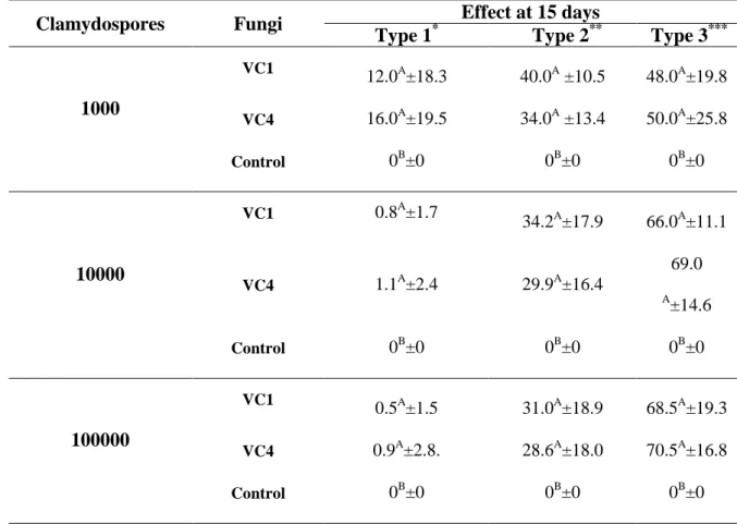
Table 1 – Percentages and standard deviation for types 1, 2 and 3 effects of ovicidal activity against Toxocara canis eggs of P. chlamydosporia (VC1 and
x
LISTA DE FIGURAS
CAPÍTULO 1
Fig. 1-Means and standard deviations of L3 of Strongyloides westeri, not preyed,
recovered from agar-water 2% (AA2%) medium by Baermann method in the seven day of treatments with the isolates Duddingtonia flagrans (AC001),
Monacrosporium thaumasium (NF34) and Arthrobotrys robusta (I-31) and in
control group (without fungus). ………...……….…22
Fig. 2-Linear regression curves of infective larvae (L3) of Strongyloides westeri
recovered from Petri dishes in treatments with the isolate Duddingtonia flagrans
(AC001), Monacrosporium thaumasium (NF34) and Arthrobotrys robusta (I-31)
and control (without fungus) due to time. ………...………..…………....23
Fig. 3-Linear regression curves of infective larvae (L3) of Strongyloides westeri
recovered from Petri dishes regarding collections (12 h, 24 h, 48 h and 72 h) in treatments with the isolate Duddingtonia flagrans (AC001), Monacrosporium thaumasium (NF34) and control (without fungus) due to time. ………..…….…24
CAPÍTULO 2
Figure 1. Mean numbers of infective larvae (L3) of Strongyloides westeri that
were recovered from 2% water-agar medium by means of the Baermann method on the fifteenth day of treatment with the fungal isolates Duddingtonia
flagrans (AC001) and Monacrosporium thaumasium (NF34), and in the control
xi CAPÍTULO 3
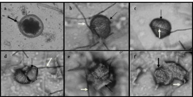
Fig. 1. (A) Toxocara canis eggs (black arrow), control. (B) Hyphae of the fungus Pochonia chlamydosporia (white arrow) attached to the eggshell, a type 1
effect. (C) P. chlamydosporia hyphae of the fungus (white arrow) causing
deformity in the T. canis egg (black arrow), a type 2 effect. (D)–(F) T. canis
eggs (black arrow) and hyphae of P. chlamydosporia destroying the eggs (white
arrow), a type 3 effect. ………..…….46
CAPÍTULO 4
Fig.1- Means of ovicidal activity for the nematophagous fungus Pochonia
chlamydosporia (VC4) and the control group against eggs of Toxocara canis at
feces collection times 6, 12, 24, 36 and 48 hours after 30 interaction days. Asterisk denote differences statistical (p<0.01)……...……….59
Fig.2A-D. Progressive destruction by hyphae from the nematophagous P.
chlamydosporia fungus (VC4), B-D (black arrows) on the surface and inside and
subsequent Toxocara canis eggs destruction, A-D (white arrows). Bars: A- 17.7
xii RESUMO
ARAUJO, Juliana Milani, D.Sc., Universidade Federal de Viçosa, julho de 2012. Avaliação de fungos nematófagos sobre larvas infectantes de Strongyloides westeri e ovos de Toxocara canis. Orientador: Jackson Victor de Araújo. Coorientadores: Laércio dos Anjos Benjamin e Giovanni Ribeiro Carvalho.
O objetivo deste trabalho foi avaliar a ação de três isolados de fungos predadores Duddingtonia flagrans (AC001), Monacrosporium thaumasium
(NF34) e Arthrobotrys robusta (I-31) em teste in vitro quanto à capacidade de
predar larvas infectantes (L3) de Strongyloides westeri e, avaliar três
concentrações de clamidósporos (1.000, 10.000 e 100.000) do fungo ovicida
Pochonia chlamydosporia (isolados VC1 e VC4) na destruição de ovos de
Toxocara canis. Em relação aos fungos predadores, quando comparado ao
grupo controle, pode-se observar que houve uma redução significativa (P<0,01) de 80,4%, 67,9%, 72,8%, nas médias de larvas infectantes de S. westeri
recuperadas dos tratamentos com os isolados AC001, NF34 e I-31, respectivamente. Todos os isolados testados foram eficientes na captura de S. westeri (P>0,01) no teste in vitro. No teste da avaliação ovicida com o fungo P.
chlamydosporia sobre ovos de T. canis, cada placa de Petri continha mil ovos
de T. canis com apenas uma das concentrações de clamidósporos de VC1 ou
de VC4, em ágar-água 2% (AA 2%) e, mil ovos em AA 2% nos grupos controle. No intervalo de 15 dias, cem ovos foram retirados de cada placa de Petri e a atividade ovicida foi avaliada quanto as alterações morfológicas. A maior destruição dos ovos (efeito do tipo 3) foi observada na concentração de 100.000 clamidósporos para ambos os isolados. Após avaliação in vitro, os
fungos predadores e ovicida, foram avaliados in vivo, quanto à sua capacidade
de suportar a passagem pelo trato gastrintestinal de eqüídeos (AC001 e NF34) e predar L3 de S. westeri, e pelo trato gastrintestinal cães (VC4) quanto sua
ação ovicida sobre ovos de T. canis. Os fungos predadores sobreviveram à
passagem pelo trato gastrintestinal dos eqüídeos e foram eficientes em predar as L3 de S. westeri desde a primeira coleta (12h) (P<0,01) em relação ao grupo
controle (sem fungo). O fungo ovicida também sobreviveu a passagem pelo trato gastrintestinal de cães mantendo sua atividade ovicida sobre os ovos de
xiii
thaumasium se mostraram promissores para serem utilizados no controle
biológico de S. westeri assim como, resultados obtidos sugere-se que o fungo P. chlamydosporia (VC4) poderia ser utilizado como uma ferramenta no
xiv ABSTRACT
ARAUJO, Juliana Milani, D.Sc., Universidade Federal de Viçosa, July, 2012. Evaluation of nematophagous fungi on infective larvae of the Strongyloides westeri and Toxocara canis eggs. Adviser: Jackson Victor de Araújo. Co-advisores: Laércio dos Anjos Benjamin and Giovanni Ribeiro Carvalho.
The objective of this work was to evaluate the action of three isolates of predatory fungi Duddingtonia flagrans (AC001), Monacrosporium thaumasium
(NF34) and Arthrobotrys robusta (I-31) in in vitro assay regarding their capacity
to prey infective larvae (L3) of Strongyloides westeri and, to evaluate in vitro
three concentrations of chlamydospores (1,000, 10,000 and 100,000) of the nematophagous fungus Pochonia chlamydosporia (isolates VC1 and VC4) in
the destruction of Toxocara canis eggs. In relation to the fungi predators when
compared to the control group, one can observe that there was a significant reduction (P<0.01) of 80.4%, 67.9%, 72.8%, in the means of recovered infective larvae of S. westeri from treatments with the isolates AC001, NF34 and I-31,
respectively. All tested isolates were efficient at capturing S. westeri (P>0.01)
when tested in vitro. In the test of ovicidal evaluation with the fungus P.
chlamydosporia on eggs of T. canis, each Petri dish contained thousand eggs
of T. canis with only one concentration of chlamydospores of VC1 or VC4 in
water-agar 2% (WA 2%), and only one thousand eggs in WA 2% in the control groups. Within 15 days, one hundred eggs were removed from each Petri dish and the ovicidal effect was evaluated according to morphological alterations. The higher percentage to effect of the type 3 was observed in concentration of 100,000 chlamydospores to both isolates. After in vitro evaluation, predatory
and ovicidal fungi were evaluated in vivo regarding their capacity to withstand
the passage through the gastrointestinal tract of horses (Duddingtonia flagrans
and Monacrosporium thaumasium) on S. westeri and dogs (Pochonia
chlamydosporia) on T. canis eggs. The predatory fungi survived the passage
through the gastrointestinal tract of horses and were efficient in preying upon the L3 since the first collection (12h) (P <0.01) compared to control (without fungi). The ovicidal fungus also survived the passage through the gastrointestinal tract of dogs keeping its ovicidal activity on T. canis eggs.
xv
promising to be used for biological control of S. westeri as well as from the
results obtained suggest that the fungus P chlamydosporia (VC4) could be used
1
1. INTRODUÇÃO GERAL
Os estudos sobre parasitismo em animais de estimação vêm despertando crescente interesse, frente à associação restrita e íntima entre o homem e os animais e sua conseqüência em saúde pública (Vasconcellos et al., 2006). Além disso, as helmintoses gastrintestinais influenciam no desenvolvimento da criação de animais domésticos de companhia e também daqueles com interesse zootécnico, em todo o mundo. Os prejuízos causados por essas infecções envolvem queda da produção, retardo no crescimento do animal, recursos terapêuticos, custos com tratamentos e em muitas vezes a morte dos animais (Araújo et al., 2004).
Dentre os grupos de animais domésticos, os cães e os equídeos têm relevante importância em todo território brasileiro. Contudo, em relação às helmintoses gastrintestinais existem diferenças na especificidade parasitária.
Os eqüídeos é um grupo muito prevalente no Brasil, apresentando a terceira maior criação no mundo (Bezerra et al., 2007). Além disso, representam uma importante espécie entre os animais domésticos em virtude da utilização destes como meio de transporte, tração, entretenimento, esporte, na recuperação de crianças especiais ou mesmo para o consumo humano de produtos e subprodutos cárneos. Em relação a forma de criação, esta pode ser extensiva ou intensiva, no entanto, sabe-se que as mesmas favorecem a grande incidência de infecções parasitárias já nas primeiras semanas de vida (Braga et al., 2009). Estes animais são hospedeiros de uma vasta quantidade de helmintos, onde os de importância mais relevada são os nematóides (Assis & Araújo, 2003; Braga et al., 2011). Por outro, a fauna parasitária é vasta e destacam-se vários parasitos dentre os quais, o Strongyloides westeri.
O nematóide S. westeri pertence a ordem Rhabditida, Superfamília
Rhabditoidea, Família Strongyloididae (Monteiro, 2010). É um parasito que afeta principalmente os animais jovens. Contudo, alguns autores mencionam que a infecção é limitada a estes animais e, que o hospedeiro se torna resistente a re-infecção. São parasitos que vivem embebidos no epitélio glandular da mucosa do intestino delgado, particularmente do duodeno e do jejuno, onde realizam a ovipostura (Bowman, 2010; Monteiro, 2010).
O gênero Strongyloides possui morfologia diferenciada com o esôfago
2
comprimento total do corpo. As fêmeas são partenogênicas e, é o único gênero entre os parasitos de animais domésticos com geração parasitária e de vida livre alternadas. De maneira resumida, o ciclo evolutivo deste parasito, comporta-se da seguinte forma: as fêmeas parasitárias filariformes (encontradas no intestino do hospedeiro) produzem ovos por partenogênese mitótica que são eliminados com as fezes no solo, e as larvas desses ovos são chamadas de geração homogônica para diferenciá-las da geração heterogônica.(vida livre – sexuada). As larvas no ambiente podem se desenvolver para larvas infectantes (L3) ou em machos e fêmeas de vida livre. Se a larva infectante encontra o hospedeiro adequado, esta penetra através da pele e se tornam adultas no intestino do hospedeiro. Caso contrário, as larvas vão se desenvolver através de mudas e se transformar em adultos no solo. Estes copulam e produzem larvas heterogônicas que podem desenvolver em larvas infectantes (Bowman, 2010; Monteiro, 2010).
Os ovos embrionados, a larva rabditiforme e a larva infectante filariforme de terceiro estágio são os estágios mais importantes nos procedimentos diagnósticos (Bowman, 2010; Monteiro, 2010). Os ovos são encontrados quase que exclusivamente em potros lactentes e recém-desmamados. A eliminação dos ovos nas fezes começa a partir de 10 dias a duas semanas após o nascimento (Bowman, 2010). A principal via de transmissão do S. westeri é a
transmamária, onde, após uma infecção inicial ter se estabelecido, larvas adicionais tendem a migrar para os tecidos corporais mais profundos, onde infectam os recém-nascidos através do leite. Também pode ocorrer a infecção através da pele ou membranas da mucosa oral, podendo causar infecções percutâneas (Bowman, 2010).
Outro grupo de animais domésticos que desempenham um papel importante na sociedade em todo o mundo são os cães. Nenhuma outra espécie animal ocupa tantos e tão diversos papéis na sociedade, trazendo benefícios inestimáveis para a melhoria das condições fisiológicas, emocionais e sociais em especial para crianças e idosos (Carvalho et al., 2010). Entretanto, o aumento crescente desses animais, principalmente nos centros urbanos vem aumentando a exposição humana a agentes zoonóticos (Carvalho et al., 2010), entre os quais estão algumas espécies de helmintos.
3
gênero Toxocara, particularmente a espécie Toxocara canis (Monteiro, 2010).
A infecção por T. canis pode ser adquirida pelo homem através da ingestão de
ovos larvados presente no ambiente onde, cães infectados por T. canis
eliminam os ovos do parasito nas fezes que, em condições ambientais apropriadas, tornam-se infectantes e podem permanecer viáveis por longo período no ambiente (Rey, 2008). Nos humanos o T. canis não atinge a
maturidade, mas as larvas permanecem vivas migrando erraticamente nos órgãos internos, produzindo uma patologia denominada Larva Migrans Visceral (LMV - onde os órgãos mais afetados são pulmão, fígado e cérebro), ou invadindo o globo ocular e causando a Larva Migrans Ocular (LMO) (Rey, 2008).
Os parasitos adultos vivem no intestino delgado de cães e gatos. Os ovos de T. canis são eliminados nas fezes, sendo as fêmeas muito fecundas.
As fêmeas podem produzir 2 milhões de ovos por dia, no período mais fértil de sua existência (entre a sétima e a 28ª semana), caindo para uma média de 200.000 ovos, até o oitavo mês (Rey, 2008).
Apenas ovos embrionados são infectantes, e quando ingeridos pelos cães, esses eclodem no intestino, invadem a mucosa e através da circulação porta fazem o ciclo fígado – coração – pulmão, regressando ao tubo digestivo via brônquios, traquéia e esôfago. O tempo mínimo para o ciclo se completar é de 30 dias, e em seguida, começam a aparecer ovos nas fezes (Rey, 2008). Contudo, a principal via de infecção nestes animais ocorre por migração transplacentária e, pode ocorrer também a infecção transmamária de larvas pelo colostro ou pelo leite, ingestão destas em estádio avançado de evolução, e em menor escala, mediante parasitas imaturos eliminados pelas fezes ou vômito de cães (Rey, 2008; Bowman, 2010).
4
Em relação ao controle das verminoses em equídeos, este é feito por anti-helmínticos, onde estes não tem sido de total eficácia devido à ação restrita aos parasitos adultos (Assis & Araújo, 2003). Por outro lado, de uma maneira geral ainda existem outras desvantagens como resíduos na carne, no leite, a possibilidade do impacto ambiental e também o desenvolvimento eminente de resistência dos parasitos (Braga et al., 2011).
Os anti-helmínticos são a principal forma de controle de verminoses em animais domésticos, em especial aqui de cães e equídeos. Por outro lado, é cada vez mais crescente a “corrente” da sociedade de se tentar minimizar o uso destas drogas que podem interferir em um aspecto global na contaminação ambiental e riscos para a saúde (Araújo et al., 2004; Braga et al., 2010). Dessa forma, a utilização de alternativas em conjunto ao controle químico, como o controle biológico utilizando fungos nematófagos, pode vir a ser uma ferramenta promissora para a diminuição dos impactos ambientais e mesmo da saúde de animais e humanos (Braga et al., 2011).
O controle biológico realizado com fungos nematófagos aparece como uma alternativa promissora e viável, que tem por finalidade o sinergismo com o controle químico (Araujo et al, 2010; Braga et al., 2010). Estes fungos vêm se destacando entre os mais variados antagonistas de nematóides, apresentando melhores desempenhos em pesquisa de controle biológico de nematóides (Araújo et al, 2004; Braga et al., 2010). De acordo com Gronvold et al., (1996), os fungos são mais promissores para o controle biológico em relação a outros organismos, pois estão em abundância no solo e podem utilizar vários substratos orgânicos e micro-habitats que promovem oportunidades para a interação com nematóides.
Na prática, o controle biológico não atua sobre estágios internos do parasito. Os fungos nematófagos atuam sobre os hospedeiros intermediários, paratênico, vetores e estágios larvais de vida livre diminuindo assim, a fonte de infecção aos hospedeiros finais, além de causar menos efeitos negativos no ambiente que os métodos químicos (Araújo et al., 2004; Braga et al., 2010). Esses fungos são antagonistas naturais de nematóides, capazes de promover a captura, a morte ou mesmo a destruição desses organismos (Braga et al., 2009).
5
naturalmente no solo, e em material orgânico em decomposição. No grupo dos predadores os gêneros Arthrobotrys, Duddingtonia e Monacrosporium se
destacam pelo controle de larvas de nematóides no ambiente. Estes produzem estruturas em forma de anéis constritores e não constritores, hifas, botões e redes tridimensionais adesivas ao longo do micélio. Após o aprisionamento pela armadilha, ocorre a penetração das hifas na cutícula do nematóide, seguido de crescimento das hifas no interior do nematóide e digestão dos conteúdos internos (Araújo et al., 2004). No grupo dos endoparasitos, esses fungos são capazes de infectar os nematóides através de esporos, que uma vez ingeridos, desenvolvem hifas responsáveis pela absorção do conteúdo interno do nematóide. Estes não produzem hifas vegetativas fora do corpo do hospedeiro, mas hifas férteis ou conidióforos contendo esporos (Araújo et a., 2004). Já no grupo dos fungos oportunistas vem se destacando o fungo
Pochonia chlamydosporia e têm ação nos ovos de helmintos. A ação deste
fungo é baseada na formação de um apressório que se desenvolve por uma hifa não diferenciada. Este fungo coloniza a superfície dos ovos e penetram no interior destes por meio de ação enzimática de proteases, lípases e quitinases, bem como a ação mecânica (Araujo et al, 2009; Braga et al., 2011).
6
2. OBJETIVOS GERAIS
Avaliar a ação in vitro dos isolados fúngicos de Duddingtonia
flagrans, Monacrosporium thaumasium e Arthrobotrys robusta sobre
larvas infectantes de Strongyloides westeri.
Avaliar a ação in vitro de dois isolados do fungo de Pochonia chlamydosporia (VC1 e VC4) sobre ovos de Toxocara canis.
Avaliar após a passagem pelo trato gastrintestinal de eqüinos e jumentas, a viabilidade e ação dos fungos Duddingtonia flagrans e
Monacrosporium thaumasium, quanto a sua atividade predatória
sobre larvas de S. westeri em diversos intervalos de tempo, e se há
diferença de predação entre esses isolados.
Avaliar se o fungo P. chlamydosporia (isolado VC4) resiste a
7
CAPÍTULO I
IN VITRO PREDATORY ACTIVITY OF NEMATOPHAGOUS FUNGI AND AFTER PASSING THROUGH GASTROINTESTINAL TRACT OF EQUINE ON
8
IN VITRO PREDATORY ACTIVITY OF NEMATOPHAGOUS FUNGI AND AFTER PASSING THROUGH GASTROINTESTINAL TRACT OF EQUINE ON INFECTIVE LARVAE OF Strongyloides westeri
Araujo, Juliana Milani; Araújo, Jackson Victor*; Braga, Fabio Ribeiro; Carvalho, Rogério Oliva.
Departamento de Veterinária – Universidade Federal de Viçosa, Viçosa Minas Gerais, Brazil. Cep: 36570000. * - Scholarship CNPq and Corresponding author: jvictor@ufv.br
Abstract
Three isolates of predator fungi Duddingtonia flagrans (AC001), Monacrosporium thaumasium (NF34) and Arthrobotrys robusta (I-31) were
assessed in vitro test regarding the capacity of prey infective larvae (L3) Strongyloides westeri. Comparing to control, without fungus, there was a
significant decrease (P<0.01) of 80.4%, 67.9%, 72.8% in means of infective larvae S. westeri recovered from treatments with isolates AC001, NF34 and
I-31, respectively. All tested isolates were efficient in the capture of S. westeri
(P>0.01) in vitro test. Linear regression coefficients of treated and control
groups were -0.21 for control, -0.32 for D. flagrans, -0.34 for M. thaumasium
and -0.22 for A. robusta. Following, isolates AC001 and NF34 were assessed in
vivo regarding the capacity of supporting the passage through equine
gastrointestinal tract, without loss of ability of preying infective larvae S. westeri.
Fungal isolates survived the passage and were efficient in preying L3 since the first 12h of collection (P<0.01) in relation to control group (without fungus). Comparing to control, there was a significant decrease (P<0.01) of 76.4% and 76.7% (12 hours), 86.4% and 85.9% (24 hours), 88.3% and 87.7% (48 hours), 89.9% and 87.2% (72 hours) in means of infective larvae S. westeri recovered
from treatments with isolates AC001 and NF34, respectively. Linear regression coefficients of L3 of recovered S. westeri regarding the collections due to time
were 1.93 for control, -3.52 for AC001 and -2.64 for NF34. Fungi D. flagrans
and M. thaumasium (NF34) have demonstrated to be promising for use in
biological control of equine parasite S. westeri.
9 1. Introduction
According to Bezerra et al. (2007), equine raising in Brazil represents the third greatest in the world, with about 36 million of animals. Those animals are hosts of a wide quantity of gastrointestinal parasite helminthes (Assis and Araújo 2003). Strongyles nematodes are common in equine, representing a group of large importance, since great part of the cattle is infected.
Strongyloides westeri is the nematode of largest prevalence among foals
with age up to four months. It causes lesions in the small intestine and has been considered as possible cause of diarrhea of the rut of foal (Fenger 2000; Melo et al. 2007).
Matthews et al. (2004) and Kaplan (2002) report that the control of worms in equines usually is performed using anti-helminthic, which have not been totally effective for control of these nematodes due to its restricted action to adult parasites and occurrence of resistance.
However, the integration of other forms of parasite control that aim at decreasing the number of infective larvae in pastures, providing the decrease of parasite load in animals and consequently decrease in number of treatments with anti-helminthic are welcome (Araújo et al. 2004; Braga et al. 2009a).
In this way, the application of biological control with nematophagous fungi has became a viable alternative, and has presenting promising results in vitro and in vivo (Larsen 1999, Braga et al. 2009a). Nematophagous fungi are
saprophytic organisms worldly studied, with capacity of preying nematodes. In predator fungi group, the species Duddingtonia flagrans detaches as the most
promising for control of gastrointestinal nematodiosis of domestic animals (Terril et al. 2004; Dias et al. 2007). Species Arthrobotrys robusta and
Monacrosporium thaumasium have predatory activity against larvae of
gastrointestinal helminthes of domestic animals (Assis and Araújo 2003; Castro et al. 2003).
However, for a nematophagous fungus to be employed as agent of biological control of nematodes, it is necessary that it mainly has skills for capturing nematodes and that resists to the passage through gastrointestinal tract (Araújo t al. 2004).
10
These formulations have demonstrated good results on field and lab conditions (Araújo and Sampaio 2000; Araújo et al. 2000).
The objective of this study was to assess the in vitro predatory capacity
of isolates nematophagous fungi D. flagrans (AC001), M. thaumasium (NF34)
and A. robusta (I-31) about infective larvae (L3) of S. westeri, equine parasites.
Also, to assess the capacity of AC001 and NF34 after the passage through equines gastrointestinal tract, without loss of predatory efficacy on L3 of S.
westeri in feces.
2. Material and Methods
2.1. Experimental test A - In vitro efficacy of nematophagous fungi on infective
larvae (L3) of Strongyloides westeri.
Panagrellus sp. (free nematodes) were maintained on Petri dishes with
oats medium, moistened and mashed. These nematodes were extracted from culture medium by immersion of small quantities of oat in distilled water in Baermann apparatus and collected in hemolysis tubes after six hours of decantation.
Brazilian isolates of predatory nematode fungi of Duddingtonia flagrans
(AC001), Monacrosporium thaumasium (NF34), and Arthrobotrys robusta (I-31)
were kept in assay tubes with 2% corn-meal-agar (2% CMA), at 4oC and in dark.
To induce the production and collection of conidia by fungal isolates, culture discs of approximately 4 mm of diameter of each fungus were transferred to 8.5 cm diameter Petri dishes with 2% water-agar (2% WA), kept at 25oC and in dark. To these plates, 1 mL of suspension with 1000 Panagrellus
sp., every three days for 21 days, was added, until complete growth of fungal mycelium in the plates was done, and all nematodes were preyed. After this period, 5 mL of distilled water were added to each Petri dish, and with the help of brush conidia mycelium fragments were removed and stored at 4oC.
L3 of S. westeri were obtained from feces of naturally infected foals,
through stool culture in vermiculite, for 15 days, and posterior use of Baermann funnel, with water at 42-45oC and decantation time of 12 h.
To study the predatory activity of fungi over L3 of S. westeri a treated
11
average recovered from control – average recovered from treatment average recovered from control
1,000 L3, and a control group with 1,000 L3, being 10 repetitions performed for each group.
Fungi with L3 were deposited on 9.0cm diameter Petri dishes, with 2% water-agar (2% WA) medium kept in stove at 25oC and in dark. Daily, for a period of 7 days, plates were observed at light optical microscope (10x objective), randomly choosing 10 field per plate, being taken the number of L3 free from predation by fungi, obtaining the mean of L3 per field.
On the seventh day, L3 were recovered with the aid of Baermann funnel, with water at 42-45oC and waiting 12 hours for decantation. L3 were counted, obtaining the mean of recovered larvae in the control group, and means of not preyed larvae of treated groups.
2.2. Statistic analysis
Data were submitted to variance analysis (F test) and Tukey test at levels of 1 or 5% of probability and linear regression with levels of 5% of probability. The estimation of L3 mean decrease percentage in treated groups was performed by the equation:
Reduction (%) = ( ) x 100
2.3. Experimental test B- Efficacy test about L3 of Strongyloides westeri after
passage through equine gastrointestinal tract.
Fungal isolates D. flagrans (AC001) and M. thaumasium (NF34) were
tested regarding quantity of passage of each isolate by equine gastrointestinal tract.
The experiment was developed in the Veterinary Department of Universidade Federal de Viçosa, located in the city of Viçosa, Minas Gerais, Brazil, latitude 20º45’20” and longitude 42º52’40”.
12
mycelia mass of isolates from D. flagrans (AC001) and M. thaumasium (NF34)
and pellets without fungus. In the treated group, each animal received 100
grams of pellets, single dose, with mycelia mass of mixed fungi in 100 grams of commercial food for equine. Animals of control group received a single administration of 100 grams of sodium alginate pellets without fungus.
In order to induce the formation of fungal mycelium, culture discs approximately 4 mm of diameter, in 2% WA, were transferred to 250 mL Erlenmeyer's flasks, with 150ml of GPY liquid medium (glucose, sodium peptone and yeast extract), under 120 rpm stirring, in dark and temperature of 26oC for 10 days. After this period, mycelia were removed, filtered and heavy in analytical balance (Walker and Connick, 1983).
Fecal samples from animals were collect at 12, 24, 48 and 72 hours after the administration of fungi. Samples of each group were homogenized, and 4 g of feces were removed and placed in 9 cm diameter Petri dishes with 2% WA, stored in stove at 25oC and in dark. In these plates, 1,000 L3 of S. westeri were spread. From each established time, five repetitions were performed for each treatment group and for control. Daily, plates were observed for research of conidia and conidiophores characteristic of tested isolates, analyzing according to classification-keys proposed by Van Oorschot (1985) and Liu and Zang (1994). In the 15th day, L3 not preyed were recovered from the Petri dishes by Baermann technique, obtaining the mean of not preyed larvae per plate.
2.4. Statistic analysis
Data were submitted for variance and regression analysis. Means were compared by using the Tukey test in level of 1 or 5% of probability (Ayres et al. 2003.
3. Results
3.1. Experimental test A
Isolates of predatory fungi of tested nematodes, D. flagrans (AC001), M.
thaumasium (NF34) and A. robusta (I-31) were able to prey L3 of S. westeri in
the in vitro experimental test. Comparing the capture and destruction of L3 of S.
13
(NF34), and A. robusta (I-31) during the experimental test, no difference
(P>0.01) was observed, Table 1.
Regarding the decrease percentage of L3 of S. westeri at the end of the
experimental test, the following decrease percentage was recorded: 80.4% (AC001), 67.9% (NF34), 72.8% (I-31).
Difference (p<0.01) was observed among means of L3 of not preyed S.
westeri per field of 4 mm diameter of the control group plates in relation to
means of L3 recorded in the plates of groups treated with fungi during the experimental test.
In the plates of the control group, no presence of nematophagous fungi during the experiment was observed. Evidence of predation was verified by the means of recovered L3 of S. westeri in the seventh day by Baermann method,
at the end of the experiment (Figure 1).
Linear regression coefficients calculated through analyses of mean L3 per field of 4 mm diameter of the treated and control groups were -0.21 for control, -0.32 for D. flagrans (AC001), -0.34 for M. thaumasium (NF34), and
-0.22 for A. robusta (I-31) (Figure 2).
3.2. Experimental test B
Fungi D. flagrans (AC001) and M. thaumasium (NF34) had capacity of
preying infective larvae of equines S. westeri, after the passage through
gastrointestinal tract, not losing its viability and were efficient on preying L3. Regarding the decrease percentage, results demonstrated by AC001 and NF34 were 76.4% and 76.7% (12 hours), 86.4% and 85.9% (24 hours), 88.3% e 87.7% (48 hours), 89.9% and 87.2% (72 hours) of L3 (P<0.01), respectively, when compared to control group.
In the plates of control group the presence of nematophagous fungi was not detected, only observing the same saprophytic fungi of treated groups.
Throughout the experiment, the mean number of L3 of control group was larger in relation to that of treated (P<0.01). Mean values of the infective larvae number of recovered S. westeri of Petri dishes incubated with four grams of
14
The growth of saprophytic fungal, not predator, species made visualization of conidia difficult, which could only be identified in the 12th day of observation, in plates referring to collections of 12 and 48 hours for all tested isolates. In plates referring to collections of 24 and 72 hours conidia were visualized from 11th day on. For AC001 it was observed that the 48-hour period was that in which this isolate had a lesser mean number of recovered L3, meaning a higher predatory activity. On the other hand, for the isolate NF34 the best period was 72 hours.
Coefficients of linear regression curves of L3 of S. westeri recovered from
Petri dishes regarding the collections due to time were 1.93 for control, -3.52 for
D. flagrans (AC001) and -2.64 for M. thaumasium (NF34)(Figure 3).
4. Discussion
Nematophagous fungi have predatory capacity over L3 of gastrointestinal nematode parasites in domestic animals, with a highlight for genus
Duddingtonia, Monacrosporium and Artrobotrys (Araújo et al. 2004; Campos et
al. 2008; Silva et al. 2009). These genus are acknowledged just as predators,
and have in the species D. flagrans and M. thaumasium and A. robusta its
predatory capacity already discussed and proved in control of L3 of gastrointestinal nematode parasites of ruminants (Chandrawathani et al. 2003; Castro et al. 2003; Dias et al. 2007). However, the predatory capacity of these species was never tested on L3 of S. westeri. Few works have mentioned the in
vitro and in vivo predatory activity of different nematophagous fungi on
nematode larvae parasites of equines (Castro et al. 2003; Braga et al. 2009a). Braga et al. (2009b), working with species D. flagrans (AC001), M.
thaumasium (NF34) and A. robusta (I-31) recorded its efficacy on infective
larvae of gastrointestinal nematode Angiostrongylus vasorum at the end of
experimental test, at the end of seven days. In that work, although no difference (P>0.01) among tested isolates was observed, AC001 had larger efficacy in the predation of larvae (80.3%). In the current work, at the end of seven days, all isolates were effective in predation of larvae of S. westeri (p>0.01), but the
isolate AC001 (80.4%) was more effective.
15
substances; still occur in consequence of limiting nutritional conditions and/or water scarcity. Also, according to Nansen et al. (1998), the higher is the mobility of nematodes, the higher is the stimulus to fungus for producing traps. In the present work, the presence of L3 of S. westeri in Petri dishes with 2% WA was
fundamental to formation of traps by fungal isolates, in a poor nutrient medium as 2% water-agar.
Regarding the results obtained in vitro test on Petri dishes (assay A), for
linear regression coefficients, it is noted that negative values indicate a descending behavior of regression curves of treatments, due to a decrease of not preyed L3 means per field of 4 mm diameter throughout time, by capture of L3 in fungal traps. On the other hand, in the control group, the decrease of means of L3 per field of 4 mm diameter throughout the seven days of observation is due to the fact of migration of part of larvae to the periphery of plates, where there is more humidity. These results are consistent with the works of Carvalho et al. (2009a, b) about infective larvae of Ancylostoma spp.
In the in vivo test (assay B), results of regression curves, represented in
Figure 3, demonstrated that two tested fungal isolates (AC001 and NF34) had negative linear correlation coefficient. According to Carvalho et al. (2009a), the negative value indicates the existence of reverse correlation between variables, what proves the viability of predatory capacity of fungal isolates after passage through gastrointestinal tract of domestic animals. This information is consistent with the present work, where a decrease of number of infective larvae recovered from equine feces in the studied timelines was observed (12, 24, 48 and 72 hours).
The use of nematophagous fungi in biological control of animals parasitized by helminthes can reduce soil contamination, directly acting in the infective larvae present in the environment. In recent work, Braga et al. (2009a) demonstrated the efficacy of fungus D. flagrans (AC001) by using in pellets of
sodium alginate matrix through the passage by gastrointestinal tract of equines. In that work, the authors registered a mean decrease in larvae recovered from stool culture of 40.9% at the end of the experiment for animals of treated group versus the animals of control group. This information is consistent with results demonstrated in the present work for the isolate AC001 over L3 of S. westeri.
16
gastrointestinal tract of equines without loss of viability to prey infective larvae of cyathostomes. These results are also consistent with the present work.
5. Conclusion
All or tested isolates (AC001, NF34 and I-31) had in vitro action against
L3 of S. westeri. Fungi D. flagrans (AC001) and M. thaumasium (NF34) have
shown to be promising to be used in the biological control of S. westeri after the
passage through gastrointestinal tract of equines without loss of its viability.
Acknowledgements
17 References
Araújo JV, Sampaio WM (2000) Effects of temperature, mineral salt and passage through gastrointestinal tract of calves on alginate formulation of
Arthrobotrys robusta. Rev Bras Parasitol Vet 9:55-59
Araújo JV, Sampaio WM, Vasconcelos RS, Campos AK (2000) Effects of different temperatures and mineral salt on pellets of Monacrosporium
thaumasium - a nematode-trapping fungus. Veterinarski Arhiv 80:181-190
Araújo JV, Mota MA, Campos AK (2004) Controle biológico de helmintos parasitos de animais por fungos nematófagos. Ver Bras Parasitol Vet 13:165–170
Assis RCL, Araújo JV, (2003) Avaliação da viabilidade de duas espécies de fungos predadores do gênero Monacrosporium sobre ciatostomíneos após a
passagem pelo trato gastrintestinal de eqüinos em formulação de alginato de sódio. Rev Bras Parasitol Vet 12:109-113
Ayres M, Ayres JRM, Ayres DL, Santos AS (2003) Aplicações estatísticas nas áreas de ciências biológicas. Belém: Sociedade civil mamirauá: Brasília CNPq, 290p.
Bezerra SQ, Couto MCM, Souza TM, Bevilaqua CML, Anjos DHS, Sampaio IBM, Rodrigues MLA (2007) Cyathostominae(strongylidae-cyathostominae) horse parasites: experimental ecology of free living stages on pasture tifton 85 (cynodon spp. cv. tifton 85) in baixada fluminense, RJ, Brazil. Rev
Latinoameric 62:27-34
18
Braga FR, Araújo JV, Silva AR, Araujo JM, Carvalho RO, Campos AK, Tavela AO, Ribeiro GC (2009a) Biological control of horse cyathostomin (Nematoda: Cyathostominae) with the nematophagous fungus Duddingtonia flagrans in tropical southeast Brazil Vet Pararasitol 163: 335-340
Campos AK, Araújo JV, Guimarães MP (2008) Interaction between the nematophagous fungus Duddingtonia flagrans and infective larvae of
Haemonchus contortus (Nematoda: Trichostrongyloidea). J Helminthol
82:337-341
Carvalho RO, Araújo JV, Braga FR, Araujo JM, Silva AR, Tavela AO (2009a) Predatory activity of nematophagous fungi on Ancylostoma ssp.
infective larvae: evaluation in vitro and after passing through gastrointestinal tract of dogs. J Helminthol 15:1-5
Carvalho RO, Araújo JV, Braga FR, Araujo JM, Silva AR, Ferreira SR, Frassy LN, Alves CDF (2009b) Biological control of Ancylostomosis in dogs with the nematode-trapping fungus Monacrosporium thaumasium in
southeastern Brazil. Vet Parasitol 165:179-183
Castro AA, Oliveira CRC, Anjos DHS, Ornellas EI, Bittencourt VREP, Araújo JV, Sampaio B M, Rodrigues MLA (2003) Potencial dos fungos nematófagos Arthrobotrys sp. E Monacrosporium thaumasium para o
controle de larvas de ciatostomíneos de eqüinos (Nematoda: Cyathostominae). Rev Bras Parasitol Vet 12:49-53
Chandrawathani P, Jamnah O, Waller PJ, Larsen M, Gillespie AT, Zahari WM (2003) Biological control of nematode parasites of small ruminants in Malaysia using the nematophagous fungi Duddingtonia flagrans. Vet
Parasitol 117:173-183
Dias AS, Araújo JV, Campos AK, Braga FR, Fonseca TA (2007) Application of a formulation of the nematophagous fungus Duddingtonia flagrans in the
19
Fenger CK (2000) Doenças dos potros. In: REED,S. M.; BAYLY, W. M. Medicina interna eqüina. Rio de Janeiro: Guanabara Koogan, 803-839.
Gronvold J, Nansen P, Henriksen SA, Larsen M, Wolstrup J, Breciani J, Rawate H, Fribert L (1996) Induction of traps by Ostertagia ostertagi larvae, chlamydospore production and growth rate in the nematode-trapping fungus Duddingtonia flagrans. J Helminthol 70:291-297
Kaplan RM (2002) Antihelmintic resistance in nematodes of horses. Vet Res Communications 33:491-507
Larsen M (1999) Biological control of helminths. International J Parasitol 29:139-146
Liu X, Zhang K (1994) Nematode-trapping species of Monacrosporium with special reference to two new species. Mycol Res; 98: 862-868
Matthews JB, Hodgkinson JE, Dowdall SMJ, Proudman CJ (2004) Recent developments in research into the Cyathostominae and Anoplocephala perfoliata. Vet Res 35:371-381
Melo UP, Ferreira C, Palhares MS (2007) Doenças gastrintestinais em potros: etiologia e tratamento. Ciência Animal Bras 4:733-744
Nansen P, Foldager J, Hansen J, Henriksen SA, Jorgensen RJ (1988) Grasing and acquisition of Ostertagia ostertagi in calves. Inter J Parasitol
27:325-335
20
Scholler M, Rubner A (1994) Predacious Activity Of The Nematode Destroying Fungus Arthrobotrys Oligospora In Dependence Of The Medium
Composition. Microbiol Res 149: 145-149
Silva AR, Araújo JV, Braga FR, Carvalho RO, Tavela AO, Frassy LN, Castejon FV (2009) Biological control of sheep gastrointestinal nematodiosis in a tropical region of the southeast of Brazil with the nematode predatory fungi Duddingtonia flagrans and Monacrosporium thaumasium. Parasitol Res 105:1707–1713
Terril TH, Larsen M, Samples O, Hsted S, Miller JE, Kaplan RM, Gelaye S (2004) Capability of the nematode-trapping fungus Duddingtonia flagrans to
reduce infective larvae of gastrointestinal nematodes in goat feces in the southeastern United States: dose titration and dose time interval studies. Vet Parasitol 120:285-296
Van Oorschot CAN (1985) Taxonomy of the Dactylaria complex. A review of Arthrobotrys and allied genera. Mycol 26:61-95
21
Table 1- Daily means of infective not preyed larvae of L3 of Strongyloides westeri per field of 4 mm diameter in 2% water-agar (2% WA) medium during a
period of seven days in treatments with isolates Duddingtonia flagrans (AC001), Monacrosporium thaumasium (NF34) and Artrobotrys robusta (I-31) and in
control group (without fungus).
Time Treatments (infective not preyed larvae)
22
Fig. 1-Means and standard deviations of L3 of Strongyloides westeri, not preyed,
recovered from 2% water-agar (2% WA) medium by Baermann method after seven day of treatments with the isolates Duddingtonia flagrans (AC001), Monacrosporium thaumasium (NF34) and Arthrobotrys robusta (I-31) and in
control group (without fungus).
AC 001 NF 34 I 31 CONTROL
0 100 200 300 400 500 600 700 800 Trearment S tr o n g y lo id e s w e st e ri ( L 3 )
Means followed by at least one letter, capital letter, in line, not differing among them at level of 1% of probability by Tukey test.
A
A
A
23
Fig. 2-Linear regression curves of infective larvae (L3) of Strongyloides westeri
recovered from Petri dishes in treatments with the isolate Duddingtonia flagrans
(AC001), Monacrosporium thaumasium (NF34) and Arthrobotrys robusta (I-31)
and control (without fungus) due to time.
y = -0.2232x + 2.5386 R2 = 0.5439
y = -0.3279x + 2.5129 R2 = 0.868 y = -0.3461x + 2.4586
R2 = 0.875 y = -0.2193x + 7.1057
R2 = 0.0979
0 1 2 3 4 5 6 7 8 9
1 2 3 4 5 6 7
Days
la
rv
ae
Control AC001 NF34a
I-31 Linear (I-31) Linear (AC001)
24
Table 2- Mean values of infective larvae number of Strongyloides westeri
recovered from Petri dishes, filled with equine feces, sampled in timelines 12, 24, 48, 72 hours after the treatment with isolates Duddingtonia flagrans
(AC001), Monacrosporium thaumasium (NF34), and in control (without fungi).
Means followed by at least one letter, capital letter, in line, not differing among them at level of 1% of probability by Tukey test.
Fig. 3-Linear regression curves of infective larvae (L3) of Strongyloides westeri
recovered from Petri dishes regarding collections (12 h, 24 h, 48 h and 72 h) in treatments with the isolate Duddingtonia flagrans (AC001), Monacrosporium
thaumasium (NF34) and control (without fungus) due to time.
y = 1.93x + 83.6 R2 = 0.6835
y = -3.52x + 21.8 R2 = 0.7898
y = -2.64x + 20.3 R2 = 0.6533
0 10 20 30 40 50 60 70 80 90 100 Time L ar va e
NF34 AC001 CONTROL
Linear (CONTROL) Linear (AC001) Linear (NF34)
Isolates Time
12 h. 24 h. 48 h. 72 h.
25 CAPÍTULO II
CONTROL OF Strongyloides westeri LARVAE BY NEMATOPHAGOUS FUNGI AFTER PASSAGE THROUGH THE GASTROINTESTINAL TRACT OF
DONKEYS
26
RESEARCH NOTE CONTROL OF Strongyloides westeri LARVAE BY NEMATOPHAGOUS FUNGI AFTER PASSAGE THROUGH THE GASTROINTESTINAL TRACT OF DONKEYS
CONTROLE DE LARVAS DE Strongyloides westeri POR FUNGOS NEMATÓFAGOS APÓS TRÂNSITO GASTRINTESTINAL EM JUMENTAS
Juliana Milani Araujo1a; Jackson Victor de Araújo1; Fabio Ribeiro Braga1; Alexandre de Oliveira Tavela1; Sebastião Rodrigo Ferreira1; Filippe Elias de
Freitas Soares2 Giovanni Ribeiro Carvalho3
ABSTRACT
Strongyloides westeri is the most prevalent nematode among equines aged up
to four months and causes gastrointestinal disorders. The objective of this study was to observe the control of infective S. westeri larvae (L3) by
the nematophagous fungi Duddingtonia flagrans (AC001) and Monacrosporium thaumasium (NF34) after passage through the gastrointestinal tract of in
donkeys. Twelve dewormed donkeys that were kept in stables were used. Two treatment groups each comprising four animals received orally 100 g of pellets made of sodium alginate matrix containing a mycelial mass of either D. flagrans
(AC001) or M. thaumasium (NF34). The control group consisted of four animals
that received pellets without fungus. Feces samples were then collected from the animal groups at different times (after 12, 24, 48 and 72 hours). These feces were placed in Petri dishes containing 2% water-agar medium and 1000 L3 of S.
westeri. AC001 and NF34 isolates showed the ability to destroy the L3, after
gastrointestinal transit, thus demonstrating their viability and predatory activity. KEY WORDS: Nematophagous fungi, Duddingtonia flagrans, Monacrosporium
thaumasium, Strongyloides westeri, Donkeys.
_________
1- Department of Veterinary Medicine, Federal University of Viçosa, Viçosa, Brazil.
2- Department of Biochemistry and Molecular Biology, Federal University of Viçosa, Viçosa, Brazil.
3- Department of Zootechnics.
27 RESUMO
O Strongyloides westeri é o nematóide de maior prevalência entre equideos
com idade até quatro meses, causando distúrbios gastrintestinais. O objetivo do presente trabalho foi observar o controle de larvas infectantes (L3) de
Strongyloides westeri pelos fungos nematófagos Duddingtonia flagrans
(AC001) e Monacrosporium thaumasium (NF34) após trânsito gastrintestinal
em jumentas. Foram utilizados 12 jumentas, estabulados e previamente vermifugados. A seguir dois grupos tratados contendo cada um 4 animais receberam por via oral 100 g de péletes em matriz de alginato de sódio contendo massa miceliana dos fungos D. flagrans (AC001) ou M. thaumasium
(NF34). O grupo controle foi constituído de 4 animais que receberam péletes sem fungo. A seguir, amostras de fezes dos grupos de animais foram coletadas
em distintos intervalos de horas (12, 24, 48 e 72). Estas fezes foram vertidas em placas de Petri contendo meio sólido ágar-água 2% e 1000 L3 de S.
westeri. Os isolados AC001 e NF34 apresentaram capacidade de destruir as
L3, após o trânsito, demonstrando sua viabilidade e atividade predatória.
PALAVRAS-CHAVES: Fungos nematófagos, Duddingtonia flagrans,
Monacrosporium thaumasium, Strongyloides westeri, jumentas.
According to FAO (2008), as cited by Morrow et al. (2011), there are approximately 43 million donkeys (Equus asinus) in the world, serving mainly as
a transportation resource in developing countries (PRITCHARD et al., 2005). Getachew et al. (2010) reported that many helminth species are found in these animals and cause direct damage to their health. Nevertheless, there are insufficient studies with data on strategic control of gastrointestinal helminthic parasites that affect this equine species (MATTHEE et al., 2002; VENEZIANO et al., 2011). Within this context, Strongyloides westeri is a relatively frequent
28
Thus, integrated control of helminthoses can be seen as a new approach, since biological control can be used synergistically with chemical control (BRAGA et al., 2009). Biological control is centered on using nematophagous fungi that can act as predators, ovicidal agents and endoparasites. These fungi, which are mainly predators, have been shown to be effective for reducing nematode populations, both in laboratories and under field conditions. Duddingtonia flagrans and Monacrosporium thaumasium are
predators and have been used worldwide to combat the infective larvae of nematode parasites of productive domestic animals (PAZ-SILVA et al., 2011; BRAGA et al., 2011).
The objective of the present study was to evaluate the control of infective
Strongyloides westeri larvae (L3) by nematophagous fungi after gastrointestinal
transit in donkeys (Equus asinus).
To obtain S. westeri L3, fecal cultures were performed on positive feces
from naturally infected young donkeys. Next, these larvae were classified in accordance with the criteria established by Soulsby et al. (1982). In order to induce formation of fungal mycelia of D. flagrans (AC001) and M. thaumasium
(NF34), culture discs of approximately 4 mm in diameter, in 2% water-agar, were transferred to 250 ml Erlenmeyer flasks containing 150 ml of liquid YPG medium (glucose, peptone and sodium yeast extract), and were kept under continual stirring at 120 rpm in the dark and at a temperature of 26 °C for 10 days. After this period, the mycelia were removed, filtered and weighed on an analytical balance.
In the in vivo assay, 12 female donkeys were used, with a mean weight
of 240 kg. They had previously been dewormed by means of vermifuge for equines at an oral dose of 200 μg/kg live weight of 1% ivermectin and 6.6 mg/kg live weight of pyrantel pamoate (Centurion Vallé®, Montes Claros, Minas Gerais, Brazil). This was done 14 days before they received pellets containing a mycelial mass of isolates of D. flagrans (AC001) or M. thaumasium (NF34), or
pellets without fungus (control).
29
commercial feed for horses. The control group received a single administration of 100 g of pellets without fungus.
After administration of the fungi, fecal samples were collected from the animal groups at different times (after 12, 24, 48 and 72 hours). These samples were then homogenized, and 4 g aliquots of feces were placed in Petri dishes of 9 cm in diameter, containing 2% water-agar (2% WA). These dishes were placed in an incubator at 25 °C, in the dark. Each Petri dish of the groups tested (treated and control groups) contained 1,000 L3 of S. westeri. At each collection
time, five repetitions were performed. To prove that the fungi tested actually passed through the gastrointestinal tract, and to identify them, the classification keys for fungal structures (conidia and/or chlamydospores) proposed by Van Oorschot (1985) and Liu and Zhang (1994) were used. The Petri dishes were viewed every day. Subsequently, on the fifteenth day, the L3 that had not been destroyed by the fungus were recovered from the Petri dishes by means of the Baermann technique. The data obtained were subjected to analysis of variance (F test) and then regression analysis. The means were compared using the Tukey test at the 1% level of probability.
It was observed that the fungi D. flagrans (AC001) and M. thaumasium
(NF34) destroyed the S. westeri L3, after passage through the gastrointestinal
tract of the donkeys. At each of the collection times studied, the following percentage reductions were found: 81.2% and 81.1% (12 hours); 62.7% and 87.2% (24 hours); 78.6% and 76.7% (48 hours); and 85.3% and 92.2% (72 hours), for the isolates AC001 and NF34, respectively, in comparison with the control group (Figure 1). At the collection time of 72 hours, both isolates (AC001 and NF34) showed higher predatory activity and, consequently higher percentage reductions in the L3 recovered from S. westeri.
On the other hand, the coefficients of the linear regression curves for S.
westeri L3 recovered from Petri dishes, relating to the collections as a function
of time were: 5.54 for the controls, -1.25 for D. flagrans (AC001) and -1.06 for
M. thaumasium (NF34). In the feces of the treated groups, conidia and
chlamydospores were identified according to the fungal species tested (D.
flagrans and M. thaumasium).
30
environment. On the other hand, although horses and donkeys generally harbor the same genera of gastrointestinal helminthic parasites, there are no studies with enough data regarding strategic control of helminths in donkeys. In this context, Araujo et al. (2010) reported that three fungal genera (Arthrobotrys, Duddingtonia and Monacrosporium) were efficient at destroying S. westeri L3 in
an in vitro assay. In this paper, the results showed that there was no difference
(p > 0.05) in predatory activity between the fungi tested and therefore either of the isolates could be used in in vivo tests. This premise provided the justification
for conducting the present study, and showed the need for knowledge regarding alternative approaches towards control of helminth parasites in donkeys. In addition, Araujo et al. (2010) used horses for the in vivo test, thus showing that the fungi D. flagrans and M. thaumasium have also been effective in passing
through the gastrointestinal tract of these animals. Nonetheless, despite being in the same group of animals (equines), donkeys are a different species and thus, studies that can demonstrate alternative control methods for endoparasites, in particular in relation to S. westeri, are important.
Several studies have been conducted with regard to biological control of nematode parasites of horses, both in vitro and under natural conditions, which
once again denotes the need to extrapolate these studies to other species of equines, such as donkeys. Tavela et al. (2011) studied the fungus M. thaumasium (NF34) in the field, administered to horses, and demonstrated that
it was effective on the larvae of cyathostomins through decreasing the recurrence of helminth infections. Similar results, thereby confirming the action of this fungus, were also found in the present work, which proved that this isolate remained viable after passage through the gastrointestinal tract of donkeys.
The efficacy of the fungus D. flagrans (AC001) was also demonstrated in
a study by Braga et al. (2009) that consisted of a six-month field test using weekly doses of pellets containing this fungus, among horses. A difference in parasite loads (p < 0.05) was recorded between the animals in the treated group and those in the control group. This result is also in accordance with the findings from the present study relating to the action of the isolate AC001 on S.
westeri L3.