Normal Platelet Integrin Function in Mice
Lacking Hydrogen Peroxide-Induced Clone-5
(Hic-5)
Michael Popp, Ina Thielmann, Bernhard Nieswandt, David Stegner*
Department of Experimental Biomedicine–Vascular Medicine, University Hospital and Rudolf Virchow
Center, University of Würzburg, Würzburg, Germany
Abstract
IntegrinαIIbβ3 plays a central role in the adhesion and aggregation of platelets and thus is essential for hemostasis and thrombosis. Integrin activation requires the transmission of a signal from the small cytoplasmic tails of theαorβsubunit to the large extracellular domains resulting in conformational changes of the extracellular domains to enable ligand binding. Hydrogen peroxide-inducible clone-5 (Hic-5), a member of the paxillin family, serves as a focal adhesion adaptor protein associated withαIIbβ3 at its cytoplasmic tails. Previous stud-ies suggested Hic-5 as a novel regulator of integrinαIIbβ3 activation and platelet aggrega-tion in mice. To assess this in more detail, we generatedHic-5-nullmice and analyzed
activation and aggregation of their plateletsin vitroandin vivo. Surprisingly, lack of Hic-5
had no detectable effect on platelet integrin activation and functionin vitroandin vivounder
all tested conditions. These results indicate that Hic-5 is dispensable for integrinαIIbβ3 acti-vation and consequently for arterial thrombosis and hemostasis in mice.
Introduction
Vessel wall injury results in the exposure of the subendothelial extracellular matrix which initiates stable platelet adhesion and aggregation [1,2]. These processes are crucial for normal hemostasis, but in diseased vessels they may lead to pathological thrombus formation and infarction of vital organs [3]. Platelet adhesion and aggregation are mediated by integrins, heterodimeric transmembrane receptors composed ofαandβsubunits that are expressed in a low affinity state under resting conditions. The major platelet integrinαIIbβ3 is present in high copy numbers (up to 100,000 per platelet) and its function is well characterized. Integrin
αIIbβ3 binds several ligands each containing an arginine-glycine-aspartic acid (RGD) sequence, such as fibrinogen, fibrin, vWF, fibronectin, thrombospondin and vitronectin [3]. After activation mediated by other platelet receptors, integrins shift to a high affinity state and efficiently bind their ligands [4]. This process requires the transmission of signals from the small cytoplasmic tails to the large extracellular domains of the integrin subunits [5]. Several proteins, including talin and kindlin-3, have been proposed to be involved in regulation of
OPEN ACCESS
Citation:Popp M, Thielmann I, Nieswandt B, Stegner D (2015) Normal Platelet Integrin Function in Mice Lacking Hydrogen Peroxide-Induced Clone-5 (Hic-5). PLoS ONE 10(7): e0133429. doi:10.1371/journal. pone.0133429
Editor:Toshiyuki Miyata, National Cerebral and Cardiovascular Center, JAPAN
Received:April 14, 2015
Accepted:June 26, 2015
Published:July 14, 2015
Copyright:© 2015 Popp et al. This is an open access article distributed under the terms of the
Creative Commons Attribution License, which permits unrestricted use, distribution, and reproduction in any medium, provided the original author and source are credited.
Data Availability Statement:All relevant data are within the paper and its Supporting Information files.
Funding:This work was supported by the Rudolf Virchow Center and the Deutsche
Forschungsgemeinschaft (SFB688). This publication was funded by the German Research Foundation (DFG) and the University of Wuerzburg in the funding programme Open Access Publishing.
integrin activation [6–12]. A functional role for these proteins in integrin activationin vivo
could not be confirmed in all cases as, for example, in the case of RIAM [13]. Recently, Kim-Kaneyama and colleagues reported thathydrogen peroxide-induced clone-5(Hic-5) acts as a novel regulatory factor for integrinαIIbβ3 activation and platelet aggregation in mice [14]. Hic-5 was identified as a gene inducible by transforming growth factorβ1 (TGFβ1) as well as hydrogen peroxide in a differential screen of cDNA libraries from the mouse osteoblastic cell line MC3T3-E1. The authors of that study speculated that Hic-5 has a role in the growth-inhib-itory pathway associated within vitrosenescence and that down-regulation of Hic-5 contrib-utes to tumorigenesis [15]. Hic-5 contains four LIM domains at the C-terminus and shares high homology with paxillin that has been shown to localize to focal adhesions and to interact with talin in platelets [16,17]. In addition, it has been recently shown that paxillin negatively regulates platelet signaling pathways resulting in augmentedαIIbβ3 activation upon stimula-tion of glycoprotein VI (GPVI) or G protein-coupled receptors (GPCR) [18].
We generated Hic-5-null (Tgfb1i1-/-) mice and found, in stark contrast to the study by Kim-Kaneyamaet al. [14], unaltered platelet integrin functionin vitroandin vivo.
Material and Methods
Animals
Animal studies were approved by the district government of Lower Franconia (Bezirksregier-ung Unterfranken; AZ 54/11).Tgfb1i1+/-mice were generated by injection of embryonic stem cell clone EPD0817_1_D04 (KOMP) into blastocysts of pseudo-pregnant C57BL/6 females to generate germ line chimeras. Male chimeras were bred to C57BL/6 females to generate
Tgfb1i1+/–mice, which were intercrossed to produceTgfb1i1–/–mice and littermate wild-type
controls. Mice were genotyped by PCR, with 5’GGGACGGGGCGTAGATAAAG 3’and 5’
GTGCAGCCCAGATTGTCTCT 3’for the wild-type, and 5’TAGATAGAGATGGCGCAACG 3’and 5’ACACCCATTCACACACTGGA 3’for theHic-5-null allele.
Chemicals and antibodies
RT-PCR analysis
Murine platelet mRNA was isolated with Trizol reagent, reverse transcribed and detected by RT-PCR, according to the manufacturer’s protocol (Invitrogen, Karlsruhe, Germany). The fol-lowing primers were used to detect theHic-5transcript: 5’TTTTGGCCGCTGCCTTTAAC 3’
and 5’AGGCTTGCATACTGTGCTGT 3’.
Immunoblotting
Proteins of lysed platelets were separated by SDS-PAGE and blotted onto polyvinylidene difluoride membranes. After blocking, the membrane was incubated with antibody overnight at 4°C. HRP-conjugated antibodies were incubated for 1 h at room temperature, and enhanced chemilumincescence was used for visualization.
Platelet preparation, aggregation and flow cytometry
Washed platelets were prepared as described previously [20]. For aggregometry, washed plate-lets (160μL with 1.5x10⁸platelets mL-1) were analyzed in the presence (CRP) or absence
(thrombin) of 70μg mL-1human fibrinogen. Light transmission was recorded on a
four-chan-nel aggregometer (Fibrintimer; APACT, Hamburg, Germany) for 10 min and expressed in arbitrary units, with buffer representing 100% light transmission. For flow cytometry, heparin-ized whole blood was diluted 1:20 in Tyrode’s-HEPES buffer, incubated with saturating amounts of fluorophore-conjugated mAbs for 15 min at room temperature, and analyzed on a FACSCalibur (BD Biosciences, Heidelberg, Germany).
Adhesion under flow conditions to collagen or vWF
For adhesion to collagen, cover slips were coated with 100μg mL-1collagen I at 37°C o/n. For
adhesion to vWF, cover slips were coated with rabbit anti-human vWF antibody at 4°C o/n, washed with PBS, blocked for 1 h with 1% BSA in H2O and incubated with 100μl murine
serum obtained from control mice.
Blood (700μl) was collected into 300μl heparin (20 U/ml in TBS, pH 7.3). Whole blood was
diluted 2:1 in Tyrode’s buffer containing Ca2+and filled into a 1 ml syringe. For adhesion to collagen, before perfusion, anticoagulated blood was incubated with Dylight-488–conjugated anti- GPIX derivative (0.2μg/mL) at 37°C for 5 minutes.
Transparent flow chambers with a slit depth of 50μm, equipped with the coated coverslips,
were connected to a syringe that was filled with diluted whole blood. Perfusion was performed using a pulse-free pump under high shear stress equivalent to a wall shear rate of 1000 s-1 (col-lagen I) or 1,700 s-1(vWF).
Aggregate formation was visualized with a Zeiss Axiovert 200 inverted microscope (40x/ 0.60 objective). Phase-contrast and fluorescence pictures were recorded with a CoolSNAP-EZ camera, and analyzed off-line using MetaVue software.
Adhesion of platelets to bound vWF started 90 sec after perfusion of the slide. Thereafter, cover slipes were washed by a 4 min perfusion with Tyrode’s buffer at the same shear rate and phase-contrast images were recorded from at least five different microscopic fields (40x/0.60 objective). Image analysis was performed off-line using MetaVue software.
Platelet spreading on fibrinogen
Cover slips were coated with 100μg mL-1human fibrinogen and blocked with 1% BSA in PBS.
After rinsing with Tyrode-HEPES buffer, washed platelets (100μL with 0.03x10⁶plateletsμL-1)
(ATP scavenger; 2U/ml) and indomethacin (inhibitor of COX-1/2; 1.4μM) and allowed to
spread on the cover slips. At the indicated time points, platelets were fixed with PBS 4% PFA and platelets were visualized with a Zeiss Axiovert 200 inverted microscope (100x/1.4 oil objec-tive). Digital images were recorded using a CoolSNAP-EZ camera (Visitron) and analyzed off-line using MetaVue software.
Platelet spreading on vWF
Glass coverslips were coated with a polyclonal rabbit anti-human vWF antibody at 4°C o/n, washed with PBS, blocked for 1 h with 1% BSA in H2O and incubated with 100μl murine
serum obtained from control mice. Washed platelets at 0.3x10⁶platelets/μl were diluted 1:2.3
in Tyrode’s with Ca2+, incubated with integrilin (40μg/ml) and botrocetin (2μg/ml) and were
allowed to adhere on the prepared cover slips. At the indicated time points, platelets were fixed with PBS 4% PFA and platelets were visualized with a Zeiss Axiovert 200 inverted microscope (100x/1.4 oil objective). Digital images were recorded using a CoolSNAP-EZ camera (Visitron) and analyzed off-line using MetaVue software.
Clot retraction
Clot retraction studies were performed at 37°C in an aggregometer tube containing diluted PRP (platelet rich plasma) (3×10⁵plateletsμL-1), thrombin (5 U mL-1), and CaCl2(20 mmol
L-1). Clot retraction was recorded with a digital camera over a time span of 4.5 hours after activation.
Intravital microscopy of thrombus formation in FeCl
3-injured mesenteric
arterioles
Four-week-old mice were anesthetized, and the mesentery was exteriorized through a midline abdominal incision [21]. Arterioles (35–60μm diameter) were visualized with a Zeiss Axiovert
200 inverted microscope (10x /0.3 NA objective) (CarlZeiss, Göttingen, Germany) equipped with a 100-W HBO fluorescent lamp source, and a CoolSNAP-EZ camera (Visitron). Digital images were recorded and analyzed off-line with MetaVue software. Injury was induced by top-ical application of a 3 mm2filter paper saturated with FeCl3(20%). Adhesion and aggregation
of fluorescently labeled platelets (DyLight-488-conjugated anti-GPIX IgG-derivative) in arteri-oles were monitored for 40 min or until complete occlusion occurred (blood flow stopped for>1 min).
Bleeding time
Mice were anesthetized, and a 1 mm segment of the tail tip was removed with a scalpel. Tail bleeding was monitored by gently absorbing blood with filter paper at 20 s intervals without making contact with the wound site. When no blood was observed on the paper, bleeding was determined to have ceased. Experiments were stopped after 20 min. For the saline model, 1 mm of the tail tip was amputated, and the tails were immersed in 0.9% isotonic saline at 37°C. The time to complete arrest of bleeding (no blood flow for 1 minute) was determined.
Data analysis
Results and Discussion
We used embryonic stem cells (clone EPD0817_1_D04) provided by CSD, a collaborative team at the Children's Hospital Oakland Research Institute, and the University of California at Davis, within the Knockout Mouse Program (KOMP) with a knockout first approach targeting exon 1 of theTgfb1i1gene leading to efficient loss of Hic-5 protein inTgfb1i1+/-mice.Tgfb1i1 +-/-mice were intercrossed to obtainTgfb1i1-/-mice (further referred to as Hic-5-null mice) and
the respective control mice. Hic-5-null mice were born at normal Mendelian ratio, were viable and fertile and appeared overall healthy. The absence of Hic-5 in platelets was confirmed by Western blot analysis (Fig 1A) and RT-PCR (S1 Fig). The expression levels of the other paxillin family members leupaxin and paxillin were unaltered in Hic-5-null platelets compared to wild-type platelets (Fig 1A). The loss of Hic-5 did not alter platelet count or size and also red and white blood cell counts were indistinguishable from controls (Table 1). Flow cytometric mea-surements of major platelet surface receptors did not show significant alterations in receptor expression levels (Table 2) in Hic-5-null platelets compared to wild-type platelets. The unal-tered basic blood and platelet parameters in the mutant mice indicate that Hic-5 plays no role for hematopoiesis and platelet morphology, in line with previous results of Kim-Kaneyama
et al. [14].
To clarify the role of Hic-5 deficiency on integrin activation and degranulation, activation of the major platelet integrinαIIbβ3 and surface exposure ofα-granular P-selectin in response to different agonists was analyzed by flow cytometry. Stimulation of G-protein coupled recep-tors with ADP, U46619, a combination of both, or thrombin resulted in comparable results for wild-type and Hic-5-null platelets (Fig 1C and 1D). Similarly, Hic-5-null platelets reacted nor-mally upon activation of the GPVI-ITAM (immunoreceptor tyrosine-based activation motif) pathway by CRP and in response to stimulation of the hemITAM receptor CLEC-2 (C-type lectin-like receptor 2) by rhodocytin (Fig 1C and 1D).β1-integrin activation was assessed by
Fig 1. Hic-5 deficiency has no effects on inside-out activation of platelet integrins, degranulation and platelet aggregation.(A) Hic-5, leupaxin and paxillin expression was assessed by Western blot analysis. GAPDH served as loading control. (B) Flow cytometric analysis ofβ1 integrin activation (9EG7-FITC). (C, D) Flow cytometric analyses of (C)αIIbβ3 integrin activation (JON/A–phycoerythrin [PE]) and (D) degranulation-dependent P-selectin
exposure in response to the indicated agonists. Results are mean fluorescence intensity (MFI)±standard deviation (SD) of five mice per group. (E) Washed platelets were stimulated with the indicated agonists, and light transmission was recorded using a four channel aggregometer. Representative aggregation curves of three individual experiments are shown. CRP, collagen-related peptide, FITC, fluorescein isothiocyanate; Thr., Thrombin; RC, rhodocytin; U46, U46619; WT, wild-type.
flow cytometry using the 9EG7-antibody, which selectively binds to the activated conformation ofβ1-integrins [22], and found to be indistinguishable between Hic-5-null and wild-type plate-lets (Fig 1B). In addition, standard aggregometry was performed, but no effects of Hic-5-defi-ciency on the aggregation response could be observed upon stimulation with any of the tested agonists (Fig 1E). In line with the previous data, Hic-5-null platelets showed normal adhesion (Fig 2A) and aggregate formation (Fig 2B) on collagen in a flow adhesion assay. These pro-cesses are strongly dependent on functional GPVI andα2β1 integrins [6,8].
These results demonstrate that Hic-5 is dispensable for platelet integrin activation down-stream of both G protein–coupled receptors and for (hem)ITAM receptors. Notably, the unal-tered integrinαIIbβ3 activation stands in contrast to the results of Kim-Kaneyamaet al. [14], who found impaired integrinαIIbβ3 activation and reduced aggregation of Hic-5-null platelets in response to thrombin. In line with our study, Kim-Kaneyamaet al. observed unaltered plate-let degranulation in Hic-5-deficient plateplate-lets [14].
Ligand-occupied integrinαIIbβ3 mediates outside-in signaling, leading to cytoskeletal reor-ganization and platelet spreading [23]. Kim-Kaneyamaet al. reported reduced filopodia length in Hic-5-deficient platelets [14]. In our experimental settings, Hic-5-null and wild-type plate-lets were allowed to spread on a fibrinogen-coated surface in the presence or absence of low concentrations of thrombin and found to form filopodia and lamellipodia with similar kinetics, and after 30 minutes, the number of fully spread platelets was comparable between both groups (Fig 2C and 2DandS2 Fig). Moreover, we did not observe differences in filopodia length. To further assess integrin outside-in signaling, we analyzed clot retraction [24] and found no dif-ferences in the kinetics and extent of this response between wild-type and Hic-5-null mice (Fig 2E and 2F). Together, these data exclude a major role of Hic-5 for integrinαIIbβ3 outside-in signaling in mouse platelets.
Table 1. Basic blood and platelet parameters. Platelet count and size, as well as white blood cell count and red blood cell count were analyzed using a blood cell counter.P0.05, n.s. WT, wild-type.
WT Hic-5-null P
PlateletsμL-1[x106] 0.94±0.13 0.93±0.14 n.s.
Platelet volume [fL] 5.48±0.15 5.63±0.05 n.s.
White blood cellsμL-1[x103] 11.52±2.44 11.20±1.26 n.s.
Red blood cellsμL-1[x106] 7.23±0.12 7.52±0.48 n.s.
doi:10.1371/journal.pone.0133429.t001
Table 2. Surface expression levels of platelet glycoproteins in wild-type and Hic-5-null mice. Expres-sion of glycoproteins on the platelet surface was determined by flow cytometry. Diluted whole blood from the indicated mice was incubated with FITC-labeled antibodies at saturating concentrations for 15 minutes at room temperature, and platelets were analyzed directly. Data are expressed as mean fluorescence intensity±SD.P0.05, n.s. WT, wild-type; CLEC-2, C-type lectin-like receptor 2.
WT Hic-5-null P
GPV 215±6 218±3 n.s.
GPIb 184±4 192±6 n.s.
GPIX 351±16 354±8 n.s.
GPVI 38±2 37±1 n.s.
αIIbβ3 420±23 434±17 n.s.
α2 44±2 47±7 n.s.
β1 117±4 118±3 n.s.
CD9 810±14 781±42 n.s.
CLEC-2 105±3 106±4 n.s.
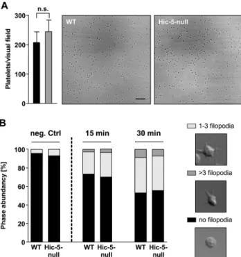
At sites of vascular injury, vWF is exposed on the subendothelial extracellular matrix (ECM). The interaction between the platelet GPIb-V-IX receptor complex and vWF is impor-tant for platelet tethering, the initial step during the formation of a hemostatic plug. A potential role of Hic-5 in GPIb-mediated processes was assessed by monitoring the adhesion of platelets on immobilized vWF under high shear conditions. Hic-5-null and wild-type platelets attached to the immobilized vWF and in part firmly adhered to the surface to comparable extent and with similar kinetics (Fig 3A). In addition, we analyzed platelet spreading on a vWF-coated matrix under conditions ofαIIbβ3 integrin-blockade. The stimulation of platelets by a GPIb specific signal results in a shape change, which is limited to contraction of the cell body and filopodia formation [25]. Both, Hic-5-null and wild-type platelets extended comparable num-bers of filopodia (Fig 3B). Together, these results indicate that GPIb function is intact in Hic-5-deficient platelets.
To investigate the role of Hic-5 in platelet functionin vivo, we utilized two widely used tail bleeding time assays (filter paper and warm saline) and found comparable bleeding times for control and Hic-5-null mice (Fig 4AandS3 Figrespectively). This result stands in contrast to the study of Kim-Kaneyamaet al. who found prolonged bleeding times in Hic-5-null mice using the saline method [14].
Kim-Kaneyamaet al. reported that Hic-5-null mice are more resistant to experimental thromboembolism than control mice [14]. Consistent with ourin vitroresults, pathological thrombus formation, as assessed by intravital microscopy of FeCl3-injured mesenteric
arteri-oles, was indistinguishable between wild-type and Hic-5-null mice, resulting in similar
Fig 2. Lack of Hic-5 has no influence on platelet adhesion to collagen, spreading on fibrinogen and platelet integrin outside-in signalingin vitro.(A, B) Adhesion (A) and thrombus formation (B) of platelets on collagen was assessed in a flow adhesion assay at a wall shear rate of 1000 s-1. Scale bar, 25μm. (C, D) Washed platelets of WT and Hic-5-null mice were allowed to spread on fibrinogen for up to 30 minutes after stimulation with 0.01 U mL-1
thrombin. (C) Representative images and (D) statistical evaluation of the percentage of spread platelets at different spreading stages are shown. Scale bar, 5μm. (E, F) Clot formation in platelet-rich plasma was induced by the addition of thrombin (5 U mL-1) and 20 mmol L-1CaCl
2and clot retraction was
monitored over time (E). (F) Residual volume of serum after clot retraction was measured.
occlusion times for both groups (Fig 4B and 4C). These data indicate a dispensable role of Hic-5 in thrombus formationin vivo.
In this study, constitutive knockout mice for Hic-5 were used to assess the role of this focal adhesion protein in platelet functionin vitroandin vivo. We did not detect any alterations in integrin inside-out or outside-in signaling in the absence of Hic-5, arguing against an essential role of the adaptor in these processes. Consequently, hemostasis and experimental thrombus formation were not affected by the lack of Hic-5. It is difficult to explain the discrepancy between our study and that of Kim-Kaneyamaet al. [14], but differences in the genetic back-ground might contribute, as previously proposed as a possible explanation for differences observed in the phenotype of GPVI-deficient mice [26]. However, based on our results we sup-pose that Hic-5 plays only a minor role in platelet integrin signalingin vitroandin vivo. Pre-sumably, Hic-5 deficiency can be compensated by the other paxillin family members, paxillin and leupaxin, which are both expressed in murine platelets [27]. This is supported by a study from Rathore and colleagues [27]. They could show, that in aggregating human platelets, Hic-5 was tyrosine phosphorylated and recruited Csk (C-terminal Src kinase) via its SH2 domains to regulate SFK (Src family kinases) activity. However, in aggregating mouse platelets, Csk bound preferentilly to paxillin instead of Hic-5, although both are abundantly expressed.
It is possible that Hic-5 and the other paxillin family members share some tissue specific functions and that Hic-5 could play important roles in cells or tissues other than platelets. As an example, Hic-5 has been shown to have a role in the development of abdominal aortic aneu-rysms [28].
Notably, however, in contrast to mouse platelets human platelets express Hic-5 as the only paxillin family member [27]. Under these conditions, loss of Hic-5 could exert effects on
Fig 3. Hic-5-deficient platelets have unaltered GPIb function.(A) Whole blood of wild-type and Hic-5-null mice was perfused over a vWF-coated surface at a wall shear rate of 1,700 sec-1and the number of adherent
platelets was quantified. (B) Washed platelets were treated with integrilin (40μg/ml) and botrocetin (2μg/ml) and allowed to adhere to vWF-coated cover slips. Filopodia formation was quantified according to the number of extensions per platelet at indicated the time points.
human platelet function in thrombosis and hemostasis. To investigate this in mice, a mouse strain lacking all three paxillin members would be required.
Supporting Information
S1 Fig. Expression ofTgfb1i1mRNA.Analysis of the presence ofTgfb1i1mRNA in platelets by RT-PCR. GAPDH mRNA served as positive control. cDNA free sample was used as nega-tive control.
(TIF)
S2 Fig. Spreading on fibrinogen of unstimulated platelets.Washed platelets of WT and Hic-5-null mice were allowed to spread on fibrinogen for up to 60 minutes in the presence of apy-rase (2 U/ml) and indomethacin (1.4μM).
(TIF)
S3 Fig. Normal hemostasis in Hic-5-null mice.Tail bleeding times in saline of wild-type and Hic-5-null mice. Each symbol represents 1 animal.
(TIF)
Author Contributions
Conceived and designed the experiments: BN DS. Performed the experiments: MP IT DS. Ana-lyzed the data: MP IT DS. Wrote the paper: MP BN DS.
Fig 4. Unalteredin vivothrombus formation in Hic-5-deficient mice.(A) Tail bleeding times of wild-type and Hic-5-null mice. Each symbol represents 1 animal. (B, C) Thrombus formation in small mesenteric arterioles was induced by topical application of 20% FeCl3. For monitoring of thrombus formation by intravital
microscopy, platelets were labeled fluorescently. Time to stable occlusion (B) and representative pictures (C) of wild-type and Hic-5-null mice are shown. Each symbol represents one individual.
References
1. Ruggeri ZM. Platelets in atherothrombosis. Nat Med. 2002; 8: 1227–1234. PMID:12411949
2. Nieswandt B, Watson SP. Platelet-collagen interaction: is GPVI the central receptor? Blood. 2003; 102: 449–61. doi:10.1182/blood-2002-12-3882PMID:12649139
3. Nieswandt B, Pleines I, Bender M. Platelet adhesion and activation mechanisms in arterial thrombosis and ischaemic stroke. J Thromb Haemost. 2011; 9: 92–104. doi:10.1111/j.1538-7836.2011.04361.x 4. Shattil SJ, Kashiwagi H, Pampori N. Integrin signaling: the platelet paradigm. Blood. 1998; 91: 2645–
57. PMID:9531572
5. Ginsberg MH, Partridge A, Shattil SJ. Integrin regulation. Curr Opin Cell Biol. 2005; 17: 509–16. doi:10.
1016/j.ceb.2005.08.010PMID:16099636
6. Nieswandt B, Moser M, Pleines I, Varga-Szabo D, Monkley S, Critchley D, et al. Loss of talin1 in plate-lets abrogates integrin activation, platelet aggregation, and thrombus formation in vitro and in vivo. J Exp Med. 2007; 204: 3113–8. doi:10.1084/jem.20071827PMID:18086864
7. Petrich BG, Marchese P, Ruggeri ZM, Spiess S, Weichert RAM, Ye F, et al. Talin is required for integ-rin-mediated platelet function in hemostasis and thrombosis. J Exp Med. 2007; 204: 3103–11. doi:10.
1084/jem.20071800PMID:18086863
8. Moser M, Nieswandt B, Ussar S, Pozgajova M, Fässler R. Kindlin-3 is essential for integrin activation and platelet aggregation. Nat Med. 2008; 14: 325–30. doi:10.1038/nm1722PMID:18278053 9. Tadokoro S, Shattil SJ, Eto K, Tai V, Liddington RC, de Pereda JM, et al. Talin binding to integrin beta
tails: a final common step in integrin activation. Science. 2003; 302: 103–6. doi:10.1126/science.
1086652PMID:14526080
10. Ma Y-Q, Qin J, Wu C, Plow EF. Kindlin-2 (Mig-2): a co-activator of beta3 integrins. J Cell Biol. 2008; 181: 439–46. doi:10.1083/jcb.200710196PMID:18458155
11. Schaller MD, Otey CA, Hildebrand JD, Parsons JT. Focal adhesion kinase and paxillin bind to peptides mimicking beta integrin cytoplasmic domains. J Cell Biol. 1995; 130: 1181–7. PMID:7657702 12. Legate KR, Fässler R. Mechanisms that regulate adaptor binding to beta-integrin cytoplasmic tails. J
Cell Sci. 2009; 122: 187–198. doi:10.1242/jcs.041624PMID:19118211
13. Stritt S, Wolf K, Lorenz V, Vögtle T, Gupta S, Bösl M, et al. Rap1-GTP–interacting adaptor molecule
(RIAM) is dispensable for platelet integrin activation and function in mice. Blood. 2015; 125: 219–223.
doi:10.1182/blood-2014-08-597542PMID:25336629
14. Kim-Kaneyama JR, Miyauchi A, Lei XF, Arita S, Mino T, Takeda N, et al. Identification of Hic-5 as a novel regulatory factor for integrinαIIbβ3 activation and platelet aggregation in mice. J Thromb Hae-most. 2012; 10: 1867–74. doi:10.1111/j.1538-7836.2012.04856.xPMID:22812543
15. Shibanuma M, Mashimo J, Kuroki T, Nose K. Characterization of the TGF beta 1-inducible hic-5 gene that encodes a putative novel zinc finger protein and its possible involvement in cellular senescence. J Biol Chem. 1994; 269: 26767–26774. PMID:7929412
16. Tumbarello DA, Turner CE. Hic-5 Contributes to Transformation Through a RhoA / ROCK-dependent Pathway. J Cell Physiol. 2006; 211: 736–747. doi:10.1002/JCP
17. Hagmann J, Grob M, Welman A, van Willigen G, Burger MM. Recruitment of the LIM protein hic-5 to focal contacts of human platelets. J Cell Sci. 1998; 111: 2181–8. PMID:9664039
18. Sakata A, Ohmori T, Nishimura S, Suzuki H, Madoiwa S, Mimuro J, et al. Paxillin is an intrinsic negative regulator of platelet activation in mice. Thromb J. 2014; 12: 1. doi:10.1186/1477-9560-12-1PMID: 24383745
19. Knight CG, Morton LF, Onley DJ, Peachey AR, Ichinohe T, Okuma M, et al. Collagen-platelet interac-tion: Gly-Pro-Hyp is uniquely specific for platelet Gp VI and mediates platelet activation by collagen. Cardiovasc Res. 1999; 41: 450–7. PMID:10341844
20. Pleines I, Eckly A, Elvers M, Hagedorn I, Eliautou S, Bender M, et al. Multiple alterations of platelet functions dominated by increased secretion in mice lacking Cdc42 in platelets. Blood. 2010; 115: 3364–73. doi:10.1182/blood-2009-09-242271PMID:20139097
21. Grosse J, Braun A, Varga-Szabo D, Beyersdorf N, Schneider B, Zeitlmann L, et al. An EF hand muta-tion in Stim1 causes premature platelet activamuta-tion and bleeding in mice. J Clin Invest. 2007; 117: 3540–
50. doi:10.1172/JCI32312.3540PMID:17965774
22. Lenter M, Uhligt H, Hamannt ALF, Jenot P, Imhof B, Vestweber D. A monoclonal antibody against an activation epitope on mouse integrin chain beta 1 blocks adhesion of lymphocytes to the endothelial integrin alpha 6 beta 1. Proc Natl Acad Sci U S A. 2000; 90: 9051–9055.
24. Morgenstern E, Ruf A, Patscheke H. Ultrastructure of the interaction between human platelets and poly-merizing fibrin within the first minutes of clot formation. Blood Coagul Fibrinolysis. 1990; 1: 547–549. 25. Pleines I, Eckly A, Elvers M, Hagedorn I, Eliautou S, Bender M, et al. Multiple alterations of platelet
functions dominated by increased secretion in mice lacking Cdc42 in platelets. Blood. 2010; 115: 3364–3373. doi:10.1182/blood-2009-09-242271PMID:20139097
26. Cheli Y, Jensen D, Marchese P, Habart D, Wiltshire T, Cooke M, et al. The Modifier of hemostasis (Mh) locus on chromosome 4 controls in vivo hemostasis of Gp6 - /- mice. Blood. 2008; 111: 1266–1274.
doi:10.1182/blood-2007-09-111369The PMID:17991808
27. Rathore VB, Okada M, Newman PJ, Newman DK. Paxillin family members function as Csk-binding pro-teins that regulate Lyn activity in human and murine platelets. Biochem J. 2007; 403: 275–81. doi:10.
1042/BJ20061618PMID:17233630