Platelet Function and Thrombus Formation
Kellie J. Hall1, Matthew T. Harper1, Karen Gilio2, Judith M. Cosemans2, Johan W. M. Heemskerk2, Alastair W. Poole1*
1Department of Physiology and Pharmacology, University of Bristol, Bristol, United Kingdom,2Department of Biochemistry, University of Maastricht, Maastricht, The Netherlands
Abstract
Background:PKChis a novel protein kinase C isozyme, predominately expressed in T cells and platelets. PKCh2/2T cells
exhibit reduced activation and PKCh2/2mice are resistant to autoimmune disease, making PKChan attractive therapeutic
target for immune modulation. Collagen is a major agonist for platelets, operating through an immunoreceptor-like signalling pathway from its receptor GPVI. Although it has recently been shown that PKChpositively regulates outside-in signalling through integrinaIIbb3in platelets, the role of PKChin GPVI-dependent signalling and functional activation of
platelets has not been assessed.
Methodology/Principal Findings: In the present study we assessed static adhesion, cell spreading, granule secretion, integrin aIIbb3 activation and platelet aggregation in washed mouse platelets lacking PKCh. Thrombus formation on a
collagen-coated surface was assessed in vitro under flow. PKCh2/2platelets exhibited reduced static adhesion and filopodia
generation on fibrinogen, suggesting that PKChpositively regulates outside-in signalling, in agreement with a previous report. In contrast, PKCh2/2 platelets also exhibited markedly enhanced GPVI-dependent a-granule secretion, although
dense granule secretion was unaffected, suggesting that PKCh differentially regulates these two granules. Inside-out regulation ofaIIbb3activation was also enhanced downstream of GPVI stimulation. Although this did not result in increased
aggregation, importantly thrombus formation on collagen under high shear (1000 s21) was enhanced.
Conclusions/Significance:These data suggest that PKChis an important negative regulator of thrombus formation on collagen, potentially mediated bya-granule secretion andaIIbb3activation. PKChtherefore may act to restrict thrombus
growth, a finding that has important implications for the development and safe clinical use of PKChinhibitors.
Citation:Hall KJ, Harper MT, Gilio K, Cosemans JM, Heemskerk JWM, et al. (2008) Genetic Analysis of the Role of Protein Kinase Chin Platelet Function and Thrombus Formation. PLoS ONE 3(9): e3277. doi:10.1371/journal.pone.0003277
Editor:Karl-Wilhelm Koch, University of Oldenburg, Germany
ReceivedAugust 1, 2008;AcceptedSeptember 5, 2008;PublishedSeptember 25, 2008
Copyright:ß2008 Hall et al. This is an open-access article distributed under the terms of the Creative Commons Attribution License, which permits unrestricted use, distribution, and reproduction in any medium, provided the original author and source are credited.
Funding:Supported by a British Heart Foundation programme grant to AWP (grant no. RG/05/015) and a grant from the Netherlands Foundation for Scientific research to JWMH (11-400-076). AWP is a BBSRC Research Development Fellow (BB/E024637/1). The funders had no role in study design, data collection and analysis, decision to publish, or preparation of the manuscript.
Competing Interests:The authors have declared that no competing interests exist. * E-mail: [email protected]
Introduction
The protein kinase C (PKC) family critically regulates platelet activation. Many platelet functional responses, including secretion and aggregation are reduced or abolished by broad-spectrum PKC inhibitors and enhanced by PKC activators [1], suggesting a positive role for the PKC family in general in platelet activation. However, calcium responses are clearly negatively regulated by PKC isoforms [2], and we have shown by pharmacological and genetic approaches that PKCdis a negative regulator of platelet aggregation by modulating actin dynamics through VASP [3,4]. Individual PKC isoforms therefore play distinct roles, both positive and negative, during platelet activation, and the effect of broad-spectrum PKC inhibition or activation reflects a balance of effects on positive and negative regulatory pathways [1].
Human platelets express predominantly four PKC isoforms:a,
b,dandh. In addition to these, mouse platelets express PKCe[3–
9]. The specific importance of each isoform is hard to assess by pharmacological approaches owing to the lack of isoform
specificity of these agents. The availability of biochemical and genetic tools has allowed the functions of specific isoforms to be addressed. Using such approaches, we and others have recently demonstrated highly specific roles for individual PKC isoforms in regulating platelet function: PKCais critically required for granule secretion and secretion-dependent aggregation [10,11]; PKCbis recruited to integrin aIIbb3 and positively regulates outside-in signalling [12]; PKCd, in contrast, negatively regulates filopodia formation, and lack of PKCd leads to enhanced platelet aggregation [13].
PKCh is a novel (i.e. DAG-sensitive, Ca2+-insensitive) PKC isoform, predominantly expressed in T-cells, muscle cells and platelets [14,15]. PKCh2/2mice exhibit reduced T cell activation, proliferation and IL-2 production downstream of T-cell receptor stimulation, owing to markedly reduced activation of multiple transcription factors [16,17] and, as a result, these mice are resistant to some models of autoimmune disease [18–20]. PKCh
activation during the very early stages of thrombosis, and the parallels between signalling downstream of the collagen receptor GPVI and that downstream of immunoreceptors, it was now important to determine the role played by PKCh in collagen-induced platelet activation and thrombus formation. We report that PKCh negatively regulates GPVI-dependent a-granule secretion and integrin aIIbb3 activation and thereby is the only PKC isoform yet described with this function. Furthermore, loss of this negative regulation in PKCh2/2platelets leads to enhanced thrombus formation under flowin vitro. These results reveal a novel
negative regulatory pathway in platelet activation, and have relevance to the current clinical and pharmaceutical interest in PKChinhibitors.
Methods
Materials
Unless stated, all reagents were from Sigma Aldrich (Poole, Dorset, U.K.). Cross-linked collagen-related peptide (CRP) was from Professor Richard Farndale (Biochemistry, University of Cambridge, U.K.). Horm collagen was from Axis Shield (Bicton, Cambs., U.K.). Phycoerythrin (PE)-labelled JON/A and fluorescein isothiocyanate (FITC)-labelled Wug.E9 (anti-P-selectin) antibodies were from Emfret Analytics (Eibelstadt, Germany). Anti-PKCa, -PKCb, -PKCd, -PKCeand anti-tubulin antibodies were from BD
Transduction Laboratories (Oxford, U.K). Anti-PKCh antibody was from Cell Signaling Technology (New England BioLabs, Hitchin, U.K.). horseradish-peroxidase (HRP)-conjugated anti-mouse IgG and anti-rabbit IgG secondary antibodies, and enhanced chemiluminescent (ECL) reagents were from Amersham (Little Chalfont, Bucks., U.K.). Luciferin-luciferase reagent was from Chronolog (LabMedics, Manchester, U.K.).
Washed platelet preparation
PKCh2/2 C57BL6/J mice have been described previously [17]. Wildtype C57BL6/J mice were used as control. Use of mouse platelets was approved by local research ethics committee at the University of Bristol, U.K. and mice were bred for this purpose under UK Home Office licence (PPL 30/2386) held by AWP. Washed platelets were prepared as previously described [13]. Of note, platelets were treated with indomethacin (10mM). Platelets were rested for 30 min after centrifugation.
Electrophoresis and Western blotting
Washed platelets (26108/ml) were lysed in Laemmli sample
buffer. Proteins were resolved by electrophoresis in 9% SDS-polyacrylamide gels. Samples were then transferred to polyviny-lidene difluoride membranes, blocked with 10% bovine serum albumin, and subjected to immunoblotting with specific antibodies to various PKC isoforms, as described in the text. Primary
Analysis ofaIIbb3activation anda-granule secretion by flow cytometry
Washed platelets (46107/ml) were aliquoted into tubes containing optimal concentrations of PE-JON/A or FITC-anti-CD62P, which bind to active integrinaIIbb3and surface-exposed P-selectin (CD62P), respectively, and CRP at the final concentra-tions indicated, for 15 min. Analysis of 20,000 events was performed using a Becton Dickinson FACScan. The platelet population as identified by forward and side scatter profile. Data were analysed using WinMDI version 2.8.
DIC imaging of platelet adhesion and spreading
Measurement of static platelet adhesion and spreading was performed as previously described [13]. Glass coverslips were coated with fibrinogen, CRP or collagen and mounted in a live-cell chamber. Adhesion and spreading of washed platelets (26107/
ml) was followed by differential interference contrast (DIC) microscopy with a wide-field microscope DM IRB attached to an ORCA ER camera (63x/1.40 NA oil objective) (Leica Microsystems, Milton Keynes, UK). Images were processed with OpenLab 4.03 (Improvision). The surface area of adherent platelets was measured using Volocity software (Improvision), while the number of adherent platelets was counted manually.
In vitro thrombus formation
Flow-induced thrombus formation was assessed basically as described before [25]. A Leica wide-field microscope DM IRB (63x/1.40 NA oil objective), attached to an ORCA ER camera was used for image capture (Leica Microsystems, Milton Keynes, UK). Heparin/PPACK-anticoagulated mouse blood was flowed over immobilised collagen through a parallel plate perfusion chamber, at a fixed shear rate of 1000 s21
for 4 minutes. For each experiment, at least 10 random phase-contrast images were captured, which were then averaged. Recorded images were analyzed with ImagePro software.
Statistics
Statistical analyses were performed using GraphPad Prism software, unless stated otherwise, using two-way ANOVA with Bonferroni post-test; p,0.05 was considered significant. Bar charts show mean data6SEM (where ‘n’ denotes the number of individual mice used).
Results
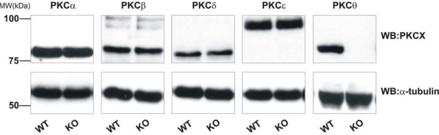
PKCh2/2platelets exhibit normal expression of other PKC isoforms
and not due to altered expression of other PKC isoforms, we assessed the expression of the major PKC isoforms in platelets by western blotting. In addition to PKCh, mouse platelets strongly express PKCa, -b, -d, and -e. No difference in expression of these
isoforms was seen in PKCh2/2 platelets relative to WT platelets (Fig. 1). The blotting membranes were stripped and re-probed for
a-tubulin, to ensure equal loading of proteins between samples (Fig. 1, lower panels).
PKChhas a small positive effect on platelet spreading on fibrinogen
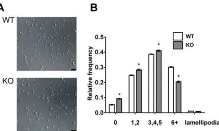
Others have reported that platelet spreading on fibrinogen was partially defective in PKCh2/2 platelets [24]. We were able to confirm and extend this result, demonstrating that both adhesion of platelets and specifically the degree of filopodia generation, rather than lamellipodia, 45 minutes after static deposition on fibrinogen-coated coverslips, were reduced in PKCh2/2platelets (Table 1). We analysed the kinetics of the spreading process to determine any further qualitative differences in spreading. Platelets during/after spreading were scored for number of filopodia and categorized as having none, few (1 or 2), some (3, 4 or 5) or many (6 or more) filopodia. The relative frequency of each morphology was determined and is shown in Fig. 2. 45 minutes after deposition on the coverslip, most WT platelets had formed at least a few
filopodia, although very few platelets formed lamellipodia, consistent with other reports [26]. PKCh2/2 mice had a significantly different distribution of filopodial number, with a lower proportion forming 6 or more filopodia (p,0.001). As a consequence, a greater proportion of PKCh2/2 platelets formed 0, 1 or 2 filopodia than WT platelets (p,0.001). Thus, PKChhas a small, positive regulatory role in filopodia generation on fibrinogen.
PKChdoes not regulate adhesion or spreading on CRP or collagen
Since PKChhad a role in platelet adhesion and spreading on fibrinogen, its role in adhesion and spreading on CRP and collagen was also assessed. CRP is a selective GPVI agonist, whereas collagen activates both GPVI and integrin a2b1. In contrast to fibrinogen, no significant effect was seen on adhesion or total platelet surface area on either of these substrates (Table 1). Platelet interaction with collagen is therefore not affected by absence of PKCh.
PKCh negatively regulates CRP-induced platelet activation
We further investigated whether PKCh regulates platelet activation following GPVI stimulation. Activation of GPVI leads to secretion ofa-granules and dense granules, and activation of integrinaIIbb3. The latter is known as inside-out signalling and is necessary for platelet aggregation.
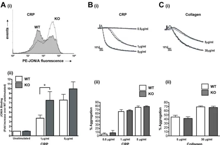
CRP-induced surface expression of P-selectin, a marker of a -granule release was enhanced in the absence of PKCh. In WT platelets, 1mg/ml CRP induced a 3.160.5 –fold increase over basal in FITC-P-selectin fluorescence, which was increased to 10.263.0 –fold in PKCh2/2 platelets (n = 8; p
,0.05; Fig. 3A), suggesting that PKCh negatively regulates the release of these granules. Interestingly, however, no difference in ATP secretion was seen between PKCh2/2and WT platelets in response to CRP (Fig. 3B) or collagen (Fig. 3C).
aIIbb3activation was determined by flow cytometry using JON/ A, an antibody that recognises the active conformation of this integrin. Importantly, JON/A binding was almost doubled in platelets activated by 1mg/ml CRP, from 3.860.7 –fold over basal in WT to 7.561.8-fold in PKCh2/2platelets (n = 8; p,0.05; Fig. 4A). In contrast, a higher concentration of CRP (5mg/ml) was not significantly affected (7.661.2 –fold in WT compared to
Figure 1. PKC isoforms are not upregulated in PKCh2/2mice.Platelets lysates from wild-type (WT) or PKCh2/2(KO) mice were assessed for PKC isoform expression by SDS-PAGE and western blotting using specific antibodies for PKCa, -b, -d, -hand -e. Membranes were stripped and
re-probed fora-tubulin as indicated to ensure equal loading of protein. Blots are representative of three independent experiments. doi:10.1371/journal.pone.0003277.g001
Table 1.PKChdoes not regulate adhesion or spreading on CRP or collagen.
Adhesion Surface area (mm2)
WT KO WT KO
Fibrinogen 111.164.5 82.7612.0 * 24.961.9 21.660.9 * CRP 83.767.3 79.662.2 ns 25.861.0 26.561.7 ns Collagen 130.4634.6 137.2641.8 ns 14.560.6 14.460.8 ns
Platelets were deposited on fibrinogen, CRP or collagen-coated coverslips in a live-cell chamber for 45 min and visualized by DIC microscopy. Five fields of view were selected at random and the number of adherent platelets was counted (adhesion) and spread surface area measured. Adhesion is total number of platelets adherent to the surface within a single 1000mm2field of
view. Shown are combined data from three independent experiments (mean6SEM;*indicates p
10.061.5 –fold in PKCh2/2; n = 8; p = 0.81; Fig. 4A). These data suggest that PKChnegatively regulates GPVI-dependent aIIbb3 activation, but that at high concentrations this inhibition can be overcome. Interestingly however, platelet aggregation was not affected at either of these concentrations of CRP (Fig. 4B), nor was collagen-induced aggregation affected (Fig. 4C).
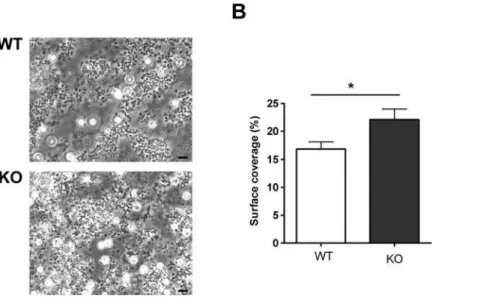
PKCh negatively regulates thrombus formationin vitro
Since PKCh2/2aggregated normally, despite increaseda IIbb3 activation and a-granule secretion, we investigated whether the role of PKCh might become more apparent during thrombus formation in the more physiological setting of flow conditions. Anticoagulated whole blood was passed over a collagen-coated
Figure 2. PKChpositively regulates filopodia number when platelets spread on fibrinogen.Platelets were deposited on fibrinogen-coated coverslips in a live-cell chamber for 45 min and visualized by DIC microscopy. Five fields of view were selected at random, and one such field is shown in (A) for WT and PKCh2/2platelets. In (B), filopodia number was counted for each visible platelet and the number of platelets with none, few (1–2), some (3–5) or many (6 or more) filopodia were expressed as a proportion of the total number of platelets in view. Shown are combined data from three independent experiments. Bar indicates 10mm.
doi:10.1371/journal.pone.0003277.g002
Figure 3. PKChnegatively regulatesa-granule secretion.A:Washed platelets from WT or KO mice were stimulated with CRP (1 or 5mg/ml) in
the presence of FITC-labelled anti-P-selectin antibody for 15 minutes, and surface labelling measured by flow cytometry. Representative histograms are shown inA(i)(1mg/ml), and fold-increase in geometric mean compared to unstimulated platelets is shown inA(ii)(mean6SEM; n = 8).B, C: ATP secretion from dense granules in response to CRP (B) or collagen (C) was monitored in a luminometer using the luciferin-luciferase reaction. Data are presented as mean6SEM (n = 4).
coverslip through a parallel-plate flow chamber at a shear rate of 1000 s21
, and thrombi observed under phase contrast after 4 min. Figure 5 shows that platelets from WT mice formed substantial thrombi on the collagen surface, However, platelets from PKCh2/ 2formed significantly larger thrombi, suggesting that the negative role of PKCh is necessary to restrict thrombus size under flow conditions.
Discussion
PKC activation is generally considered to positively regulate platelet signalling, since platelet activation is inhibited by broad-spectrum PKC inhibitors, and PKC activators can enhance platelet activation. However, here we show that the role of PKCh
is more complicated than this, as it negatively regulatesa-granule secretion and inside-out signalling to integrinaIIbb3, yet positively regulates outside-in integrin signalling. In the absence of PKCh, thrombus formation was markedly enhanced, suggesting that PKChrestricts thrombus size.
First, we observed a significant reduction in PKCh2/2platelet adhesion and reduced spreading on fibrinogen compared to WT platelets, in agreement with Soriani et al. [24]. Interestingly, Soriani’s study showed an approximately 50% reduction in spread platelet surface area whereas our study only showed a 13% reduction. This apparent quantitative (though not qualitative)
discrepancy could result from technical differences between our experiments. We used DIC microscopy to image platelet spreading, and the surface area of platelets was measured by manually outlining each cell (approximately 25mm2). Another study by McCarty et al [26] that used this approach saw a similar surface area. In both McCarty’s study and ours, mouse platelets rarely formed large lamellipodia when spreading on fibrinogen, (in contrast to human platelets, which form full lamellipodia on fibrinogen) and filopodia were still apparent even after 45 minutes. In contrast, Soriani et al. [24] measured the surface area by confocal microscopy of rhodamine-phalloidin stained platelets, and reported a much lower surface area (approximately 8mm2). Rather than measure the surface area directly, this method measures F-actin coverage, perhaps suggesting that PKChregulates actin polymer-ization. WT platelets spread on fibrinogen and imaged using this method do not appear to exhibit the spiky morphology we and others [26,27] observe using DIC microscopy. Our analysis suggests that PKChpositively regulates filopodia formation, since a smaller proportion of PKCh2/2 platelets showed many (
.5) filopodia compared to WT. Regardless of these quantitative differences, both of our studies qualitatively agree that PKChis a positive regulator of outside-in signalling by integrinaIIbb3.
In contrast, PKCh negatively regulates GPVI-induced aIIbb3 activation. The selective GPVI agonist, CRP, induced a concentration-dependent increase in binding of JON/A, an
Figure 4. PKChnegatively regulates CRP-stimulated integrinaIIbb3activation but not platelet aggregation.A: Washed platelets from
WT or KO mice were stimulated with CRP (1 or 5mg/ml) in the presence of PE-labelled JON/A for 15 minutes, and surface labelling measured by flow
cytometry. Representative histograms are shown in(i)(1mg/ml), and fold-increase in geometric mean compared to unstimulated platelets is shown in(ii)(mean6SEM; n = 8).B, C: Platelet aggregation in response to CRP (B) or collagen (C) was monitored by turbidometry. Traces, shown inB(i)and C(i), are representative of at least three separate experiments. Mean extent of aggregation at 5 min (6SEM; n = 3) is shown inB(ii)andC(ii), for CRP and collagen, respectively.
activation state-specificaIIbb3antibody. In PKCh2/2platelets this was markedly enhanced compared to WT at an intermediate concentration of CRP, though not at a higher concentration, suggesting that PKChreduces expression of activeaIIbb3on the platelet surface, although inhibition can be overcome as agonist stimulation increases. It has been previously reported that PKCh
does not regulate aIIbb3 activation in response to ADP or AYPGKF [24], both of which act through G protein-coupled receptors, suggesting that the regulatory role of PKChmay be specific to GPVI signalling.
CRP-induced aggregation was not affected by the absence of PKCh, however. Similarly, collagen-induced aggregation was also the same in WT and PKCh2/2platelets. The lack of any effect on the rate or extent of aggregation was surprising, especially in response to 1mg/ml CRP. At this concentration, the rate of aggregation was submaximal and yet the extent of integrin activation strongly enhanced. It might be expected, therefore, that the increased integrin activation would accelerate aggregation. However, since the extent of aggregation in response to 1mg/ml CRP was almost maximal, further enhancement of aIIbb3 in PKCh2/2 platelets can have little further effect. The apparent disparity between absolute levels of integrin activation and extent of aggregation highlights the large level in integrin reserve believed to exist in platelets.b3+/2platelets, with only 50% of the WT levels of
b3 on their surface, have almost identical bleeding times and aggregation responses to PMA, ADP, thrombin and arachidonic acid compared to WT platelets [28]. In like manner, although WT platelets show approximately 50 % less integrin activation than PKCh2/2 platelets at 1mg/ml CRP, we should not expect this necessarily to relate to a difference in the extent of aggregation.
PKChalso negatively regulatesa-granule secretion, although no difference in dense granule secretion was observed. This suggests that the release of different platelet granules is regulated by distinct mechanisms. The PKC family in general is a critical positive regulator of platelet granule secretion [2,10,11], although this positive function is likely to be mediated through conventional (Ca2+-dependent) isoforms [5,10,11]. Thus, it appears that the different PKC isoforms have contrasting roles in plateleta-granule secretion: PKCais critically required fora-granule secretion, and
PKChacts to counter this action. PKCais also critically important for dense granule secretion, which is not countered by PKCh. It has been suggested that PKCd, closely related to PKCh, may negatively regulate GPVI-dependent dense granule secretion [5]. This interpretation was based on the use of rottlerin, a supposedly specific PKCd inhibitor (though several PKCd-independent targets have been reported [29–31]). However, we have previously reported that rottlerin enhances GPVI-dependent dense granule release even in PKCd2/2mice [13]. Thus, negative regulation of GPVI-dependent dense granule release does not appear to be mediated by either PKCdor PKCh.
PKChnegatively regulates thrombus formation under flow over a collagen-coated surface. Binding to collagen activates GPVI, leading to integrin aIIbb3 activation, which is enhanced in PKCh2/2 platelets. The increased number of adhesive contacts between platelets may accelerate the growth of the thrombus. Thus, negative regulation of inside-out signalling by PKChmay be an important brake on thrombus growth at a site of injury. This effect is in contrast to the lack of effect seen in aggregation, highlighting the importance of physiological flow conditions [32]. In standard aggregometry, platelets exhibit a very large integrin reserve, whereas under flow, with higher shear force on any platelet-platelet interactions, integrin activation may be a limiting factor. Increased aIIbb3 activation would therefore enhance thrombus growth. This effect may be partially countered by the slightly reduced platelet adhesion to fibrinogen and reduced subsequent spreading, perhaps leading to fewer platelet-platelet contacts. Given the large effect on integrin activation compared to the smaller effect on spreading, however, the balance of these appears to favour increased thrombus size in PKCh2/2platelets. In summary, we have shown that PKChnegatively regulates GPVI-dependent inside-out signalling, in contrast to the positive role generally ascribed to the PKC family in general. Although enhanced integrin aIIbb3 activation does not lead to increased aggregation in an aggregometer tube, PKCh2/2platelets display enhanced thrombus formation on collagen under flow, suggesting that, under more physiological conditions, the regulatory role of PKChmay restrict thrombus size. This may impact on the clinical safety of PKChinhibitors.
Figure 5. PKChnegatively regulates thrombus formation on collagen under flowin vitro.Whole blood was passed over a collagen-coated coverslip at 1000 s21for four minutes then observed by phase contrast microscopy (A). Surface coverage was measured and is shown inBas
Acknowledgments
We are grateful to Professor Dan Littman (Skirball Institute of Biomolecular Medicine, New York, USA) for generating the PKCh2/2
mice used in this study, and to Professor Isabella Screpanti (University ‘‘La Sapienza’’, Rome, Italy) for supplying them. We thank Elizabeth Aitken for expert technical assistance supporting this work. We are grateful to Dr Mark Jepson and Alan Leard for their assistance within the School of
Medical Sciences Wolfson Bioimaging Facility. AWP is a BBSRC Research Development Fellow.
Author Contributions
Conceived and designed the experiments: KJH MTH KG JMC JH AWP. Performed the experiments: KJH MTH KG JMC JH. Analyzed the data: KJH MTH KG JMC JH AWP. Wrote the paper: KJH MTH JH AWP.
References
1. Harper MT, Poole AW (2007) Isoform-specific functions of PKC: the platelet paradigm. Biochem Soc Trans 35: 1005–1008.
2. Strehl A, Munnix ICA, Kuijpers MJE, van der Meijden PEJ, Cosemans JMEM, et al. (2007) Dual role of platelet protein kinase C in thrombus formation: stimulation of pro-aggregatory and suppression of procoagulant activity in platelets. J Biol Chem 282: 7046–7055.
3. Crosby D, Poole AW (2003) Physical and functional interaction between protein kinase C delta and Fyn tyrosine kinase in human platelets. J Biol Chem 278: 24533–24541.
4. Crosby D, Poole AW (2002) Interaction of Bruton’s tyrosine kinase and protein kinase Ctheta in platelets. Cross-talk between tyrosine and serine/threonine kinases. J Biol Chem 277: 9958–9965.
5. Murugappan S, Tuluc F, Dorsam RT, Shankar H, Kunapuli SP (2004) Differential role of PKC-delta isoform in agonist-induced dense granule secretion in human platelets. J Biol Chem 279: 2360–2367.
6. Grabarek J, Raychowdhury M, Ravid K, Kent KC, Newman PJ, et al. (1992) Identification and functional characterization of protein kinase C isozymes in platelets and HEL cells. J Biol Chem 267: 10011–10017.
7. Crabos M, Imber R, Woodtli T, Fabbro D, Erne P (1991) Different translocation of three distinct PKC isoforms with tumor-promoting phorbol ester in human platelets. Biochem Biophys Res Commun 178: 878–883. 8. Baldassare JJ, Henderson PA, Burns D, Loomis C, Fisher GJ (1992)
Translocation of protein kinase C isozymes in thrombin-stimulated human platelets. Correlation with 1,2-diacylglycerol levels. J Biol Chem 267: 15585–15590.
9. Wang F, Naik UP, Ehrlich YH, Freyberg Z, Osada S, et al. (1993) A new protein kinase C, nPKC eta, and nPKC theta are expressed in human platelets: involvement of nPKC eta and nPKC theta in signal transduction stimulated by PAF. Biochem Biophys Res Commun 191: 240–246.
10. Yoshioka A, Shirakawa R, Nishioka H, Tabuchi A, Higashi T, et al. (2001) Identification of protein kinase C-alpha as an essential, but not sufficient, cytosolic factor for Ca2+-induced alpha- and dense-core granule secretion in platelets. J Biol Chem 276: 39379–39385.
11. Tabuchi A, Yoshioka A, Higashi T, Shirakawa R, Nishioka H, et al. (2003) Direct demonstration of involvement of protein kinase C-alpha in the Ca2+ -induced platelet aggregation. J Biol Chem 278: 26374–26379.
12. Buensuceso CS, Obergfell A, Soriani A, Eto K, Kiosses WB, et al. (2005) Regulation of outside-in signaling in platelets by integrin-associated protein kinase C-beta. J Biol Chem 280: 644–653.
13. Pula G, Schuh K, Nakayama K, Nakayama KI, Walter U, et al. (2006) PKC-delta regulates collagen-induced platelet aggregation through inhibition of VASP-mediated filopodia formation. Blood 108: 4035–4044.
14. Baier G, Telford D, Giampa L, Coggeshall KM, Baier-Bitterlich G, et al. (1993) Molecular cloning and characterization of PKC theta, a novel member of the protein kinase C (PKC) gene family expressed predominantly in hematopoietic cells. J Biol Chem 268: 4997–5004.
15. Chang JD, Xu Y, Raychowdhury MK, Ware JA (1993) Molecular cloning and expression of a cDNA encoding a novel isoenzyme of protein kinase C (nPKC). A new member of the nPKC family expressed in skeletal muscle, megakaryo-blastic cells, and platelets. J Biol Chem 268: 14208–14214.
16. Pfeifhofer C, Kofler K, Gruber T, Tabrizi NG, Lutz C, et al. (2003) Protein kinase C theta affects Ca2+mobilization and NFAT cell activation in primary mouse T cells. J Exp Med 197: 1525–1535.
17. Sun Z, Arendt CW, Ellmeier W, Schaeffer EM, Sunshine MJ, et al. (2000) PKC-theta is required for TCR-induced NF-kappaB activation in mature but not immature T lymphocytes. Nature 404: 402–407.
18. Anderson K, Fitzgerald M, Dupont M, Wang T, Paz N, et al. (2006) Mice deficient in PKC theta demonstrate impaired in vivo T cell activation and protection from T cell-mediated inflammatory diseases. Autoimmunity 39: 469–478.
19. Healy AM, Izmailova E, Fitzgerald M, Walker R, Hattersley M, et al. (2006) PKC-theta-deficient mice are protected from Th1-dependent antigen-induced arthritis. J Immunol 177: 1886–1893.
20. Tan SL, Zhao J, Bi C, Chen XC, Hepburn DL, et al. (2006) Resistance to experimental autoimmune encephalomyelitis and impaired IL-17 production in protein kinase C theta-deficient mice. J Immunol 176: 2872–2879.
21. Kim JK, Fillmore JJ, Sunshine MJ, Albrecht B, Higashimori T, et al. (2004) PKC-theta knockout mice are protected from fat-induced insulin resistance. J Clin Invest 114: 823–827.
22. Cywin CL, Dahmann G, Prokopowicz A Sr, Young ER, Magolda RL, et al. (2007) Discovery of potent and selective PKC-theta inhibitors. Bioorg Med Chem Lett 17: 225–230.
23. Mosyak L, Xu Z, Joseph-McCarthy D, Brooijmans N, Somers W, et al. (2007) Structure-based optimization of PKCtheta inhibitors. Biochem Soc Trans 35: 1027–1031.
24. Soriani A, Moran B, De Virgilio M, Kawakami T, Altman A, et al. (2006) A role for PKC-theta in outside-in aIIbb3 signaling. J Thromb Haemost 4: 648–655. 25. Kuijpers MJE, Schulte V, Bergmeier W, Lindhout T, Brakebusch C, et al. (2001)
Complementary roles of platelet glycoprotein VI and integrin a2b1 in collagen-induced thrombus formation in flowing whole blood ex vivo. FASEB J 17: 685–687.
26. McCarty OJ, Larson MK, Auger JM, Kalia N, Atkinson BT, et al. (2005) Rac1 is essential for platelet lamellipodia formation and aggregate stability under flow. J Biol Chem 280: 39474–39484.
27. Thornber K, McCarty OJ, Watson SP, Pears CJ (2006) Distinct but critical roles for integrin aIIbb3 in platelet lamellipodia formation on fibrinogen, collagen-related peptide and thrombin. FEBS J 273: 5032–5043.
28. Hodivala-Dilke KM, McHugh KP, Tsakiris DA, Rayburn H, Crowley D, et al. (1999) Beta3-integrin-deficient mice are a model for Glanzmann thrombasthenia showing placental defects and reduced survival. J Clin Invest 103: 229–238. 29. Soltoff SP (2000) Rottlerin is a mitochondrial uncoupler that decreases cellular
ATP levels and indirectly blocks protein kinase Cdelta tyrosine phosphorylation. J Biol Chem 276: 37986–37992.
30. Davies SP, Reddy H, Caivano M, Cohen P (2000) Specificity and mechanism of action of some commonly used protein kinase inhibitors. Biochem J 351: 95–105.
31. McGovern SL, Shoichet BK (2003) Kinase inhibitors: not just for kinases anymore. J Med Chem 46: 1478–1483.