1
UNIVERSIDADE FEDERAL DO RIO GRANDE DO NORTE INSTITUTO DO CÉREBRO
PROGRAMA DE PÓS-GRADUAÇÃO EM NEUROCIÊNCIAS
MALEK CHOUCHANE
REPROGRAMMING OF DISTINCT ASTROGLIAL POPULATIONS INTO SPECIFIC NEURONAL SUBTYPES IN VITRO AND IN VIVO
NATAL
2
"REPROGRAMMING OF DISTINCT ASTROGLIAL POPULATIONS INTO SPECIFIC NEURONAL SUBTYPES IN VITRO AND IN VIVO"
MALEK CHOUCHANE
ORIENTADOR: PROF. DR. MARCOS ROMUALDO COSTA
NATAL
FEVEREIRO DE 2016
Tese de doutorado apresentada ao Programa de Pós-graduação
em Neurociências da
Chouchane, Malek.
Reprogramming of Distinct Astroglial Populations Into Specific Neuronal Subtypes In Vitro and In Vivo / Malek Chouchane. - Natal, 2016.
102f: il.
Orientador: Marcos Romualdo Costa.
Universidade Federal do Rio Grande do Norte. Instituto do Cérebro.
PGNeuro.
1. Lineage-reprogramming. 2. Astroglial cells. 3. Induced neurons. I. Costa, Marcos Romualdo. II. Título.
RN/UF/BSICe CDU 612.8
Catalogação da Publicação na Fonte
3
ACKNOWLEDGEMENTS
I would like to express my gratitude to Prof. Dr. Marcos Costa for his support, dedication
and especially for his great positivity that critically motivated my work in this PhD years. I also would like to thank Cecilia Hedin for her advice and support during all these years. Beside my thesis committee, I would like to thank all the Neurocell group that has always been of great
support.
I thank also my family, especially my parents, for always encouraging me even in the
craziest adventures and special thanks to Lollo who has been of great help in stressing times at the end of my thesis.
Finally, I would like to thank life for giving me the chance to discover Brazil and make
4
SUMMARY
Recently, the field of cellular reprogramming has been revolutionized by works showing
the potential to directly lineage-reprogram somatic cells into neurons upon overexpression of specific transcription factors. This technique offers a promising strategy to study the molecular mechanisms of neuronal specification, identify potential therapeutic targets for neurological
diseases and eventually repair the central nervous system damaged by neurological conditions. Notably, studies with cortical astroglia revealed the high potential of these cells to reprogram
into neurons using a single neuronal transcription factor. However, it remains unknown whether astroglia isolated from different regions of the central nervous system have the same neurogenic potential and if they will generate the same phenotype of induced neurons. In this study we
investigate the potential to reprogram astroglial cells isolated from the postnatal cerebellum into functional neurons using the proneural transcription factors Neurogenin-2 (Neurog2) and
Achaete scute homolog-1 (Ascl1). We also evaluated the capacity of astroglia isolated from the cerebral cortex or cerebellum and reprogrammed into induced neurons to integrate into a
neurogenic (subventricular zone – SVZ) or non-neurogenic (cerebral cortex) milieu in vivo. We observed that cortical astroglia underwent a full process of neuronal reprogramming in the brain, independently of the region of transplant and the transcription factor used. However, induced
neurons behaved differently when transplanted in the SVZ or cerebral cortex. While astroglia overexpressing Ascl1 or Neurog2 reprogrammed into induced neurons migrated through the
rostral migratory stream and integrated in the olfactory bulb (OB), astroglia overexpressing Neurog2 and transplanted in the cerebral cortex converted into spiny pyramidal neurons. Collectively, our results indicate that astroglial cells isolated from different regions undergo a
5
6
INDEX
ACKNOWLEDGEMENTS ... 3
SUMMARY ... 4
INDEX ... 6
INTRODUCTION ... 9
From cell discovery to the notion of genetic equivalence: a brief historical perspective ... 9
Cell discovery: the fundamental unit of life ... 9
Birth of experimental embryology ... 10
Nuclear tra sfer: the fa tastic experi e t ... 11
Nuclear transfer: initial steps towards cell reprogramming ... 12
Cellular reprogramming ... 13
Artificial transcriptional regulation ... 13
Direct cell reprogramming ... 13
Direct generation of neurons from somatic cells ... 14
Direct lineage reprogramming of astrocytes into neurons ... 15
Astrocytes and neurons: a common ancestor ... 15
Proneural transcription factors: explaining cell diversity in the CNS ... 18
Astrocytes reprogramming into neurons: development inspired reprogramming ... 22
Astrocytes induced neurons: alternative for cell therapy? ... 23
GENERAL QUESTIONS AND OBJECTIVES ... 25
MATERIAL AND METHODS ... 26
Animals ... 26
Astroglia culture ... 26
Astroglia transfection ... 27
Co-culture of iNs with hippocampal neurons ... 28
Transplantation of transfected astroglia ... 28
Tissue preparation and histology ... 29
Quantifications and statistical analysis ... 30
MANUSCRIPT #1 ... 32
Cell therapy for stroke: use of local astrocytes ... 32
Abstract ... 33
7
Astroglial cells in the adult brain possess neurogenic potential ... 35
Efficient reprogramming of astrocytes into glutamatergic and GABAergic neurons ... 36
Reprogramming of postnatal astrocytes into dopaminergic neurons ... 38
Astrocytes in the adult brain resume proliferation and acquire neurogenic potential after lesion ... 39
Reprogramming of human astrocytes into neurons ... 40
Reprogramming of astrocytes into subtype-specific neurons ... 41
Targeting astrocytes for reprogramming in vivo ... 42
Conclusions ... 43
Figures and legends ... 45
MANUSCRIPT #2 ... 47
Lineage reprogramming of astroglial cells from different origins into distinct neuronal subtypes ... 47
SUMMARY ... 48
INTRODUCTION ... 49
MATERIALS AND METHODS ... 51
RESULTS ... 54
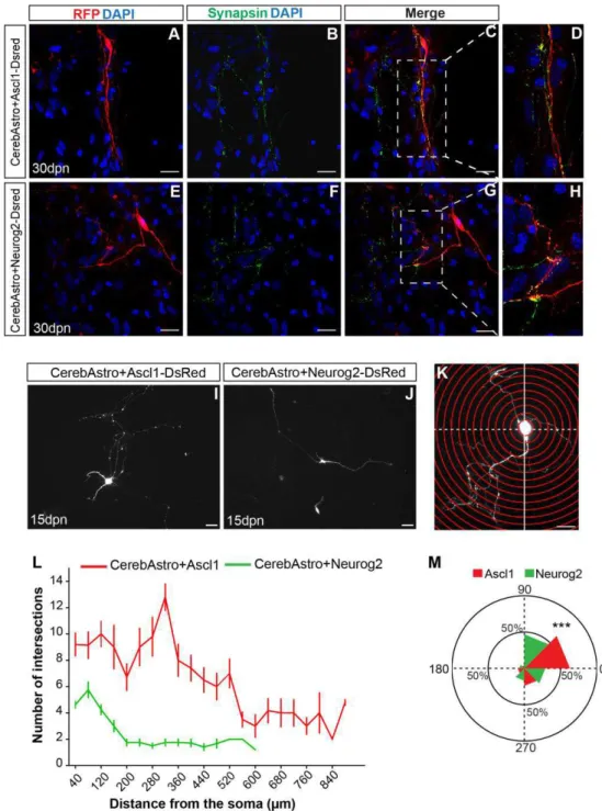
Expression of Ascl1 or Neurog2 efficiently reprograms cerebellum astroglia in iNs ... 54
Neurog2 and Ascl1 induces distinct morphological features in iNs... 57
Ascl1 and Neurog2 induce different neurotransmitter identity in astroglia of different origin ... 60
Ascl1 and Neurog2 generate iNs population expressing calcium binding proteins ... 62
CtxAstro nucleofected with Neurog2 differentiate into pyramidal cell like neurons after transplantation in the postnatal mouse cortex. ... 64
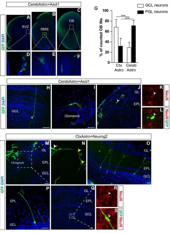
CerebAstro and CtxAstro iNs integrate as olfactory bulb interneurons upon transplantation in the SVZ ... 67
DISCUSSION ... 72
Supplemental Figures and Legends ... 78
Supplemental Experimental Procedures ... 86
GENERAL DISCUSSION ... 90
Discovering the neurogenic potential of cerebellum astroglia ... 90
Astroglia reprogramming in vivo: an incomplete puzzle ... 91
Conclusion: how similar is the copy to the authentic? ... 95
9 INTRODUCTION
From cell discovery to the notion of genetic equivalence: a brief historical perspective
“There exists a general principle of construction for all organic products, and this principle of construction is cell formation”
(Theodor Schwann, 1839)
Cell discovery: the fundamental unit of life
Thesmallest and most basic form of life was observed for the first time under the light
microscope of Robert Hooke in 1665. In his famous work Micrographia originally published in
the same year, as illustrating thin slices of cork tissue, he observed several tiny pores and termed
them “cells” which were boxlike cavities that reminded him of the cell of a monastery (Hooke,
1665). Observation of plant cells was then only the beginning of many important discoveries.
Just after, Theodor Schwann extended cell observation to animals in 1839 while observing
animal cartilage. In his book Microscopical researches into the accordance in the structure and
growth of animals and plants (Schwann, 1847) he defined the cell as having three essential
elements which are a nucleus, a fluid content and a wall. Subsequently, he stated the theory that
cell is the basic unit of structure for all organisms. In other words, plants and animals consist of
combinations of these basic units, which are arranged in accordance with definite rules. Despite
the fact that cell theory enjoyed a general consensus, the question of how cells were formed was
still highly controversial. Faithfull to the long dating theory of spontaneous generation, Schwann
suggested that cells were formed by crystallization of inanimate material inside the cell.
However, soon, Robert Remak put this theory to the question after his work on red blood cells.
10
creation as postulated by Schwann cannot be proved… The cells of which the animal germ
consists, multiply by continuous division, which starts as the nucleus as I had observed it”
(Remak, 1855). Despite his brilliant statement, Remak’s theory required support from more
popular scientists to be accepted. Luckily, the very famous German scientist Rudolf Virchow
backed his statement and added a new doctrine to the cell theory “Omnis cellula e cellula”; or all
cells develop only from existing cells. Since then, cell theory became widely accepted as an
explanation of the relation between cells and living things: cells are the basic unit of life building
up all organisms and old cells dividing into two create new cells.
Birth of experimental embryology
Subsequent to the cell discovery, many questions arise on how cells, such microscopical
tiny things, organize themselves to develop into an entire and complex organism. These
questions opened a new field in biology: development. Until the 1880´s, observation and
histology were the only available techniques limiting studies to be mainly descriptive. Even though a lot can be learned by these simple techniques, many rising questions could not be
answered. By the end of the XIX century, a group of scientists came out with the idea to directly manipulate vertebrate embryos in order to understand the rules that govern normal development, introducing by this way for the first time the field of experimental embryology. Subsequently,
control of cellular differentiation became one of the most fundamental question in developmental biology. When do cells become unequal during development? Do they get permanent heritable
alteration as they differentiate and adopt a specific fate? Does it happen sometime during cell division? Or is it determined at the very beginning in the substance of the undivided egg? Many pioneer scientific groups started to respond these questions by using amphibians at first stages of
11
manipulation, published in 1888 results showing that killing one cell of the two cell stage amphibian embryo led to the development of only half of the animal. This work corroborated
with the hypothesis of Weisman stating that development worked by “qualitative division” among daughter cells (Weismann et al., 1893). Indeed, some scientists started to believe that after
division, each cell receives different subsets of heritable material to specify their unique traits. On the other hand, other biologists, amongst whom Hans Driesch, who was performing similar experiments on sea urchins embryos, observed different outcomes. Instead of killing one of the
cells, he was able to separate the two cells of an embryo and keep them both alive. In 1882, he related that after separation of sea urchin embryos at the two cells stage, both cells were capable of
forming a complete but half sized embryo. These conflicting results raised more questions than they answered. However, at that time, many scientists considered Driesch´s experiment, stating that Roux´s outcomes may result from killing one of the two cells. Driesch´s experiments
suggested that each cell in the early embryo was capable of generating all the organs and tissues of the fully formed organism. It proposed that at least during embryonic development, each cell
retains the same and the entire genetic material. Indeed, this statement enhanced the notion of nuclear equivalence at least at the two-cell stage.
Nuclear transfer: the “fantastic experiment”
Soon after Driesch´s observations, many scientists started to investigate the role of genetic material in cellular differentiation. Indeed, the question was whether development and
differentiation were achieved by loss of information and/or irreversible changes contained within the genes. Hans Spemann, Nobel laureate for his work on the organizer and embryonic induction, was one of the first scientists interested in nuclear equivalence. After his retirement, he published
12
proposed the “fantastical experiment”: to transfer the nuclei from advanced developmental stages back to the zygote from which the genetic material have been removed (Sutovsky, 2007).
However, unfortunately, this procedure was not feasible at his time and was only completed after his death.
Nuclear transfer: initial steps towards cell reprogramming
Later on, thanks to technical advances allowing better nuclear cell transfers, many scientist could obtain enlightening responses regarding the overriding question of whether the process of
development and cell differentiation requires a loss or stable change in the genetic constitution of cells (Gurdon and Byrne, 2003). The immense contribution to our actual understanding of nuclear
equivalence dates from the second half of the XX century, thanks to the to the seminal work by Gurdon with nuclear transfer in Xenopus and other important researchers in the field (Gurdon and Byrne, 2003). Gurdon’s key contribution to cellular biology was the remarkable experiment
showing that normal development to adulthood could be achieved by transferring a fully differentiated nucleus from an intestinal cell of feeding-stage larvae into an irradiated egg (Gurdon
1962; Gurdon and 1966). These results led to the assumption that some highly differentiated cells still retain all the important hereditary information as the zygote. Hence, it seems probable that neither discard of nuclear material nor irreversible genetic alteration take place during cell
differentiation. In other words, nuclear content of undifferentiated and fully differentiated cells are equivalent. This principle of nuclear equivalence was further extended to adult mammalian somatic
13
reprogramming activity of egg and oocyte cytoplasm” and endeavored to find out the mechanisms
responsible for such nuclear reprogramming (Byrne 2003).
Cellular reprogramming
Artificial transcriptional regulation
In 2012, Gurdon’s work on nuclear transfer was awarded with the Nobel Prize in Medicine.
The prize was shared with Yamanaka, a much younger scientist, but not less important, in the field of stem cell biology. Originally, Yamanaka´s group and many others were working on the isolation
of key transcriptional regulators responsible for the maintenance of pluripotency in human embryonic stem cells (Mitsui et al., 2003; Chen and Daley, 2008). Yamanaka came up then with
the brilliant and simple idea to test whether an adult somatic cell could be reprogrammed by artificially expressing transcriptional regulators instead of proceeding to nuclear transfer. His group investigated therefore the possibility to induce pluripotency in mouse fibroblasts by overexpression
of some TFs instructing pluripotency. Surprisingly, his group found out that four transcription factors, namely Oct4, Klf4, Sox2, and cMyc, were capable of reprogramming murine adult and
embryonic fibroblasts into pluripotent stem cells. In fact, reprogrammed cells expressed many of embryonic stem cells markers and showed the ability to differentiate into cells from all three germ layers. Subsequently, these cells were referred as induced pluripotent stem cells (iPSCs)
(Takahashi and Yamanaka, 2006). One year later, generation of iPSCs was extended to human somatic cells (Takahashi et al., 2007; Yu et al., 2007; Huangfu et al., 2008; Park et al., 2008).
Direct cell reprogramming
The possibility to convert adult somatic cells into iPSCs by expression of few
14
set to generate different cell types from iPSCs such as adipocytes, cardiomyocytes, primitive
hematopoietic cells, pancreatic beta-cells, and several different neuronal cell types (review
Amabile and Meissner, 2009). However, one important question remained unanswered: could an adult somatic cell be lineage reprogrammed into another cell type without passing through a
pluripotent state? Several laboratories independently addressed this question and could show
evidences for direct reprogramming of somatic cells into cardiomyocytes (Ieda et al., 2010), insulin producing cells B cells (Zhou et al., 2008) and even neurons (Vierbuchen et al., 2010) by
overexpressing of TFs. These works provided direct evidence that cellular fates are not
irreversible. Therefore, adult somatic cell can be directly lineage converted following expression
of transcription factors. Direct cell reprogramming became with no doubt a trendy field
concerning many cellular types. Recently, it gained ground also in neuroscience, with a growing
number of groups focusing on the generation of neurons from somatic cells.
Direct generation of neurons from somatic cells
Direct reprogramming of fibroblasts into neurons has been widely demonstrated by
several laboratories (Vierbuchen et al., 2010; Ambasudhan et al., 2011; Son et al., 2011; Pang et al., 2011; Liu et al., 2012, 2013; Victor et al., 2014; Lau et al., 2014; Zhou et al., 2014; Aravantinou-Fatorou et al., 2015; Hu et al., 2015; Li et al., 2015; Zhou et al., 2015; Blanchard et
al., 2014). In a pioneer work, Vierbuchen and colleagues showed that viral-mediated delivery of neural-lineage specific transcription factors Ascl1, Brn2 and Mytl1, is sufficient to reprogram
mouse embryonic and adult fibroblasts into functional neurons (Vierbuchen et al., 2010). However, the neural conversion efficiency of these cells is still considered low (19.5%) (Vierbuchen et al., 2010). In order to ameliorate the reprogramming rates, additional molecules
15
subtypes such as striatal (Victor et al., 2014), dopaminergic (Liu et al., 2013), cholinergic (Liu et al., 2013) and even sensory neurons from fibroblasts (Blanchard et al., 2014). Nevertheless, it
seems that a great genetic barrier has to be overcome in order to convert fibroblasts into specific neural subtypes given that it is necessary to use a complex combination of reprogramming
factors such as several TFs, small molecules or microRNAs. Albeit the advantage of being easily isolated and the possibility to use patient fibroblasts for future cell therapies, these cells are still far from being the appropriate cell type to generate neurons. For those reasons, resident central
nervous system glial cells become extremely attractive, as we shall discuss in the following section.
Direct lineage reprogramming of astrocytes into neurons
Astrocytes and neurons: a common ancestor
In the 19th century, Santiago Ramón y Cajal discovered that the brain and the rest of the
nervous system consisted not of one mass of tissue, but of various distinct cells. At that time, drawings made by His provided scientists with the first evidence of the extraordinary cell
diversity in the brain. Since then, scientists have been identifying and classifying brain cells for more than a hundred years. A very basic classification of cell type in the brain is the distinction between neurons and glia. For a long time scientists highly regarded the idea that both types of
cells originate from distinct progenitor pools. Indeed the main claim was that neurons derive from specialized progenitors called neuroblasts whereas glial cells originate from spongioblasts
(see historical facts in (Pease, 1971)). The different origin of neurons and glial cells became since then broadly approved and it is relatively recent that this concept has been demonstrated to be incorrect. Nowadays spongioblasts correspond to what we call radial glia cells (RGCs). This
16
restricted developmental periods and has been shown to be a common progenitor for both glia and neurons in the CNS (Costa et al., 2010). Important studies showed that virtually all cortical
astrocytes are derived from progenitors that contribute to the generation of neurons at early developmental stage (Costa et al., 2009; Magavi et al., 2012). The major advance in defining the
nature and function of these cells came with the introduction of new methods in developmental biology such as electron microscopy, 3H-thymidine autoradiography and immunocytochemistry which provided higher resolution of cellular mechanisms and allowed more reliable
identification of cell classes. Furthermore the use of cell culture, transgenic technology and retroviral gene transfer methods allowed a more accurate analysis of cell lineage relationship as
well as their function in the developing mammalian brain (Rakic, 2003).
Nowadays it is well established that RGCs differentiate from neuroepithelial cells of the neural tube, and acquire typical astroglial features, characterized by the presence of glycogen
storage granules and the expression of astroglial-specific proteins, such as the astrocyte-specific glutamate and aspartate transporter (GLAST), brain lipid-binding protein (BLBP) and tenascin-C (Rakic, 2003; Pinto and Götz, 2007). Although RGCs were first associated with neuronal radial
migration (Rakic, 2003), there is now a general consensus that they comprise a specialized population of neural stem cells (NSCs) during cerebral cortex development (Kriegstein and
Alvarez-Buylla, 2009). Many evidences showing that RGCs are multipotent cells generating both neuronal and glial progeny, and are capable of self-renewing by cell divisions generating
either two new RGCs (symmetric division) or one RGC and a fate-restricted progenitor (asymmetric division) (Miyata et al., 2001; Noctor et al., 2004) indicate that RGCs exhibit defining stem cell hallmarks and are thus often considered as embryonic NSCs. Despite the fact
17
(Malatesta et al., 2000, 2003; Miyata et al., 2001; Noctor et al., 2001; Tamamaki et al., 2001), at the end of this period, the remaining radial glia lose their apical and basal processes and
transform into parenchymal astrocytes (Voigt, 1989; Alves et al., 2002) which do not generate neurons in the adult brain under physiological conditions. These astrocytes will constitute a
supportive glial cell population that has a heterogeneous morphology extending numerous processes surrounding neurons and blood vessels and containing intermediate filaments (Wang and Bordey, 2008). Astrocytes are essential for maintaining a viable nervous system environment
for neurons. Among their well-established functions are: buffering excess of potassium and neurotransmitters, providing nutrients, structural support around synapses, and contributing to
the integrity of the blood–brain barrier (Wang and Bordey, 2008). Interestingly, the disappearance of radial glia cells in the cerebral cortex has been suggested to account for the incapacity to generate new neuron (Kriegstein and Alvarez-Buylla, 2009). According to this
view, the high regenerative capacity in the brain of some non-vertebrates is tightly linked to the persistence of immature RGCs that retain many developmental features (Pinto and Götz, 2007).
Noteworthy, RGCs do not only give rise to parenchymal astrocytes, but in restricted zones of the adult central nervous system transform into astroglial neural stem cells (Merkle et al., 2004). These cells reside within two neurogenic niches, namely subventricular zone (SVZ) and
subgranular zone (SGZ), and maintain the potential to generate new neurons throughout life. Interestingly, these adult NSCs display features of both RGCs and mature cortical astrocytes
(Doetsch et al., 1999; Seri et al., 2001). Curiously, when astroglial cells from the early postnatal cerebral cortex are grown in vitro in the absence of serum factors but in the presence growth factors, these cells can still give rise to self-renewing and multipotent neurospheres (Laywell et
18
the second postnatal week. This transformation appears to be a gradual process during which the neurogenic potential of the astroglial cells becomes progressively diminished. Some mechanisms
involving silencing of genes encoding the transcription of neurogenic fate determinants are thought to be involved (Hirabayashi et al., 2009). Interestingly, studies from Vaccarino‘s lab
have shown that early postnatal astroglia in the cerebral cortex may retain some capacity of neurogenesis in vivo. They showed by fate-mapping technique (which permits the follow up of astroglial cells using Cre-recombinase activity driven by the hGFAP promoter), that a very small
percentage of the fate-mapped cells gave rise to neurons one month after recombination (Ganat et al., 2006). This potential is highly evoked in the early postnatal brain by damage such as
caused by hypoxia (Fagel et al., 2006, 2009), indicating that early postnatal astroglia can respond to stimuli generating new neurons.
Proneural transcription factors: explaining cell diversity in the CNS
Nowadays, it is a general consensus that RGCs are common progenitors for neurons and astrocytes in the developing brain (Kriegstein and Alvarez-Buylla, 2009). Yet, it remains a
matter of intense debate how these cells generate the huge cell diversity characteristic of the mammalian nervous system. Telencephalon is one of the best-studied system. It harbors a broad variety of progenitors that give rise to neurons and glial cells to the cerebral cortex and basal
ganglia (striatum and pallidum). These regions are composed of subdivisions that have specific patterns of gene expression, reflecting the differential environmental influences which
progenitors are exposed to. This early developed regional identity has been shown to contribute for the generation of the huge cell diversity in the cerebral cortex (Guillemot, 2007). In accordance with thesr observations, RGCs located at distinct regions of the telencephalon
19
these TFs, some belonging to the bHLH family (basic helix-loop-helix) have been assigned an important intrinsic role in the neuronal differentiation of progenitor cells (Bertrand et al., 2002).
These proteins form a large superfamily of TFs that are found in almost all eukaryotes and have been assigned an important role in embryonic development. Due to the heterogeneity of their
DNA sequences and dimer formation, bHLH proteins are involved in very diverse functions. Some of these proteins are widely expressed in different tissues and cells, however others are more cell specific. These latter are involved in cell fate determination in many cell lineages and
have been shown to act on processes such as neurogenesis, cardiogenesis and hematopoiesis. bHLH proteins involved in neurogenesis include Ascl1 and Neurog2, belonging to the
achaete-scute and neurogenin subfamilies respectively ( For review see Jones, 2004). Several studies have shown that mice mutant for Ascl1 and Neurog2 show severe defects including a reduction of neurogenesis and premature excessive generation of astrocytic precursors (Guillemot et al., 1993; Fode et al., 1998; Horton et al., 1999; Ma et al., 1999). It has also been shown that both
TFs dimerize with E-proteins forming by this way protein-protein interaction through their HLH domains, two α-helices connected by a loop. Following dimerization, these TFs use their DNA
binding domains to bind E-box specific consensus sequences in the promoter or enhancers of their target genes (Murre et al., 1989; Johnson et al., 1992; Bertrand et al., 2002; Ross et al., 2003). E-box consensus sequences are frequent throughout the genome, nevertheless it is their
clustering that promote strong bHLH binding. Interestingly, it has been shown that sequences between these consensus sites are also important given that they can influence target gene
20
Proneural factors also affect the balance between proliferation and differentiation. Following cell division, newly generated cells can either re-enter the cell cycle continuing to
proliferate or exit the cell cycle, differentiating. Consistent with their role in promoting neuronal differentiation, proneural TFs play an important role in the control of cell cycle exit. Indeed,
Neurog2 has been shown to promote cell cycle exit in cortical progenitors and other neural cell populations (Farah et al., 2000; Mizuguchi et al., 2001; Mattar et al., 2008). On the other hand, Ascl1 role in this cellular event is not as sharp. It has been shown that it can induce either
proliferation or differentiation of progenitor cells (Castro et al., 2011). Beside their generic function, Ascl1 and Neurog2 have been also shown to display context-dependent effects
contributing to the differentiation of specific neuronal subtypes (Bertrand et al., 2002; Powell and Jarman, 2008). It is nowadays established that these genes are expressed in non-overlapping patterns in the murine central and peripheral nervous system where they drive the production of
different neuronal populations (Fode et al., 2000; Parras et al., 2002; Osório et al., 2010; Brzezinski et al., 2011). For instance, several works showed that Ascl1´s implication in the
generation of neurons is highly time and space dependent (Mizuguchi et al., 2006; Jo et al., 2007;
Peltopuro et al., 2010). Ascl1 is also involved in the development of several neuronal subtypes such as hindbrain serotoninergic neurons (Pattyn et al., 2004), central and peripheral
noradrenergic neurons (Goridis and Rohrer, 2002), mesencephalic dopaminergic neurons (Park
et al., 2008). RGCs in the ventral telencephalon express Nkx2.1, which acts upstream of Ascl1
and Dlx2 (Xu et al., 2004). Ascl1 has been shown to induce the expression of Dlx2 genes and direct the differentiation of GABAergic phenotype in the developing telencephalon (Fode et al., 2000). In a similar way, Neurog2 proneural functions are also temporally and spatially regulated
21
these TFs regulation, it has been shown that continued expression of Neurog2 in post mitotic
neurons leads to cell death or neuronal degeneration (Cai et al., 2000; Guichet et al., 2013).
Cortical dorsal radial glia express the transcription factor paired box gene 6 (Pax6), which is required for their proper development (Götz et al., 1998). Indeed, it has been shown that Pax6
acts upstream of neurogenin 2 (Neurog2), which is specifically expressed in the dorsal telencephalon and have the dual role of repressing ventral identity and activating downstream transcription factors that control neuronal differentiation (Götz et al., 1998; Heins et al., 2002).
Neurons generated from Pax6-expressing cells are distinguished by the early expression of T-box brain 1 gene (Tbr1), coding for a transcription factor involved in the specification of the
glutamatergic lineage in the telencephalon (Hevner et al., 2006). Only a handful of Neurog2-regulated cortical genes have been validated until now as direct target among which are NeuroD1, NeuroD4, Tbr2 (Mattar et al., 2004; Schuurmans et al., 2004; Seo et al., 2007; Gohlke
et al., 2008). An interesting study showed that Neurog2 activity can also be modulated by phosphorylation events. It has been shown that phosphorylation of Neurog2 by GSK inhibits
Neurog2 transcriptional activity by preventing the formation of homodimers and instead promoting the formation of another complex Neurog2-E47 heterodimer which have a reduced ability to transactivate target gene promoter (Li et al., 2012). It is therefore not a coincidence that
GSK is progressively activated in later stage cortical progenitors, but not early stage cortical progenitors where Neurog2 is most active (Li et al., 2012).
Briefly, these data suggest that cell diversity in the telencephalon is established early in development and is sustained, partly, by different expression pattern of distinct TFs in RGCs. Nevertheless, mechanisms by which these TFs function seem to be very far from simple and
22
Astrocytes reprogramming into neurons: development inspired reprogramming
A Gene transcriptional regulation is nowadays considered as the guiding path of cellular
differentiation. Developmental TFs, such as the ones belonging to the bHLH family have a considerable role in the determination of neural fate and subtype specification (Nieto et al.,
2001). Amongst the member of this important family Pax6, Neurog2, Dlx2 and Ascl1 have been tested as potential neuronal reprogramming factors in astrocytes. Indeed, an interesting study showed that retrovirus mediated expression of Pax6 in cultured astroglia isolated from the
cerebral cortex resulted in a rapid down-regulation of astroglial-specific genes such as GFAP and the up-regulation of neuronal genes such as βIII-tubulin (Heins et al., 2002). However, this study did not evaluate the functional maturation of these cells. A following study showed that not only
forced expression of Pax6, but also of the other neurogenic transcription factors, such as Neurog2 and Ascl1, reprogrammed astroglial cells into fully functional neurons that could fire
repetitive action potentials (Berninger et al., 2007). Strikingly, only Neurog2 expression induced the expression of Tbr1 in reprogrammed neurons, indicating that these neurons were following a program towards a glutamatergic fate (Berninger et al., 2007), similar to what has been described
in the developing dorsal telencephalon. Likewise, forced expression of Ascl1, a transcription factor involved in the genesis of GABAergic neurons during embryonic development and adult
neurogenesis (Petryniak et al., 2007), showed that the same cortical astroglia can be directed towards the genesis of GABAergic neurons. These data suggest that early postnatal astroglia can
be reprogrammed towards different neuronal lineages even if they are restricted to glial identity under physiological conditions. However this first study failed to demonstrate that reprogrammed astrocytes could establish functional presynaptic complexes. This failure is
23
the retroviral vector (pMXIG) used in the study (Berninger et al., 2007). To overcome this technical limitation, another study of the same group used a different type of retroviral vector
driving the neurogenic gene expression under the control of a chicken beta actin promoter pCAG optimized for long-term expression over months in the adult mouse brain (Zhao et al., 2006). In
fact, this allowed a more complete reprogramming towards a fully mature, synapse forming neuronal phenotype (Heinrich et al., 2010). Reprogrammed neurons showed not only the ability to receive synaptic inputs but also to form functional presynaptic outputs onto other
astroglia-derived neurons (Heinrich et al., 2010). It has been shown in another publication of the same group that reprogrammed astrocytes could even generate networks of spontaneously active
neurons (Blum et al., 2011). A subsequent study showed that by only adding two other neurogenic TFs, such as Nurr1 and Brn2 to Ascl1, it is sufficient to convert cortical astrocytes into dopaminergic neurons (Addis et al., 2011). All these data are suggesting that astrocytes may
be a suitable cell population for neuron generation. Not only they can be reprogrammed via a minimum number of TFs but also they can generate different subtypes of induced neurons (iNs)
depending on the TFs used.
Astrocytes induced neurons: alternative for cell therapy?
The dream of therapeutic organ replacement in man returns to antiquity. Legends back far
from the Roman and Greek civilizations recited magical substitution of lost tissues, such as restoration of limbs or eyes, or even replacement of decapitated heads. In the course of the last
four centuries, humanity witnessed from the first attempts at blood transfusion (Lower et al, 1667) to the first successful full face transplant in 2010 (Barret et al, 2011). However, organ transplant presents many real limiting problems such as immunological rejection, side effects of
24
difficulties concerning who should be treated and from whom the organs should be taken. A promising and more simplistic alternative to partially sidestep all these problems is stem cell
therapy. Many studies on animal models observed neuronal replacement and partial reconstruction of affected neuronal circuitry following stem cells grafts. Also, clinical trials with
cell replacement on diseased human brain revealed the possibility of symptomatic relief. However, many basic issues remain unresolved such as the control of undesired growth and genetic alterations, increasing the risk for tumor formation (Amariglio et al, 2009). In addition,
efficacy of functional integration and differentiation into the appropriate cell type still need to be ameliorated.
Sustainability of stem-cell tissue replacement may become less reliable as the cellular
complexity of the concerned organ turns higher. A relevant example is the central nervous system. Moreover, it is generally known that susceptibility to neuronal conditions is strongly
neuronal class specific. Therefore, generation of specific neuronal subtypes for the purpose of cell replacement therapy or in vitro disease modeling becomes important. Concerning all the studies related previously in this text, astrocytes reprogramming into neurons may be an
interesting potential for cell therapy. Their abundancy and omnipresence make them even more reachable than any other cell type in the brain. However, we are still a way far from any clinical
trials and things are not very promising if we refer to studies that tried in vivo reprogramming of astrocytes (Heinrich et al., 2015). More studies should be done in order to understand the
25 GENERAL QUESTIONS AND OBJECTIVES
Our work introduces many relevant questions concerning astroglia reprogramming and
subtype specification of induced neurons (iNs). One of our main concerns is to discuss and propose some experiments that could help to understand how finely we can tune the fate of an
iN. We believe that the final phenotype of a reprogrammed cell is not restrained by a simple factor. Instead, we think that cell reprogramming is a complex mechanism that can be controlled by harmonizing several distinct elements. Transcription factor, cell origin and environmental
context are amongst the elements that could potentially influence the fate of an iN.
Here, we present a perspective about the possible application of lineage-reprogrammed
astroglia iN for the treatment of neurological disorders, focusing on brain ischemia (manuscript 1). Next, we present our results using two different neurogenic TFs, Ascl1 and Neurog2, to convert different types of astroglial populations into iNs and assessed the phenotypes of these
cells in vitro (manuscript 2). To the best of our knowledge, this is the first description of the neurogenic potential of astroglial population isolated from the hindbrain. This work contribute to
answer (1) whether neurogenic potential can be extended to other astroglial cell types and (2) if the origin of the source cell is important in the phenotypic determination of the iN. Finally, we transplanted lineage-reprogrammed astroglia in different brain regions of the postnatal and adult
mouse (manuscript 2). This allowed us to investigate whether origin of astroglial cell, TF used for reprogramming, region of integration and age of animal affect the integration of iN in the
host animal. Altogether, our work brings new insights on the potential of astroglial cells to generate neuronal diversity and contributes to discuss some of the challenges in the field of cell lineage reprogramming for the use of such strategy in cell-based therapies to treat neurological
26 MATERIAL AND METHODS
Animals
In this work, we used C57BL/6 and GFP mice (Okabe et al., 1997) from the animal facility of the Brain Institute (UFRN, Natal). All animal procedures were done in accordance with national and international laws and were approved by the local ethical committee
(CEUA/UFRN, license # 008/2014).
Astroglia culture
Postnatal cerebellum and cortical astroglial cells were isolated from postnatal day (P) 5-7 mice, as previously described (Berninger et al., 2007). Briefly, animals were killed by decapitation and had both brain and cerebellum removed from the skull and immersed in PBS.
Using a stereoscopic microscope, meninges were removed and the cerebral cortex gray matter and entire cerebellum were removed and maintained separately. Next, tissues were mechanically
dissociated to obtain small pieces of 2-4 mm. Cerebellum and cortical tissues were then directly plated in T75 culture flasks containing Astromedium - DMEM/F12 (Gibco), 3.5 mM glucose (Sigma), 10% fetal bovine serum (Gibco), 5% horse serum (Gibco), penicillin/streptomycin
(Gibco), and supplemented with 2 % B27 (Gibco), 10 ng/mL epidermal growth factor (EGF, ) and 10ng/mL fibroblast growth factor 2 (FGF2). Cultures were incubated at 5% CO2 and 37°C
without moving. After 3-4 days, cultures were washed vigorously with PBS in order to remove unattached and weakly attached cells and medium was replaced with fresh Astromedium.
27 Astroglia transfection
Transfection of cells was proceeded via nucleofection. Astroglial cells were transfected
using the 4D nucleofector device and the nucleofection solution kit P3 (Lonza). Briefly, 106 cells were suspended in 20µl of P3 solution containing 1-2 µg of plasmid DNA. Cell/DNA suspension
was dropped in the nucleofection well to receive an electrical shock with the program EM110 for mammalian glial cells (Lonza). Astroglial cells were nucleofected with either pCAG-Neurog2-IRES-DsRed, pCAG-Ascl1-IRES-DsRed or the control plasmid pCAG-IRES-DsRed. Next, cells
were plated at a density of 70.000 to 100.000 cells/well in poly-D-lysine coated 24-well tissue plates containing a medium composed of DMEM/F12 (Gibco), 3.5 mM glucose (Sigma), 10% fetal bovine serum (Gibco), 5% horse serum (Gibco), penicillin/streptomycin (Gibco), and
supplemented with 2 % B27 (Gibco). 24 hours after nucleofection, medium was replaced with a serum free differentiation medium composed of DMEM/F12, 3.5 mM glucose,
penicillin/streptomycin and 2% B27. Brain-derived neurotrophic factor (BDNF, Sigma) was added at 20ng/mL every fifth day of the differentiation process.
Observations: It is important to prevent bubble formation while loading the cell/DNA suspension in the wells. This will prevent more cell death. We also observed that cell clusters are more resistant to nucleofection than totally dissociated cells. It is preferable to plate cells in an already
heated and equilibrated medium. Although using the same protocol, cell nucleofection efficiency and cell death vary a lot from one experiment to the other. Therefore, we always took into
account experiments with higher efficiency in our analysis.
28 Co-culture of iNs with hippocampal neurons
Due to the decreasing survival rate of iNs starting from 20 days post transfection, we
co-cultured transfected cells 5 days after nucleofection with hippocampal neurons isolated from P0 pups. Cortical and hippocampal tissues were dissected and dissociated in trypsin/ EDTA for 15
min. Cells were then centrifuged (1000 rpm, 4ºC) and suspended in a serum containing medium to stop trypsin activity. Next, cells were centrifuged again and suspended in a serum free medium to be subsequently added at a density of 50.000 to 70.000 cells/well.
Transplantation of transfected astroglia
Cerebellar and cortical astroglia were isolated from postnatal GFP animals (Okabe et al.,
1997) and cultured as mentioned in a previous section. At confluency (70-80%), cells were nucleofected with either pCAG-Neurog2-IRES-DsRed, pCAG-Ascl1-IRES-DsRed or the control plasmid pCAG-IRES-DsRed. Cells were then resuspended and mechanically dissociated in
DMEM-F12. Next we kept cells at a density of 50.000 cells/µl in ice until transplantation procedure.
Observations: Cells can survive up to 2 hours in ice. However, it is preferable to keep them less
than one hour before transplantation. It is very important that the cell suspension is well dissociated to a single cell suspension otherwise this can block the glass capillary.
P0-2 C57BL/6 mice were anesthetized by hypothermia for 10 min and positioned under a light source. Make sure to see where the midline and bregma are. Injection was targeted to
29
the pial surface with an angle of 45 º. Approximately 1 to 2 µl of cell suspension was injected for transplantation. Glass capillary must be slowly withdrawn. After the procedure, animals were
revived on a heat pad and returned to their mothers. Transplantation in adults were proceeded on P30 C57BL/6 mice under isoflurane anesthesia. Cells were strereotaxically injected using a
nanoinjector (NANOLITER 2010, WPI) coupled to a glass capillary in the following coordinates: SVZ (in mm) (AP: 1.58, ML: 3.44, DV: 1.55) and cortex (AP: 1.58, ML: 3.44, DV: 1.40). 1µl of cell suspension was injected for each transplant at a speed of 1µl/min. After the
procedure, animals received the essential care and returned to the animal facility.
Observations: Glass capillaries were prepared using a heater at 62ºC. Before transplantation capillaries were cut (1mm) in order to push up the cells.
Tissue preparation and histology
Cell cultures were fixed with 4% PFA for 10 minutes at room temperature and stored in
PBS. For anti-glutamate staining, we also added 0.3% glutaraldehyde to the fixative solution. Primary antibodies were prepared in a solution of 0.5% Triton X-100, 10% normal goat serum
(NGS) and 2% bovine serum albumin (BSA). Samples were incubated with primary antibody solution at 4ºC overnight. The following primary antibodies were used: polyclonal anti-green fluorescent protein (GFP, chicken, 1:500, Aves Labs, GFP-1020), polyclonal anti-Glial Fibrillary
Acidic Protein (GFAP, rabbit, 1:4000, DakoCytomation, Z0334), polyclonal anti-Red Fluorescent Protein (RFP, rabbit, Rockland, 1:1000, 600-401-379), monoclonal anti-Microtubule
Associated Protein 2 (MAP2, mouse IgG1, 1:200, Sigma-Aldrich, M4403), monoclonal anti-NeuN (mouse, 1:500, Millipore, MAB377), monoclonal anti-synapsin 1 (mouse IgG2, 1:2000, Synaptic Systems, 106001), polyclonal anti-Tbr1 (rabbit, 1:500, Abcam, ab51502), monoclonal
30
IgG1, 1:2000, Swant), monoclonal anti-parvalbumin (mouse IgG1, 1:2000, Sigma, p3088), monoclonal anti-Cux1 (mouse IgG1, 1:500, ABCAM), monoclonal anti-glutamate (mouse,
1:1000, Sigma, g9282), monoclonal anti-GABA (mouse, 1:2000, Swant). For some nuclear staining, TO-PRO-3 (1:2000, Invitrogen) was incubated together with secondary antibody
solution. After washing with PBS cells were incubated with appropriate species secondary AlexaFluor (Invitrogen) antibodies for 2 hours at room temperature. After 3 washes in PBS, cell nuclei were stained with DAPI and coverslips were mounted on glass slides with an anti-fading
mounting medium (Aqua Poly/Mount). Coverslips were analyzed using fluorescence (AxioImager, Zeiss) and confocal microscopy (LSM 710, Zeiss). Images were acquired using the
software ZEN 2 blue edition (Zeiss).
Tissue fixation was performed 20 to 30 days post transplantation. For that, animals were deeply anesthetized with THIOPENTAX (Cristalia) and transcardially perfused using a
ventricular catheter with 0.9% saline solution for 15 min and 4% phosphate-buffered saline-buffered paraformaldehyde (PFA) for another 15 min. Brains were removed and kept in phosphate-buffered saline (PBS) overnight. The next day, brains were kept in 30% sucrose
solution for cryoprotection before freezing procedure. Brains were cut in slices going from 40 to 50µm of thickness using a cryostat (Leica). Subsequently, slices were mounted on gelatin-coated
slides and stored at -20ºC until immunohistochemistry.
Quantifications and statistical analysis
For the in vitro study, cells were quantified for colocalization of DsRed and BIII-tubulin immunoreactivity at 7 days post nucleofection (dpn). Colocalization of DsRed, Map-2 and NeuN was done 15 dpn. Calbindin and parvalbumin expression were analyzed at 20 dpn whereas
31
(iNs) or DsRed+ neurons terms refer to DsRed positive cells that have a clear neuronal morphology. Morphological analysis was performed using the plugin “Sholl Analysis” (version
3.4.5 June 2015) in ImageJ version 1.49 (NIH) at 15 dpn. For the in vivo experiments, we studied GFP+ cells for their morphology, hodology and neurochemical markers such as NeuN
and Cux1. For each transplant, cells were quantified through the entire brain 20 or 30 days post transplantation.
Student t-test, One-way ANOVA with Tukey’s post test, or Two-way ANOVA with
Bonferoni´s post test were performed using GraphPad Prism version 5.00 for Windows,
32
MANUSCRIPT #1
Published, Frontiers in Cellular Neuroscience
Cell therapy for stroke: use of local astrocytes
Melek Chouchane and Marcos R. Costa
33 Abstract
Stroke refers to a variety of conditions caused by the occlusion or hemorrhage of blood
vessels supplying the brain, which is one of the main causes of death and the leading cause of disability worldwide. In the last years, cell-based therapies have been proposed as a new
approach to ameliorate post stroke deficits. However, the most appropriate type of cell to be used in such therapies, as well as their sources, remains a matter of intense research. A good candidate cell should, in principle, display high plasticity to generate diverse types of neurons and, at the
same type, low risk to cause undesired outcomes, such as malignant transformation. Recently, a new approach grounded on the reprogramming of endogenous astrocytes towards neuronal fates emerged as an alternative to restore neurological functions in several central nervous system
diseases. In this perspective, we review data about the potential of astrocytes to become functional neurons following expression of neurogenic genes and discuss the potential benefits
and risks of reprogramming astrocytes in the glial scar to replace neurons lost after stroke.
Background
Ischemic insults result in a severe loss of neural cells in the core of the lesion and variable effects in the surrounding area, commonly described as ischemic penumbra. While cell
death occurs during the first hours after interruption of blood supply within the core of the ischemic lesion, tissue damage in the surrounding regions is a delayed process, involving several
34
the extension of the core ischemic area (Goldstein, 2007). Currently, the only treatment available to reduce the size of the ischemic area is the use of recombinant tissue plasminogen activator
(t-PA)(1995), which is approved to be administered within 3 hours after the onset of ischemia (Goldstein, 2007). This narrow time window as well as a number of contraindications for t-PA
therapy makes such treatment accessible to an extremely low number of stroke victims (Katzan et al., 2000), boosting the necessity to develop new strategies to treat stroke patients.
In the last years, cell-based therapies have been proposed as a new approach to
ameliorate post stroke deficits. Different from t-PA, cell-based therapies could, in principle, be administered at any time following the ischemic event and contribute to replace neurons lost after ischemia and presumably restore neurological functions (Lindvall and Kokaia, 2011).
Chiefly, two main types of intervention have been proposed: i) transplantation of exogenous cells (Benchoua and Onteniente, 2011;Lindvall and Kokaia, 2011); and ii) mobilization of
endogenous stem or progenitor cells (Leker et al., 2009;Lindvall and Kokaia, 2011). Both strategies have been shown to promote some degree of improvement in animal models of stroke (Lindvall and Kokaia, 2004; 2006). Yet, important limitations regarding the number of neurons
replaced, specification of these neurons into the appropriate neurochemical subtypes, integration to the preexisting circuitry and potential side effects hamper the translation into clinical practice
of such therapies.
Amongst the potential side effects, the most feared is the generation of tumors. In fact, it
has been shown that both stem cell transplantation and stimulation of endogenous neural stem cell proliferation can lead to tumor formation in rodents and humans (Doetsch et al., 2002;Erdo et al., 2003;Amariglio et al., 2009). Therefore, the development of new strategies to replace
35
to move cell-based therapies into clinic. In this scenario, we put in perspective the potential of a new approach grounded on the reprogramming of local astrocytes into neurons.
Astroglial cells in the adult brain possess neurogenic potential
Contrary to the previous notion that neurons were not generated in the mammalian brain after birth, in the last two decades two neurogenic regions in the adult mammalian brain have
been uncovered: the subependymal zone (SEZ), located along the lateral walls of the lateral ventricles, which holds a population of astroglial neural stem cells (ANSC) that constantly
supply the olfactory bulb with interneurons (Kriegstein and Alvarez-Buylla, 2009); and the subgranular zone (SGZ) of the hippocampus, which also contains a population of ANSC capable
of generating neurons to the dentate gyrus throughout life (Gage, 2000). Besides the significance of these findings to our understanding of brain physiology (Lledo et al., 2006), they also opened a new and promising avenue to brain repair after damage (Lindvall and Kokaia, 2006).
In fact, it has been shown that global and focal ischemic injuries in rodents lead to a significant increase in the number of neurons generate from ANSC both in the SGZ and the SEZ
(Liu et al., 1998;Kee et al., 2001;Arvidsson et al., 2002;Parent et al., 2002). Some of these newly generate neurons change their route of migration and roam to lesioned areas after focal ischemia, where they acquire some characteristics of local neurons (Arvidsson et al., 2002;Parent et al.,
2002). However, survival of these neurons in the lesioned areas is extremely poor, suggesting that some survival signal might be missing in re-routed neurons, leading to their premature death.
As mentioned before, treatments with growth factors, used to increase the generation and survival of newborn neurons from endogenous neural stem cells (Leker et al., 2009), have also been related with glioma formation (Doetsch et al., 2002), making such approaches unsafe.
36
the great distance that newly generated neurons would need to transverse to reach the lesioned cortical tissue as compared to the rodent brain. Last but not least, the functionality of therapies
aiming to recruit neurons from endogenous neurogenic niches relies on the occurrence of neurogenesis in the adult human brain, what has not been observed under physiological
conditions (Sanai et al., 2004;Sanai et al., 2011).
More recently, astrocytes of the cortical parenchyma, another population of astroglial cells, were suggested as an alternative source for neuronal replacement in neurological diseases
(Robel et al., 2011). Compared to ANSC residing in neurogenic compartments, cortical astrocytes would have four main advantages: i) they are located within the lesioned site, eliminating the need of relocation; ii) their amount is significantly increased after stroke (Buffo
et al., 2008), generating a large amount of exploitable cells; iii) they can be efficiently reprogrammed into neurons using simple molecular manipulations (Berninger et al.,
2007;Heinrich et al., 2010;Blum et al., 2011); and iv) they are involved in the formation of the glial scar, which contributes to generate an anti-neurogenic environment (Pekny and Nilsson, 2005). Therefore, astrocyte reprogramming could provide at once a source of new neurons in
large numbers to replace the circuitry lost after stroke and reduce some negative effects of the glial scar (see below).
Efficient reprogramming of astrocytes into glutamatergic and GABAergic neurons
Astrocytes isolated from rodent postnatal brain are highly susceptible to neuronal
reprogramming following forced expression of a single neurogenic fate determinant, such as Neurogenin 2 (NEUROG2), Distal-less homeobox 2 (DLX2) or Achaete-scute homolog 1 (ASCLl1, also known as MASH1) (Berninger et al., 2007;Heinrich et al., 2010;Blum et al.,
37
expression of MASH1 and DLX2 induces a GABAergic phenotype, resembling the roles of those transcription factors (TFs) in the developing forebrain (Guillemot, 2005). Astrocytes can
not only be reprogrammed into neurons of specific subtypes but also acquire electrical properties compatible with a mature neuronal phenotype, such as intrinsic excitability and the ability to
generate action potentials and synaptic contacts (Berninger et al., 2007;Heinrich et al., 2010).
Reprogramming of postnatal astrocytes using neurogenic TFs is a highly efficient process. Approximately, 70% of NEUROG2-transduced astrocytes differentiated into βIII tubulin
-positive neurons after 7-10 days (Berninger et al., 2007;Heinrich et al., 2010). By 2-3 weeks post-transduction, reprogrammed neurons acquire MAP2 immunoreactivity, indicative for dendritic maturation, and express the vesicular glutamate transporter 1 (VGLUT1), present in
synaptic vesicles within presynaptic terminals of glutamatergic neurons (Heinrich et al., 2010). Thus, astrocytes reprogrammed by forced expression of a sinlge TF (NEUROG2) adopt a full
neuronal glutamatergic phenotype forming presynaptic specializations. Indeed,
electrophysiological recordings of neurons reprogrammed from astrocytes with NEUROG2 demonstrated both autaptic and synaptic currents that were blocked by CNQX (an
AMPA/kainate glutamate receptor antagonist), further confirming the glutamatergic nature of the reprogrammed neurons. Amongst all NEUROG2-transduced astrocyte-derived neurons recorded,
~60% exhibited either glutamatergic autaptic connections onto themselves or glutamatergic synapses onto nearby neurons (Heinrich et al., 2010). Calcium-imaging experiments
demonstrated that cultures of astrocytes reprogrammed with NEUROG2 are even capable of generating networks of spontaneously active neurons (Heinrich et al., 2010;Blum et al., 2011).
The efficiency of reprogramming astrocytes into GABAergic neurons using MASH1 or
38
2007;Heinrich et al., 2010). Nevertheless, neurons reprogrammed from astrocytes using DLX2 express GAD67 immunoreactivity, display autaptic responses with slow decay time kinetics
which are abolished by the GABAA receptor antagonist and show spontaneous synaptic currents exhibiting a slow decay time, characteristic of GABAergic current (Heinrich et al., 2010),
indicating that astrocytes are converted to functional GABAergic neurons. Co-expression of MASH1 and DLX2 in postnatal astrocytes increased the rate of neuronal conversion up to 90% (Heinrich et al., 2010), showing that the efficiency to reprogram astrocytes into GABAergic
neurons can be drastically improved by combining two TFs. Taken together, these data clearly indicate that astrocytes can be efficiently converted to functional glutamatergic or GABAergic
neurons through simple molecular manipulations.
Reprogramming of postnatal astrocytes into dopaminergic neurons
Postnatal cortical astrocytes can also be reprogrammed into dopaminergic neurons,
although this requires a more complex set of TFs (Addis et al., 2011). Astrocytes transduced with a polycistronic lentiviral vector encoding for MASH1, LIM homeobox transcription factor 1
(LMX1) and nuclear receptor related 1 protein (NURR1) differentiate into neurons expressing biochemical and electrophysiological characteristics analogous with midbrain dopaminergic neurons. However, the efficiency of astrocyte conversion to dopaminergic neurons (~18%) is
much lower than that described previously for glutamatergic and GABAergic neurons (Heinrich et al., 2010;Addis et al., 2011). Yet, the finding that astrocytes isolated from a region that
normally do not generate dopaminergic neurons can be reprogrammed into these types of neurons using few TFs reveals the great plasticity of astrocytes and supports the notion that these cells are good candidates to replace distinct types of neurons in damaged brain areas.
39
Astrocytes in the adult brain resume proliferation and acquire neurogenic potential after lesion
Astrocytes account for up to one-fifth of the dividing cells in the first 7 days following traumatic or ischemic brain injury (Buffo et al., 2005) and at least part of these cells are mature
astrocytes that resume proliferation after lesion (Buffo et al., 2008). These reactive, proliferating astrocytes acquire some neural stem cell-like properties after injury (Buffo et al., 2008;Robel et al., 2011) and are suitable to reprogramming into functional neurons (Heinrich et al., 2010).
Although astrocyte activation may play beneficial roles at early time-points after stroke, there is convincing evidence that astrocytes in the glial scar are detrimental for regeneration of
the adult brain (Pekny and Nilsson, 2005;Robel et al., 2011). For instance, attenuation of reactive gliosis through genetic deletion of intermediate filaments (IFs) glial fibrillary acidic protein (GFAP) and vimentin in animals subjected to traumatic brain injury improved regeneration, with
a positive effect on complete synaptic restoration (Wilhelmsson et al., 2004). In these same models, neuronal differentiation and dendritic growth of transplanted cells were enhanced after
transplantation, indicating that reactive gliosis adversely affects integration of neuronal cells (Widestrand et al., 2007). In an opposite direction, increasing reactive gliosis worsens brain injuries, as demonstrated by the finding that overexpression of S100b, an astrocyte-derived
protein, enlarged infarct size and impaired neurological outcome after ischemia (Mori et al., 2008). Therefore, reprogramming of astrocytes in the glial scar could per se improve
neurological functions after stroke.
In an ideal scenario, we should be able to find a balance between diminishing the number of detrimental astrocytes in the glial scar through reprogramming of these cells into neurons and,
40
appropriate environment for the development and functioning of new synaptic contacts between reprogrammed neurons and the pre-existing circuitry (Wang and Bordey, 2008). To this point, it
is unclear whether reactive astrocytes acquiring stem cell-like properties after injury represent a sobpopulation of astrocytes and what would be the role of such cells in the glial scar. Future
studies should help to clarify this point and indicate methods to target specific subpopulations of astrocytes to reprogramming.
Reprogramming of human astrocytes into neurons
An important question towards translation of astrocyte reprogramming into clinic would be whether human astrocytes possess the same potential to be reprogrammed into neurons. A
partial answer to this question has been recently published in a paper from Corti and colleagues (2012). By cultivating astrocytes from the human cerebral cortex and inducing the expression of transcription factors involved in pluripotency (Takahashi and Yamanaka, 2006;Wernig et al.,
2007), the authors could show that astrocytes expressing OCT4, SOX2 or NANOG generated colonies of neural stem cells (Corti et al., 2012). These colonies could be expanded and
differentiated into the three major neural cell types – neurons, astrocytes and oligodendrocytes (Corti et al., 2012). Neurons expressed typical neuronal proteins, such as MAP2, synapsin and GABA, suggesting that human astrocytes could be reprogrammed into neurons acquiring part of
the machinery to establish synaptic contacts. Expression of MASH1 in NSCs derived from human astrocytes significantly increased the frequency of neuronal differentiation (Corti et al.,
2012), further supporting the key role of neurogenic determinants to convert astrocytes into neurons.
Strikingly, human astrocytes transduced with NANOG and transplanted in the lateral
41
delivery. Some transplanted cells expressed MAP2 and displayed complex and long neuritic extensions, compatible with neuronal differentiation (Corti et al., 2012). Thus, human astrocytes
can be efficiently reprogrammed into neurons both in vitro and in vivo. Noteworthy, neuronal conversion of human astrocytes occurred without regression to a pluripotent state, what could
contribute to avoid some complications linked to that state, including the risk of malignancy.
Reprogramming of astrocytes into subtype-specific neurons
The adult human brain harbors a large variety of neuronal cell types, each exhibiting
specific structural, molecular and functional features (DeFelipe, 1993;Douglas and Martin, 2004; Markram et al., 2004;Klausberger and Somogyi, 2008). Therefore, one important step towards
the clinical translation of astrocyte reprogramming as a therapy for stroke would be to direct the specification of defined neuronal subtypes. It remains to be evaluated, for instance, whether astrocytes reprogrammed by forced expression of NEUROG2 will generate principal and local
glutamatergic neurons of different cortical layers. Similarly, it is unclear whether GABAergic neurons generated from astrocytes reprogrammed with MASH1, DLX2 or combination of these
two TFs will adopt distinct morphological and electrophysiological properties, contributing to generate distinct subtypes of GABAergic interneurons, such as basket and chandelier cells (Markram et al., 2004) (Figure 2). These questions can only be answered by experiments
assessing the differentiation of neurons reprogrammed from astrocytes in vivo, either by expressing neurogenic transcription factors in astrocytes directly in the brain or transplanting