CELLULAR REPROGRAMMING STRATEGIES FOR
DEGENERATIVE DISORDERS INVOLVING THE
RETINAL PIGMENT EPITHELIUM
CRISTIANA FERREIRA PIRES
Tese para obtenção do grau de Doutor em Ciências da Vida
na Especialidade em Biomedicina
na Faculdade de Ciências Médicas
CELLULAR REPROGRAMMING STRATEGIES FOR
DEGENERATIVE DISORDERS INVOLVING THE
RETINAL PIGMENT EPITHELIUM
Cristiana Ferreira Pires
Orientador: Miguel C. Seabra, Professor Doutor
Co-orientador: António Jacinto, Professor Doutor
Tese para obtenção do grau de Doutor em Ciências da Vida
na Especialidade em Biomedicina
iii
This work was performed by Cristiana Ferreira Pires in CEDOC – CHRONIC DISEASES RESEARCH CENTER, Molecular Mechanisms of Disease group, NOVA Medical School
– Faculdade de Ciências Médicas da Universidade Nova de Lisboa, under the supervision of Miguel C. Seabra, MD PhD, and António Jacinto, PhD.
v
Acknowledgements
“Passou a nuvem; o sol volta. A alegria girassolou.”
Fernando Pessoa
The happy feeling of fulfillment is only complete once the joy is shared with everyone that helped and supported me along this path.
My first acknowledgement goes to Miguel Seabra, my PhD supervisor, for accepting me as a PhD student and enthusiastically drawing the first sketch of this project with me over lunch. Even when physically absent, his ideas and tremendous support had a major influence in this thesis. Miguel gave me the freedom to explore on my own, whilst still being present. I am deeply grateful for his mentoring and for teaching me how to stay focused and motivated, throughout the numerous scientific adversities.
I would also like to acknowledge the GABBA Phd Program for my PhD fellowship and, more importantly, for the opportunity to learn how to think as a scientist.
I owe my sincere gratitude to my PhD co-supervisor António Jacinto, for harbouring me as his student and for his support and always helpful opinions.
I was also luckily assigned to two “informal” supervisors. José Ramalho received me in his lab and taught me all the basic notions on cell and molecular biology techniques. I always tried to be an eager student of his lessons, including the hard work rhythm. I hope he agrees with me that even our vivid and loud discussions had positive outcomes. Duarte Barral was also a helpful support, allowing me to perform some experiments in his lab and to attend lab meetings and lab retreats. I am very thankful for his readiness to help me when I needed.
I would like to express my genuine gratitude to the CRF team, Sara Maia, Martim Portal and Catarina Sequeira. I believe that a good team always facilitates the attainment of the assigned objectives and I was lucky enough to have you working on my side. Thank you for your partnership, encouragement and friendship.
I owe my sincere gratitude to Dr. Tanya Tolmachova for welcoming me at Imperial College London and for the possibility to use the animal models.
vi
I would also like to acknowledge Doutora Jacinta Serpa and Professora Doutora Ana Félix for their helpful collaboration with the teratomas’ formation and analysis.
In addition, past and present members from the lab always supported me and enriched my work with their questions and helpful suggestions. Thank you to Magui and our cigarrette/fresh air-breaks! Thank you to Zézé Sandoval, Pedro Olho Azul, “Lau-Lau” Portelinha, Cristina Casalou, Cecília (“dos cílios”), Mary, Xiquilim, Elsa Seixas, Lucio
“bacaninho” and Abulinho. I am particularly grateful for the friendship we have built over the years. A thankful word goes to Augusta, Firmina and Teresa Salreta, and also to Isabel, Isabelinha, Ana Sofia, Clara and Teresa Barona for all the joyful lunches and coffees at FCM.
A very special thank you goes to my PhD “sisterhood”, Carolina “Carolzinha” and Inês “Agnes”. I was told, even before we met, that Carolzinha and I would get along! And we
did! Oh yes we did. By the contrary, Agnes knew first that we were meant to be friends. And we did! Oh yes we did! Carolzinha and Agnes, we are a great team together. I am very proud of both of you and now it is my turn to finish this. We made it together (Carrapito power!!!), but the PhD is just a small piece of it…Thank you for smiling with me!
Thank you to Carlos, the honorary member of our “sisterhood”! You have been more than just Inês’ husband. Thank you for your friendship and support in so many occasions! The fox was right…”cativar é criar laços”.
To Alex, thank you for being my scientific mentor and my friend throughout these years. Thank you for teaching me that accepting our limitations is the first step to overcome them.
Thank you to my special GABBA friends. Di, Bi and Marghi, thank you for falling in the dance floor with me! And for always helping me to rise again and to keep on dancing! And smiling! And thank you to Bernie and Rosinha, for being my family in London (KT rules!).
Aproveito também para manifestar a minha sentida gratidão a todos os meus amigos de Aveiro, Palhaça e Família Académia alargada, aos “meus” profissionais de saúde, aos
amigos-família e a toda a minha família, por sempre me perguntarem como estavam os meus bichos, células e ratinhos. Apesar da maioria nem perceber o que realmente andei a fazer estes anos todos, o vosso apoio e amizade foi muito importante. Um obrigada
especial para todos os meus “Priminhos” por brincarem comigo, numa óptima
vii À minha irmã Diana, minha companheira de sempre! Talvez já não do banco de trás do carro, mas certamente da Vida. Ao meu cunhado Francisco, pelas longas conversas telefónicas e em viagens de carro na A1. Às minhas sobrinhas Carolina e Benedita, simplesmente por existirem e me encherem o coração de Amor. Obrigada.
E por fim, aos meus Pais, a quem dedico esta tese. Por me terem mostrado os
princípios que devem reger a vida e o trabalho (“rigor e disciplina”). Por me darem força e motivação para atingir os meus objectivos. Por fazerem sempre tudo, com o propósito último de nos sentirmos felizes e amadas! Bem-haja pelo porto de abrigo que tão bem sabem criar e manter.
Here it is.
ix
Abstract
Cellular reprogramming is an emerging research field in which a somatic cell is reprogrammed into a different cell type by forcing the expression of lineage-specific transcription factors (TFs). Cellular identities can be manipulated using experimental techniques with the attainment of pluripotency properties and the generation of induced Pluripotent Stem (iPS) cells, or the direct conversion of one somatic cell into another somatic cell type. These pioneering discoveries offer new unprecedented opportunities for the establishment of novel cell-based therapies and disease models, as well as serving as valuable tools for the study of molecular mechanisms governing cell fate establishment and developmental processes.
Several retinal degenerative disorders, inherited and acquired, lead to visual impairment due to an underlying dysfunction of the support cells of the retina, the retinal pigment epithelium (RPE). Choroideremia (CHM), an X-linked monogenic disease caused by a loss of function mutation in a key regulator of intracellular trafficking, is characterized by a progressive degeneration of the RPE and other components of the retina, such as the photoreceptors and the choroid. Evidence suggest that RPE plays an important role in CHM pathogenesis, thus implying that regenerative approaches aiming at rescuing RPE function may be of great benefit for CHM patients. Additionally, lack of appropriate in
vitro models has contributed to the still poorly-characterized molecular events in the base of CHM degenerative process. Therefore, the main focus of this work was to explore the potential applications of cellular reprogramming technology in the context of RPE-related retinal degenerations.
x
regions of RPE-specific genes were activated indicating that the transcriptional identity of the cells was being altered into the pursued cell fate.
xi
Resumo
A reprogramação celular permite que uma célula somática seja reprogramada para outra célula diferente através da expressão forçada de factores de transcrição (FTs) específicos de determinada linhagem celular, e constitui uma área de investigação emergente nos últimos anos. As células somáticas podem ser experimentalmente manipuladas de modo a obter células estaminais pluripotentes induzidas (CEPi), ou convertidas directamente noutro tipo de célula somática. Estas descobertas inovadoras oferecem oportunidades promissoras para o desenvolvimento de novas terapias de substituição celular e modelos de doença, funcionando também como ferramentas valiosas para o estudo dos mecanismos moleculares que estabelecem a identidade celular e regulam os processos de desenvolvimento.
Existem várias doenças degenerativas hereditárias e adquiridas da retina que causam deficiência visual devido a uma disfunção no tecido de suporte da retina, o epitélio pigmentar da retina (EPR). Uma destas doenças é a Coroideremia (CHM), uma doença hereditária monogénica ligada ao cromossoma X causada por mutações que implicam a perda de função duma proteína com funções importantes na regulação do tráfico intracelular. A CHM é caracterizada pela degenerescência progressiva do EPR, assim como dos foto-receptores e da coróide. Resultados experimentais sugerem que o EPR desempenha um papel importante na patogénese da CHM, o que parece indicar uma possível vantagem terapêutica na substituição do EPR nos doentes com CHM. Por outro lado, existe uma lacuna em termos de modelos in vitro de EPR para estudar a CHM, o que pode explicar o ainda desconhecimento dos mecanismos moleculares que explicam a patogénese desta doença. Assim, este trabalho focou-se principalmente na exploração das potencialidades das técnicas de reprogramação celular no contexto das doenças de degenerescência da retina, em particular no caso da CHM.
Células de murganho de estirpe selvagem, bem como células derivadas de um ratinho modelo de knockout condicional de Chm, foram convertidos com sucesso em CEPi recorrendo a um sistema lentiviral induzido que permite a expressão forçada dos 4 factores clássicos de reprogramação, a saber Oct4, Sox2, Klf4 e c-Myc. Estas células mostraram ter equivalência morfológica, molecular e funcional a células estaminais embrionárias (CES). As CEPi obtidas foram seguidamente submetidas a protocolos de diferenciação com o objectivo final de obter células do EPR. Os resultados promissores obtidos revelam a possibilidade de gerar um valioso modelo de EPR-CHM para estudos
xii
seleccionados devido ao seu papel fundamental no desenvolvimento embrionário e especificação do EPR. Conjuntos de 10 ou menos FTs foram usados para transduzir fibroblastos, que adquiriram morfologia pigmentada e expressão de alguns marcadores específicos do EPR. Adicionalmente, observou-se a activação de regiões promotoras de genes específicos de EPR, indicando que a identidade transcricional das células foi alterada no sentido pretendido.
xiii
Publications
Papers in International Scientific Periodicals with Referees
Carolina Thieleke-Matos, Mafalda Lopes da Silva, Laura Cabrita-Santos, Cristiana F. Pires, José
S. Ramalho, Ognian Ikonomov, Assia Shisheva, Miguel C. Seabra, and Duarte C. Barral. “Host
PI(3,5)P2 activity is required for Plasmodium berghei growth during liver stage infection”. Traffic.
2014; 15(10):1066-82. DOI: 10.1111/tra.12190.
Cristiana F. Pires et al., Modelling Choroideremia using induced Pluripotent Stem cell
technology. (Manuscript in preparation)
Cristiana F. Pires et al., Direct conversion of fibroblastos into RPE-like cells. (Manuscript in
preparation)
Inês P. Rodrigues, Sara Maia, Cristiana F. Pires, Martim D. Portal, Carolina Thieleke-Matos,
Olaf Strauss, Miguel C. Seabra & Jose S. Ramalho. New roles of Rab GTPases on RPE: targeting VEGF secretion. (Manuscript in preparation)
Papers in Conference Proceedings
Cristiana F. Pires, Martim D. Portal, Sara Maia, Inês P. Rodrigues, Carolina Thieleke-Matos,
Tanya Tolmachova, José S. Ramalho, António Jacinto, Miguel C. Seabra. Generation of induced Pluripotent Stem cells from a model of retinal degeneration. 2013. Pigment Cell Melanoma Res. 26, 5, E16-E17. DOI: 10.1111/pcmr.12152.
Sara Maia*, Cristiana F. Pires*, Martim D. Portal, Margarida S. Silva, Inês P. Rodrigues,
Catarina Sequeira, Teresa M. Barona, José S. Ramalho, Miguel C. Seabra. Towards the direct conversion of fibroblasts into RPE by defined factors. 2013. Pigment Cell Melanoma Res. 26, 5,
E17. (*These authors contributed equally to this work)
Martim D. Portal*, Cristiana F. Pires*, Sara Maia, Inês P. Rodrigues, Margarida S. Silva, José S.
Ramalho, Miguel C. Seabra. Molecular tools for direct differentiation of retinal pigment epithelium.
Pigment Cell Melanoma Res. 2013. 26, 5, E18. (*These authors contributed equally to this work)
Inês P. Rodrigues, Sara Maia, Cristiana F. Pires, Martim D. Portal, Carolina Thieleke-Matos,
Olaf Strauss, Miguel C. Seabra, José S. Ramalho. VEGF secretion by retinal pigment epithelium is regulated by Rab GTPases. Pigment Cell Melanoma Res. 2013. 26, 5, E17.
Cristiana F. Pires, Inês P. Rodrigues, Sara Maia, Martim D. Portal, José S. Ramalho, Miguel C.
xiv
Oral Communications by Invitation
Inês P. Rodrigues, Sara Maia, Cristiana F. Pires, Martim D. Portal, Carolina Thieleke-Matos,
Olaf Strauss, Henrique Girão, Miguel C. Seabra, José S. Ramalho. Rab GTPases as regulators of
VEGF secretion by retinal pigment epithelium. 7th Meeting on Cell Signaling – SINAL 2013.
Aveiro, Portugal. 2013.
Cristiana F. Pires, Martim D. Portal, Sara Maia, Inês P. Rodrigues, Carolina Thieleke-Matos,
Tanya Tolmachova, José S. Ramalho, António Jacinto, Miguel C. Seabra. Generation of induced
Pluripotent Stem cells from a model of retinal degeneration. 18th ESPCR Meeting. Lisboa,
Portugal. 2013.
Inês P. Rodrigues, Sara Maia, Cristiana F. Pires, Martim D. Portal, Carolina Thieleke-Matos,
Olaf Strauss, Miguel C. Seabra, José S. Ramalho. VEGF secretion by retinal pigment epithelium
is regulated by Rab GTPases. 18th ESPCR Meeting. Lisboa, Portugal. 2013.
Cristiana F. Pires, Inês P. Rodrigues, Sara Maia, Martim D. Portal, José S. Ramalho, Miguel C.
Seabra. Optimization of a Retinal Pigment Epithelium differentiation protocol driven by ectopic expression of eye transcription factors. 17th ESPCR Meeting. Genève, Switzerland. 2012.
Inês P. Rodrigues, Cristiana F. Pires, Miguel C. Seabra & José S. Ramalho. Uncovering the
VEGF Secretory Pathway in Retinal Pigment Epithelium: the Rab proteins’ role. Pan-American Research Day 2012. Fort Lauderdale, USA. 2012.
Posters in Conferences
Inês P. Rodrigues, Sara Maia, Cristiana F. Pires, Martim D. Portal, Carolina Thieleke-Matos,
Olaf Strauss, Henrique Girão, Miguel C. Seabra, José S. Ramalho. Rab GTPases as regulators of VEGF secretion by retinal pigment epithelium. V Annual Meeting of IBILI. Coimbra, Portugal. 2013.
Inês P. Rodrigues, Sara Maia, Cristiana F. Pires, Martim D. Portal, Carolina Thieleke-Matos,
Olaf Strauss, Miguel C. Seabra, José S. Ramalho. New roles of Rab GTPases on eye diseases: targeting VEGF secretion. The EMBO Meeting 2013. Amsterdam, Netherlands. 2013.
Carolina Thieleke-Matos, Mafalda Lopes da Silva, Laura Cabrita-Santos, Cristiana F. Pires, José
S. Ramalho, Ognian Ikonomov, Assia Shisheva, Miguel C. Seabra, and Duarte C. Barral. “Host
xv Cristiana F. Pires, Martim D. Portal, Sara Maia, Inês P. Rodrigues, Carolina Thieleke-Matos,
Tanya Tolmachova, José S. Ramalho, António Jacinto, Miguel C. Seabra. Generation of induced Pluripotent Stem cells from a model of retinal degeneration. 18th ESPCR Meeting. Lisboa, Portugal. 2013.
Sara Maia*, Cristiana F. Pires*, Martim D. Portal, Margarida S. Silva, Inês P. Rodrigues,
Catarina Sequeira, Teresa M. Barona, José S. Ramalho, Miguel C. Seabra. Towards the direct conversion of fibroblasts into RPE by defined factors. 18th ESPCR Meeting. Lisboa, Portugal.
2013. (*These authors contributed equally to this work)
Martim D. Portal*, Cristiana F. Pires*, Sara Maia, Inês P. Rodrigues, Margarida S. Silva, José S.
Ramalho, Miguel C. Seabra. Molecular tools for direct differentiation of retinal pigment epithelium.
18th ESPCR Meeting. Lisboa, Portugal. 2013. (*These authors contributed equally to this work)
Inês P. Rodrigues, Sara Maia, Cristiana F. Pires, Martim D. Portal, Carolina Thieleke-Matos,
Olaf Strauss, Miguel C. Seabra, José S. Ramalho. VEGF secretion by retinal pigment epithelium is regulated by Rab GTPases. 18th ESPCR Meeting. Lisboa, Portugal. 2013.
Inês P. Rodrigues, Sara Maia, Cristiana F. Pires, Martim D. Portal, Carolina Thieleke-Matos,
Olaf Strauss, Miguel C. Seabra, José S. Ramalho. A novel in vitro model to study hypoxia-induced effects in retinal pigment epithelium. Molecular Biology in Portugal and EMBL (and EMBL Alumni). Lisbon, Portugal. 2013.
Inês P. Rodrigues, Sara Maia, Cristiana F. Pires, Martim D. Portal, Carolina Thieleke-Matos,
Olaf Strauss, Miguel C. Seabra, José S. Ramalho. A novel in vitro model to study
hypoxia-induced effects in retinal pigment epithelium. EMBO Young Scientists’ Forum. Lisbon, Portugal.
2013.
Cristiana F. Pires, Sara Maia, Martim D. Portal, Inês P. Rodrigues, Tanya Tolmachova, José S.
Ramalho, António Jacinto, Miguel C. Seabra. Towards the generation of induced Pluripotent Stem cells from a model of retinal degeneration: following reprogramming events. 8th International Meeting of the Portuguese Society for Stem Cells and Cell Therapies. Faro, Portugal. 2013.
Cristiana F. Pires, Inês P. Rodrigues, Sara Maia, Martim D. Portal, José S. Ramalho, Miguel C.
Seabra. Optimization of a Retinal Pigment Epithelium differentiation protocol driven by ectopic expression of eye transcription factors. 17th ESPCR Meeting. Genève, Switzerland. 2012.
Inês P. Rodrigues, Cristiana F. Pires, Miguel C. Seabra & José S. Ramalho. Uncovering the
VEGF Secretory Pathway in Retinal Pigment Epithelium: the Rab proteins’ role. ARVO 2012
xvi
Pires, Cristiana F., Ramalho, José S., Seabra, Miguel C. Induced pluripotent stem cell-based
therapy for Choroideremia. Cell Symposia: Stem Cell Programming and Reprogramming. Lisboa, Portugal. 2011.
Inês P. Rodrigues, José S. Ramalho, Marta S. Pedro, Cristiana F. Pires, Margarida S. Silva,
Miguel C. Seabra. Tools for studying Rab proteins’ function in secretory pathways in RPE. 1st
International Symposium on “Protein Trafficking in Health and Disease”. Hamburg, Germany.
xvii
Contents
Acknowledgements ... v
Abstract ... ix
Resumo ... xi
Publications ... xiii
Contents ... xvii
List of figures ... xxi
List of tables ... xxiv
Abbreviations ... xxv
Chapter 1 : Introduction ... 3
Cellular Reprogramming ... 3
Reprogramming concepts ... 3
1. Nuclear transfer ... 4
2. Cell fusion ... 6
3. Trancription factor transduction ... 7
Somatic to pluripotent TF-mediated reprogramming ... 9
Pluripotency regulation: transcriptional network and signalling pathways ... 9
Mechanistic insights into the reprogramming process ... 14
Technological overview of reprogramming into pluripotency ... 17
Somatic to somatic TF-mediated reprogramming ... 25
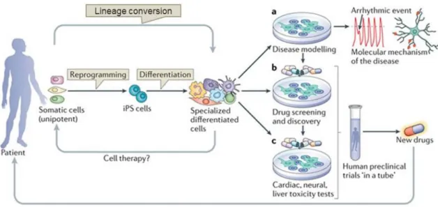
Applications ... 30
Cell transplantation therapy ... 31
Disease modelling ... 33
Drug screening and toxicological assessment ... 34
Applications in the basic sciences ... 35
Retinal Pigment Epithelium ... 37
Vertebrate eye, retina and RPE ... 37
Eye development: the view from the RPE ... 45
1. Eye field ... 46
2. Optic vesicle ... 52
3. Optic Cup ... 58
Degenerative disorders involving the RPE ... 61
xviii
Cell-based therapies for RPE disorders ... 73
Main goals and thesis overview ... 77
Chapter 2 : Materials and methods ... 81
Materials ... 81
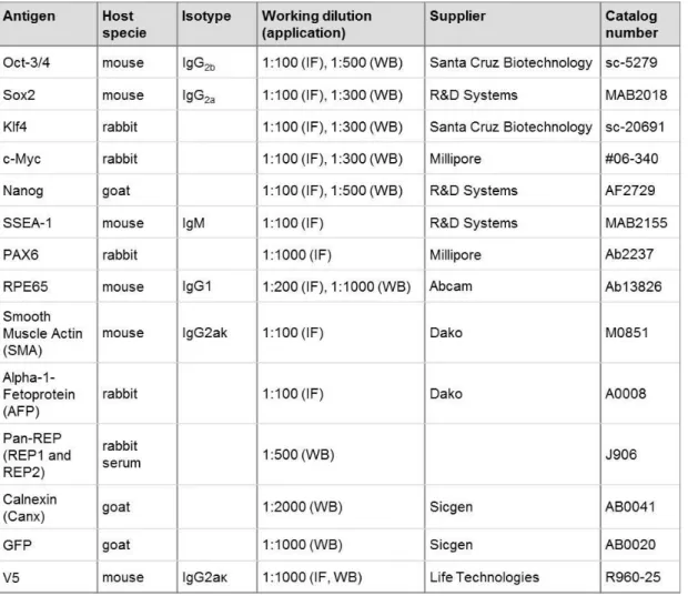
Antibodies ... 81
Animals ... 82
Cells and cell culture conditions... 82
Growth and maintenance of mammalian cell lines ... 82
Growth and maintenance of pluripotent stem cells ... 83
Mouse Embryonic Fibroblast (MEF) isolation and culture ... 85
Preparation of feeder cells to culture pluripotent stem cells ... 85
Mouse RPE primary cells’ isolation and culture ... 86
Constructs and generation of new molecular tools ... 86
Transfection of HEK-293FT Cells with plasmid DNA ... 89
Preparation and use of lentiviral transduction particles ... 89
Preparation of adenoviral transduction particles ... 90
Reprogramming somatic cells into pluripotency ... 90
Characterization of pluripotent stem cells ... 91
Alkaline phosphatase staining ... 91
Embryoid Bodies’ differentiation assay ... 92
Teratoma formation assay ... 92
Differentiation of pluripotent stem cells into retinal lineages ... 93
Differentiation protocol adapted from Zhu et al. ... 93
Differentiation protocol adapted from Eiraku et al. and Gonzalez-Cordero et al. .... 93
Differentiation protocol adapted from La Torre et al. and Osakada et al. ... 93
Cell viability assay ... 94
PCR Genotyping ... 95
RNA isolation and Reverse Transcriptase (RT) - PCR ... 95
Immunofluorescence (IF) ... 96
Image acquisition and analysis ... 96
Flow cytometry ... 97
Preparation of protein lysates and western blotting analysis ... 97
Statistics ... 98
xix
Chapter 3 : Induced Pluripotent Stem cell technology ... 105
Summary ... 105
Results ... 105
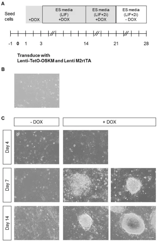
Lentiviral molecular tools efficiently transduce MEFs and allow expression of reprogramming factors ... 105
Lentiviral transduced cells display morphology and gene expression alterations during reprogramming protocol ... 107
Adenoviral molecular tools efficiently transduce MEFs and allow expression of reprogramming factors ... 113
Adenoviral transduced cells do not display typical morphological and gene expression alterations during reprogramming protocol ... 116
Lentiviral iPS cell clones exhibit typical morphology in culture, self-renewal properties, and expression of key pluripotency markers ... 118
Established iPS cell lines demonstrate a functional pluripotent capability ... 122
Discussion ... 124
Chapter 4 : Induced Pluripotent Stem cell-based applications for Choroideremia .... 131
Summary ... 131
Results ... 132
Chm MEFs primary cultures can be used as an in vitro model of Rep1 KO ... 132
Chm MEFs can be reprogrammed into pluripotency using a lentiviral based protocol ... 136
Chm iPS cell lines display morphology, self-renewal and pluripotency attributes .. 139
Chm iPS cell lines generated from Chmnull fibroblasts display efficient Rep1 KO .. 144
Chm iPS cell lines fail to differentiate into a polarized neuroepithelium when subjected to a protocol adapted from Zhu et al. ... 149
Chm iPS cell lines give rise to OV-like protusions when subjected to a differentiation protocol adapted from Eiraku et al. and Gonzalez-Cordero et al. ... 152
Chm iPS cell lines differentiate into retinal progenitor cells when subjected to a protocol from La Torre et al. and Osakada et al. ... 156
Discussion ... 160
Chapter 5 : Direct Reprogramming of fibroblasts into RPE cells ... 169
Summary ... 169
xx
MEFs have different morphology and expression profiles when compared to RPE
cells ... 170
Lentiviral molecular tools efficiently induce expression of TFs in transduced MEFs ... 172
Transduction with multiple lentiviral particles can be optimized without compromising cell viability ... 176
MEFs transduced with lentivirus encoding for 10 Eye TF gain some RPE features ... 180
Lentiviral reporter systems drive expression of GFP protein in RPE cells ... 184
Lentiviral reporter systems can be used to optimize pool of direct reprogramming TFs ... 186
Discussion ... 195
Chapter 6 : Concluding remarks and future perspectives ... 203
Chapter 7 : References ... 213
Chapter 8 : Supplementary material ... 241
xxi
List of figures
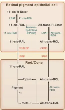
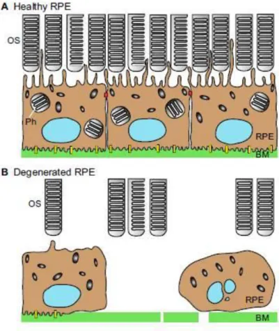
Figure 1.1: Waddington’s “epigenetic landscape” model. ... 3 Figure 1.2: Experimental approaches for cellular reprogramming to pluripotency. ... 5 Figure 1.3: Pluripotency maintenance circuitry in mouse and human ES cells. ... 11 Figure 1.4: Selected molecular events occurring during reprogramming... 15 Figure 1.5: Parameters to consider before each reprogramming experiment. ... 18 Figure 1.6: Direct lineage conversion of somatic cells. ... 26 Figure 1.7: Cellular reprogramming strategies allowing the generation of specific cell types. ... 29 Figure 1.8: Major applications of differentiated cells obtained by reprogramming strategies. ... 31 Figure 1.9: Structure of the adult human eye. ... 38 Figure 1.10: The Retinal Pigment Epithelium, located in the outer retina. ... 40 Figure 1.11: Visual cycle of retinal in RPE cells and PRs. ... 44 Figure 1.12: Embryonic development of the vertebrate eye cup. ... 47 Figure 1.13: Patterning of the OV in the presumptive NR and RPE. ... 53 Figure 1.14: Maintenance of RPE phenotype within the OC. ... 59 Figure 1.15: Healthy and degenerated RPE: implications for retina’s overall structure. ... 62
Figure 1.16: Rab proteins’ cycle showing membrane recruitment and activation. ... 67 Figure 2.1: Schematic representation of Lenti-TetO-OSKM. ... 87 Figure 3.1: MEFs transduced with inducible lentiviral particles express the 4 reprogramming factors, in the presence of DOX. ... 106 Figure 3.2: MEFs are transduced by the reprogramming lentiviral particles with 25% of efficiency. ... 107 Figure 3.3: MEFs subjected to reprogramming protocol display typical morphological alterations. ... 108 Figure 3.4: Stem cell-like colonies arise during the reprogramming protocol and express typical pluripotency markers. ... 110 Figure 3.5: Reprogramming procedure induces consistent temporal alterations in terms of gene and protein expression. ... 111 Figure 3.6: Stem cell-like colonies are isolated, subcultured and expanded to generate iPS cell clones that will be further characterized. ... 113 Figure 3.7: Adenoviral transduced cells express the 4 reprogramming factors. ... 114 Figure 3.8: MEFs are efficiently transduced by Ad-OSKM depending on the volume of transducing viral particles. ... 115 Figure 3.9: MEFs transduced with lentiviral and adenoviral particles used for delivery of reprogramming factors express Oct4 protein. ... 116 Figure 3.10: Timeline representing reprogramming protocol adapted for adenoviral delivery of reprogramming TFs. ... 117
Figure 3.11: Protocol using adenovirus does not induce alterations of pluripotency genes’
xxii
Figure 3.12: iPS cells clones demonstrate morphology in culture and AP positive staining, similar to ES cells. ... 119 Figure 3.13: iPS cell lines express endogenous pluripotency markers. ... 120 Figure 3.14: iPS cell lines express ES cell characteristic TFs and surface markers. ... 121
Figure 3.15: iPS cell clones demonstrate their functional pluripotency in in vitro differentiation
assay. ... 123
Figure 3.16: Established iPS cell lines give rise to tissues from the three germ layers, in in vivo
differentiation assay. ... 124
Figure 4.1: Breeding of Chmflox animals allows isolation of MEFs suitable as Chm model. ... 133
Figure 4.2: Treatment with 6 µM TM for 96 h induces genomic recombination in Chm MEFs without affecting cell survival... 134
Figure 4.3: Primary cultures of Chmnull MEFs have reduced levels of Rep1 expression. ... 136
Figure 4.4: Lentiviral reprogramming particles efficiently transduce Chm MEFs. ... 137
Figure 4.5: Chmnull MEFs subjected to reprogramming protocol display typical morphological
alterations. ... 138
Figure 4.6: Chmnull and Chmflox iPS cell clones demonstrate morphology in culture and AP positive
staining similar to ES cells. ... 140 Figure 4.7: iPS cell lines express endogenous pluripotency markers. ... 141 Figure 4.8: iPS cell lines express ES cell characteristic TFs and surface markers. ... 142
Figure 4.9: Chmnull and Chmflox iPS cell clones demonstrate their functional pluripotency in in vitro
differentiation assay. ... 143
Figure 4.10: Chmnull and Chmflox iPS cell clones demonstrate their functional pluripotency in in vivo
differentiation assay. ... 144
Figure 4.11: Generated Chmnull iPS cell lines display an efficient KO of Rep1 gene. ... 146
Figure 4.12: All iPS cells have equivalent growth rates, as assessed by MTT assay. ... 148
Figure 4.13: iPS cell lines submitted to differentiation protocol adapted from Zhu et al. display
morphological alterations. ... 150
Figure 4.14: iPS cell lines submitted to differentiation protocol adapted from Zhu et al. have
diminished levels of pluripotency markers expression but no detectable levels of EFTFs (except
Otx2) and RPE markers at day 5. ... 151
Figure 4.15: iPS cell lines submitted to differentiation protocol adapted from Eiraku et al. and
Gonzalez-Cordero et al. display morphological alterations. ... 153
Figure 4.16: iPS cell lines submitted to differentiation protocol adapted from Eiraku et al. and
Gonzalez-Cordero et al. have diminished levels of expression of pluripotency markers and
increased expression of EFTFs. ... 155
Figure 4.17: iPS cell lines submitted to differentiation protocol adapted from La Torre et al. and
Osakada et al. display morphological alterations. ... 157
Figure 4.18: iPS cell lines submitted to differentiation protocol adapted from La Torre et al. and
Osakada et al. have diminished levels of pluripotency markers expression and increased
xxiii
Figure 5.1: MEFs and RPE primary cultures display different morphology and gene expression profiles. ... 171 Figure 5.2: Inducible lentiviral vector allows efficient inducible expression of GFP protein. ... 173 Figure 5.3: V5-tagged versions of 10 TFs involved in eye and RPE developmental processes were successfully cloned into DOX-inducible lentiviral vectors. ... 175 Figure 5.4: Inducible lentiviral particles allow expression of 10 Eye TF in transduced MEFs... 176 Figure 5.5: Transduction with multiple lentiviral particles can be optimized without compromising cell viability ... 179 Figure 5.6: Ten-fold increase of lentiviral volumes increases percentage of transduced cells as well as their fluorescent intensity. ... 180 Figure 5.7: MEFs transduced with pool of inducible lentivirus encoding for the 10 Eye TFs display morphological alterations, in terms of pigmentation. ... 181 Figure 5.8: MEFs transduced with pool of inducible lentivirus encoding for the 10 Eye TFs display alterations in gene expression. ... 182 Figure 5.9: MEFs transduced with pool of inducible lentivirus encoding for the 10 Eye TFs express Rpe65 protein at 22 dpt. ... 184 Figure 5.10: Human RPE cell line ARPE-19 transduced with lentiviral reporter systems express GFP protein. ... 186
Figure 5.11: Untransduced MEFs’ survival is affected by increasing concentrations of blasticidin.
xxiv
List of tables
Table 1.1: Viral delivery methods of reprogramming factors. ... 22 Table 1.2: Non-viral delivery methods of reprogramming factors. ... 24 Table 2.1: List of antibodies used throughout this work, either for Immunofluorescence (IF) or western blot (WB) applications. ... 81 Table 2.2: Established iPS cell lines mentioned in this work and corresponding genotype,
according to Chm alleles and MerCreMer transgene presence/absence. ... 84
Table 2.3: PCR primers used for genotyping. ... 95
xxv
Abbreviations
2i Dual inhibitors (1 µM of MEK inhibitor PD 0325901 and 3 µM of GSK3 inhibitor
CHIR99021)
3D Three-dimensional
a.u. Arbitrary units
ABC ATP binding cassette
AMD Age-related Macular Degeneration
ANOVA Analysis of variance
AP Alkaline phosphatase
BMP Bone morphogenetic protein
cDNA Complementary DNA
CMZ Ciliary marginal zone
DAPI 4',6-diamidino-2-phenylindole
DMEM Dulbecco’s Modified Eagle Medium
DMEM/F12 DMEM and Ham’s F12 nutrient mixture 1:1
DNA Deoxyribonucleic acid
DOX Doxycyclin
DR Diabetic Retinopathy
E13.5 Embryonic day 13.5
EB Embryoid body
EF Eye field
EFTF Eye field transcription factor
EMT Epithelial-to-mesenchymal
EpiS cell Epiblast stem cell
ERK Extracellular signal regulated kinases 1 and 2
ES cell Embryonic stem cell
FBS Fetal Bovine Serum
FGF Fibroblast growth factor
GFP Green fluorescent protein
GGTase II Rab geranylgeranyl transferase
GMP Good Manufacturing Practice
HLA Human leucocyte antigen
ICM Inner cell mass
IF Immunofluorescence
IGF-1 Insulin-like growth factor-1
iPS cell induced Pluripotent Stem cell
JAK/STAT Janus kinase/signal transducers and activators of transcription
KO knockout
LCA Leber Congenital Amaurosis
LIF Leukaemia Inhibitory Factor
M2rtTA Reverse tetracycline-controllable transactivator
MAPK or MEK Mitogen-activated protein kinase
MEF Mouse embryonic fibroblast
Melan Ink4a Bl6 (Black6) Ink4a -/- Melanocytes
MET Mesenchymal-to-epithelial
xxvi
miRNA microRNA
MOI Multiplicity of infection
MTT 3-[4,5-dimethylthiazol-2-yl]-2,5 diphenyl tetrazolium bromide
NOD.Scid Non-obese diabetic/ severe combined immunodeficiency
NPC Neural progenitor cell
NR Neuroretina
OC Optic cup
OS Outer-segment
OSKM OCT4, SOX2, KLF4, c-MYC
OV Optic vesicle
PBS Phosphate buffered saline
PCR Polymerase Chain Reaction
PEDF Pigment epithelium-derived factor
PFA Paraformaldehyde
PI3K Phosphatidylinositol 3' –kinase
PR Photoreceptors
Rab GDI Rab GDP-dissociation inhibitor
RCS Royal College of Surgeons
REP1 Rab Escort Protein 1
RGC Retinal ganglion cells
RNA Ribonucleic acid
ROS Reactive oxygen species
RPE Retinal pigment epithelium
RPE Retinitis Pigmentosa
RT Room temperature
RT-qPCR Reverse Transcriptase - quantitative PCR
SCNT Somatic-cell nuclear transfer
SFEBq Serum-free floating cultures of EB-like aggregates with quick reaggregation
Smad Small Body Size / Mothers Against Decapentaplegic
SSEA-1 Stage-specific embryonic antigen-1
TetO Tetracycline operator minimal promoter
TF Transcription factor
TGF Transforming growth factor
UD Undifferentiated
VEGF Vascular endothelial growth factor
3
Chapter 1
: Introduction
Cellular Reprogramming
Reprogramming concepts
Multicellular organisms are composed of an assortment of differentiated cells responsible for different functions and whose stability is essential for the growth, survival and perpetuation of the whole organism. During development, uncommitted stem cells differentiate into various tissue-specific cell types, in a process established and maintained by a complex interplay of endogenous and exogenous factors. For a long time, lineage commitment and differentiation was believed to be unidirectional and irreversible, as Conrad Waddington represented in his model in 1957 (Figure 1.1). In this classic view of cell fate hierarchy, the undifferentiated cell resides above the different committed and differentiated states. Furthermore, it was long thought that along with the differentiation process, there was a concomitant loss of chromosomes or permanent inactivation of genes that were no longer needed.
For instance, in a mammalian organism, the unicellular totipotent zygote lies in the beginning of the developmental process given its ability to give rise to all cells of an organism, including embryonic and extra-embryonic tissues (such as the placenta). At the blastocyst stage of the early embryo, the cells from the inner mass are pluripotent: they are able to generate all the cells of the embryo, and so they form each of the three
Figure 1.1: Waddington’s “epigenetic landscape” model.
4
germ layers – endoderm, mesoderm and ectoderm. As differentiation follows, cells become progressively more committed to their cell fate and more restricted in terms of developmental potency. Cells that are committed to each of the germ layers specialize to give rise to the tissues of the adult body, which still contains multipotent and unipotent cells. The former retain the ability to differentiate into multiple cell types within the same lineage (such as hematopoietic stem cells and neural stem cells), whilst the later only have the capacity to differentiate into one type of cell as spermatogonial stem cells (Jaenisch and Young, 2008).
Contrarily to this unidirectional developmental process, studies suggesting cellular plasticity in the animal kingdom go back to 1895 when Wolff reported that, after surgical removal of lens from the adult eye of newts, a structurally and functionally complete lens regenerated from the dorsal, pigmented epithelial cells (Wolff, 1895). This example constituted the first experimental evidence of in vivo adult cellular reprogramming (or transdifferentiation). Cellular reprogramming (or nuclear reprogramming) refers to the
concept of “rewiring the epigenetic and transcriptional network of one cell state to that of
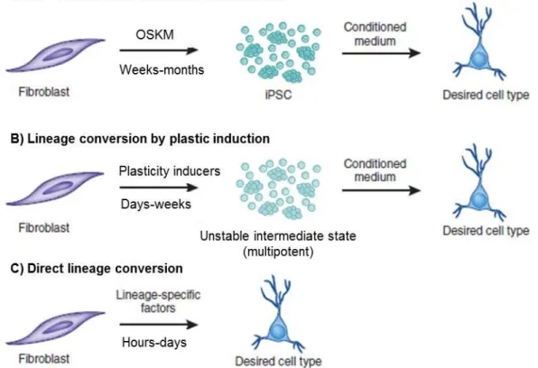
a different cell type” (Hanna et al., 2008). Three different experimental approaches have definitively confirmed that, although the differentiated state of a cell is generally stable, cellular identity is dynamically controlled and subjected to perturbations in the stoichiometry of the transcriptional and epigenetic regulators present in the cell in any given time. Nuclear transfer, cell fusion and transcription factor (TF)-transduction have provided means to induce in vitro reprogramming of defined and specialized cells either into a different somatic cell type (lineage conversion) or into an embryonic pluripotent state (Figure 1.2) (Yamanaka and Blau, 2010). On 2012, the Nobel Prize in Physiology
or Medicine was jointly attributed to John B. Gurdon and Shinya Yamanaka “for the discovery that mature cells can be reprogrammed to become pluripotent”.
1. Nuclear transfer
5 somatic-cell nuclear transfer (SCNT), or cloning, provided definite evidence that cell specialization involves changes in gene expression rather than gene content. Thus, it is a reversible process, once genes that are required to create an entire organism are still present in the nucleus of the specialized cell (despite silenced) and are activated after exposure to the existing reprogramming factors of the oocyte.
The first successfully cloned mammal was Dolly the sheep by Wilmut and colleagues, mice followed as a wide range of different species. Relevantly, the hypothesis that the reprogramming was only due to contaminating cells was put to rest when a mouse clone was produced from the nuclei of B cells in which the immunoglobulin had been rearranged (Hochedlinger and Jaenisch, 2002; Wakayama et al., 1998; Wilmut et al., 1997). Instrumental contributes to allow Figure 1.2: Experimental approaches for cellular reprogramming to pluripotency.
6
reprogramming of somatic cell into pluripotency was the derivation and stable maintenance of pluripotent cell lines in vitro, specifically the derivation of embryonic stem (ES) cells from the blastocyst inner cell mass (Evans and Kaufman, 1981). The efficiency of generating cloned mice is usually low (1-2%), but increases substantially (20%) when the cell source for nuclei is nuclear-transfer-derived ES cells suggesting that the process of nuclear reprogramming is enhanced by a passage through an ES cell state (Hochedlinger and Jaenisch, 2002). Additionally, developmental defects in cloned animals are common and believed to stem from
incomplete erasure of “epigenetic memory”, meaning that epigenetic factors, such as regulators of DNA methylation, histone modifications and replacements and ATP-dependent chromatin remodelers, have not been completely reprogrammed into a pluripotent state (Simonsson and Gurdon, 2004). Recently, optimized nuclear transfer approach, designed to circumvent early embryonic arrest on prior attempts, allowed the derivation of human nuclear-transfer-derived ES cells (Tachibana et al., 2013).
2. Cell fusion
Cell fusion involves fusing two or more cell types to form a single entity, which can result in heterokaryons (that do not proliferate and thus contain more than one nucleus) or in hybrids (that proliferate, with fusion of the nuclei of the original cell occurring due to cell division). In the 1960s, the existence of trans-acting repressors acting on DNA to regulate gene expression was uncovered by cell fusion experiments of a fibroblast with a melanocyte given rise to a hybrid with no melanin synthesis (Davidson et al., 1966). Moreover, in 1983, heterokaryons of mouse muscle cells and human amniotic cells have been shown to express human muscle proteins (such as human myosin light chains 1 and 2) indicating that previously silenced genes were activated (Blau et al., 1983). Equivalent experiments for other cell types were rapidly performed, with the relative ratio of the nuclei, or the gene dosage, of the two cell types dictating the outcome of the reprogramming, and therefore confirming that the differentiated state was continuously controlled by the balance of regulators present at any given time (Yamanaka and Blau, 2010).
7 mouse Oct4 (Tada et al., 2001). Fusion-based nuclear reprogramming was also shown to be strongly enhanced (up to 200-fold) after overexpression of the pluripotency TF Nanog (Silva et al., 2006). Moreover, experiments with heterokaryons are well suited for elucidating the molecular mechanisms required for the initiation of reprogramming into a pluripotent state, given their rapid rate of reprogramming. Loss-of-function and gain-of-function approaches have allowed to uncover essential molecular players, namely Oct4 and enzymes responsible for DNA demethylation (Bhutani et al., 2010; Pereira et al., 2008).
3. Trancription factor transduction
In the previously described molecularly undefined reprogramming methods, a milieu of components or elements (e.g., transcription factors, histone-modifying and chromatin-remodelling enzymes, and DNA demethylases) that are largely unknown contribute to achieve cellular reprogramming. Conversely, direct reprogramming methods use defined genetic or nongenetic elements to induce rewiring of the cell state (Hanna et al., 2008). Overexpression of a single tissue-specific TF in somatic cells was surprisingly able to activate genes typical of other somatic cell types and alter the cell fate. First report was by Gehring and co-workers using Drosophila melanogaster in 1987, followed by another work showing that ectopic expression of eyeless (Pax6 in mice), a master regulator of eye morphogenesis, lead to induction of ectopic eye structures on the wings, the legs and the antennae (Halder et al., 1995; Schneuwly et al., 1987). Davis and colleagues have demonstrated reprogramming of mouse fibroblasts into myoblast-like cells upon ectopic expression of the MyoD transcription factor (Davis et al., 1987). More recent work has shown that overexpression of a myeloid transcription factor CCAAT/ enhancer-binding protein α (C/EBPα) promotes conversion of
lineage-committed B and T cells into macrophage-like cells (Xie et al., 2004).
8
expression of neomycin resistance cassette was used. Drug-resistant clones were isolated and demonstrated morphology, proliferation and gene expression similar to ES cells. To confirm pluripotency, cells were injected into immunodeficient mice, forming teratomas, tumours including all three germ layers, endoderm, mesoderm and ectoderm. To further determine which of the 24 candidates were critical for the reprogramming process, the effect of withdrawal of individual factors from the pool of transduced candidate genes on the formation of drug-resistant colonies was assessed. Four TFs were identified as being essential for the reprogramming of fibroblasts into pluripotent cells, Oct4, Sox2, Klf4 and c-Myc, and resulting cells were named induced Pluripotent Stem cells (iPS cells). After Yamanaka’s seminal discovery, a panoply of subsequent works followed, demonstrating optimized ways of generating iPS cells or providing mechanistic insights of the reprogramming process. Importantly, human iPS cells were generated and proof-of-principle experiments demonstrated its therapeutic potential, as such cells can be used as a cell source for tissue repair or replacement while avoiding ethical and immunological concerns associated with the use of ES cells (Takahashi et al., 2007a; Yu et al., 2007). Besides patient-specific, disease-specific pluripotent cell lines derived from human patients with specific diseases were obtained, constituting invaluable tools and an unlimited source for biological material that can be used to study these complex diseases in the Petri dish (Park et al., 2008; Raya et al., 2009).
9 All the three mentioned approaches to cellular reprogramming display common features, such that in each case if the balance of regulators is tilted to favour pluripotency (or other somatic cell fate), the epigenome is altered and the expression of pluripotency/cell-specific factors that otherwise would be silenced in a stably differentiated cells. In order to maintain the new phenotypic identity of the reprogrammed cell, there must be activation of feedback and auto-regulatory mechanisms to attain critical threshold levels of endogenous cell-specific transcriptional regulators. Nevertheless, when comparing the 3 approaches, there are differences in terms of technical feasibility, time required for reprogramming, efficiency of the process, cell yield and probably also in the underlying molecular mechanism. In terms of cell yield, TF-transduction provides the abundant and easily reproducible across the world generation of iPS cells, which concurs to their advantageous prospective use in therapeutic settings, as well as usefulness for disease-modelling and drug testing (Hanna et al., 2008; Yamanaka and Blau, 2010). Additionally, the advent of these technological breakthroughs has put to argue the paradigm of unidirectional development. A non-hierarchical “epigenetic disc” model to explain interconversion of somatic and pluripotent cell fates has been recently proposed as an alternative to Waddington’s classical view (Ladewig et al., 2013).
Somatic to pluripotent TF-mediated reprogramming
Pluripotent stem cell lines can be obtained through the reprogramming of somatic cells, by ectopic expression of defined factors known to be important for the maintenance pluripotent stem cells identity. Generated iPS cell lines are characterized and compared with their biological counterparts (ES cells) in order to assess reprogramming efficiency and fidelity. Moreover, several studies of the molecular basis, both genetic or epigenetic, of these natural and induced pluripotent states, as well as investigations into how pluripotency is maintained and the mechanisms of lineage commitment have provided insights for improving the understanding of mammalian embryogenesis and cellular differentiation, but also for developing successful stem cell-based therapies for regenerative medicine.
Pluripotency regulation: transcriptional network and signalling pathways
10
the adult body. ES cells were first isolated from the inner cell mass (ICM) of pre-implantation mouse blastocyst embryos at embryonic day 3.5 (E3.5). Mouse ES cells are rapidly proliferating cells that form tight, dome-shaped colonies (Evans and Kaufman, 1981). To maintain their self-renewal capacity in an undifferentiated state, they require the growth factors leukaemia inhibitory factor (LIF) and bone morphogenetic protein 4 (BMP4) in mouse embryonic fibroblast (MEF)- and serum-free conditions, respectively (Figure 1.3) (Williams et al., 1988a; Ying et al., 2003). The pluripotent cells in the pre-implantation embryo are considered naïve because they have unbiased developmental potential and can give rise to germline-competent chimeras when reintroduced into a blastocyst. Contrarily, another stem cell population derived from post-implantation embryo, epiblast stem (EpiS) cells,
exhibits a “primed” state of pluripotency (Nichols and Smith, 2009; Tesar et al., 2007). Human ES cells are derived from human blastocysts but, in contrast to murine ES cells, form flat 2D colonies dependent on basic fibroblast growth factor (bFGF) and activin/transforming grothw factor-β (TGFβ) signalling (Figure 1.3) (Thomson et al., 1998). Human ES cells share several molecular features with naïve mouse ES cells but they also share a variety of epigenetic properties with
primed murine EpiS cells, displaying a “primed” pluripotency. Very recently however, derivation conditions for the establishment of human naïve pluripotent cells was also reported (Gafni et al., 2013).
There is a core regulatory circuitry composed of a set of TF that functions to maintain the pluripotent state in pluripotent stem cells, natural or induced and in humans and mice. OCT4 (also known as POU5F1), NANOG and SOX2 function together to positively regulate their own promoters, forming an interconnected autoregulatory loop. Additionally, they co-occupy promoter regions of genes that are involved in pluripotency maintenance (keeping them in an active state) and early lineage differentiation (repressing them) (Figure 1.3) (Masui et al., 2007; Silva and Smith, 2008; Young, 2011).
11 Figure 1.3: Pluripotency maintenance circuitry in mouse and human ES cells.
In both cell types, Nanog, Oct4, and Sox2 form a positive autoregulatory loop that regulates self-renewal and pluripotency. (A) Mouse ES cells require LIF and BMP4 for maintenance. LIF promotes self-renewal by activating the Janus kinase/signal transducers and activators
of transcription (JAK/STAT3) and phosphatidylinositol 3' –kinase (PI3K)/AKT signalling
pathways. There’s an upregulation of Klf4 and Tbx3, which then activate Sox2 and Nanog,
respectively. BMP4 upregulates transcription of inhibitor of differentiation (Id) genes
through activation of SMAD (Small Body Size / Mothers Against Decapentaplegic) proteins
1, 5, and 8. (B) Human ES cells and mouse EpiS cells require insulin-like growth factor
IGF/insulin and bFGF for maintenance. bFGF activates the mitogen-activated protein kinase (MAPK) as well as the Activin/Nodal signalling pathways, and IGF activates the Ras and PI3K pathways. MEFs are also stimulated by bFGF in culture to secrete IGF (dashed arrows). SMADs 2 and 3 propagate Activin/Nodal signalling as well as directly bind and
12
Sox2, a SRY-related TF containing the high-mobility group-box DNA-binding domain, has also been shown to possess a loss-of-function phenotype similar to that of Oct4 in both embryos and ES cells. Ectopic expression of wild-type levels of Oct4 can rescue the Sox2-null phenotype in ES cells, which, in addition to the similarity of phenotypes, suggests a synergistic action of the two TFs in regulating the expression of themselves and other ES cells-specific genes (Avilion et al., 2003; Masui et al., 2007).
Nanog is a homeodomain protein that was discovered in a screen for self-renewal factors that could sustain mouse ES cells in the absence of LIF signalling. Nanog is critical for mammalian development and is required for specification of the ICM in the pre-implantation embryo. Although ES cells can be propagated in the absence of Nanog, it promotes a stable undifferentiated ES cell state. Overexpression of Nanog leads to enhanced self-renewal of ES cells, illustrating a positive effect on the pluripotent network (Chambers et al., 2007; Saunders et al., 2013).
The interconnected regulatory loop of Oct4, Sox2 and Nanog promotes a bistable state for ES cells: residence in a positive-feedback-controlled gene expression program when the factors are expressed at appropriate levels, versus entrance into a differentiation program when any one of the master transcription factors is no longer functionally available. The core TFs collaboratively activate a substantial fraction of the actively transcribed protein-coding and microRNAs (miRNAs) genes in ES cells. A large proportion of these actively transcribed genes are bound and regulated by both the core transcription factors and also c-Myc, which plays important roles in ES cells proliferation and self-renewal. While Oct4, Sox2, and Nanog core regulators are involved in RNA polymerase II recruitment, c-Myc is believed to stimulate the transcriptional pause release of RNA polymerase II. Consequently, Oct4/Sox2/Nanog apparently play dominant roles in selecting the set of ES cell genes that will be actively transcribed and recruiting RNA polymerase II to these genes, while c-Myc regulates the efficiency with which these selected genes are fully transcribed (Rahl et al., 2010; Young, 2011).
Simultaneously, the core regulators repress the expression of a wide spectrum of cell-lineage-specific regulatory genes, through a process mediated by SetDB1 and Polycomb group (PcG) chromatin regulators. Once their repressive signal is lost, a rapid induction of expression occurs indicating that these genes are poised for activation. Interestingly, the chromatin conformation associated with many of these
key developmental genes is composed of ‘bivalent domains’ consisting of both
13 4 methylation marks. These bivalent domains are lost in differentiated cells. Thus, the core regulatory circuitry, and additional collaborative regulators of gene expression, are responsible for maintaining ES cells in a stable pluripotent state whilst remaining poised to differentiation (Bernstein et al., 2006).
In addition to Oct4, Sox2, Nanog, and c-Myc, the transcription factors Tcf3, Smad1, Stat3, Esrrb, Sall4, Tbx3, Zfx, Ronin, Klf2, Klf4, Klf5, and PRDM14 have been shown to play important roles in control of ES cell state. Transcriptional regulation of pluripotency state is also dependent on cofactors, protein complexes that contribute to activation (coactivators) and repression (corepressors) of expression but do not have DNA-binding properties of their own. Chromatin regulators, such as cohesin/condensin protein complexes, histone-modifying enzymes, ATP-dependent chromatin-remodeling complexes and DNA methyltransferases, also play a role in maintaining the ES cell viability and stability. A variety of non-coding RNA species have also been implicated in control of ES cell state, including miRNAs, which can regulate the stability and translatability of mRNAs, and longer non-coding RNAs, which have been implicated in recruitment of chromatin regulators (Yeo and Ng, 2013; Young, 2011).
As mentioned earlier, signal transduction pathways are involved in cells’ response to their surrounding cellular and biochemical environment. For ES cells, maintenance of the pluripotent state is dependent on the absence or inhibition of signals that stimulate differentiation. Traditionally, mouse ES cells were cultured and kept pluripotent on a layer of mitotically inactivated feeder cells in serum-supplemented media. The combinatorial use of LIF and BMP4 allowed the establishment of a defined feeder- and serum-free culture sufficient to derive and maintain germ-line transmittable mouse ES cells. LIF and BMP4 induce phosphorylation and activation of their downstream TFs Stat3 and Smad1, respectively, which in turn co-bind at Oct4, Sox2 and Nanog regions and thus sustain the core ES cell transcriptional network (Niwa et al., 1998; Pera and Tam, 2010; Ying et al., 2003).
14
Tcf3, the most abundantly expressed member of TCF/LEF family of TFs in mouse
ES cells. This nuclear effector of Wnt/β-catenin signalling, Tcf3, is bound to the same regulatory regions as Oct4 and Nanog, and has been known to negatively balance their effects in the maintenance of the pluripotent state (Wray et al., 2011).
Recently, β-catenin was also shown to contribute to pluripotency acting through an Oct4 complex, on a transcriptional-independent manner (Faunes et al., 2013).
Hallmarks of naïve pluripotency include driving Oct4 transcription by its distal enhancer, retaining a pre-inactivation X chromosome state, and global reduction in DNA methylation and in H3K27me3 repressive chromatin mark deposition on developmental regulatory gene promoters. In recent years a combination of small-molecule inhibitors has been identified that greatly facilitates murine ES cells derivation and maintenance in a naïve pluripotent state. This so-called 2i inhibitor cocktail consists of a MEK/ERK inhibitor (PD0325901) and a GSK3 inhibitor (CHIR99021). 2i culture conditions promotes the achievement of a “ground state”
pluripotency by blocking the pro-differentiation effect of the FGF–MEK–ERK signalling pathway and simultaneously inhibiting glycogen synthase kinase GSK3b, thereby promoting the self-renewal positive effect of Wnt/β-catenin signalling. Moreover, factors associated with lineage-specification are repressed under 2i culture conditions, at an epigenetic level. A more homogeneous expression of key pluripotency regulators is also induced in 2i culture conditions. Particularly, biallelic expression of the key pluripotency regulator Nanog is achieved (whilst in serum conditions Nanog is only expressed by one allele), which has been shown to be important for the survival of the peri-implantation inner cell mass (Marks et al., 2012; Miyanari and Torres-Padilla, 2012; Silva et al., 2008; Ying et al., 2008).
Mechanistic insights into the reprogramming process
15 Figure 1.4: Selected molecular events occurring during reprogramming.
16
Reprogramming process is initiated by the forced expression of traditional OSKM factors. However, in the nucleus, the majority of DNA is packed into nucleosomes, occluded by higher order chromatin structure and repressors. Cell proliferation may facilitate reprogramming by allowing TF access to otherwise occluded cis-regulatory regions through nucleosome displacement during DNA replication. In the absence of cell division, several models have been proposed to account for the
access of transcription factors to their relevant binding sites, including the ‘pioneer’
TF model. Contrarily to other factors, pioneer factors can access their target sites in repressed regions of the genome, through inducing local chromatin opening, nucleosome repositioning, and recruitment of chromatin modifiers and co-regulators. OCT4, SOX2 and KLF4 might act as pioneer factors, facilitated by C-MYC proliferative action. This initiation is then followed by feed-forward induction of additional TFs to execute the reprogramming process (Soufi et al., 2012; Taberlay et al., 2011; Vierbuchen and Wernig, 2012).
In the early stages of reprogramming, gene expression is stochastic with resulting differential expression of genes involved in cell-division cycle, DNA replication and a process called the mesenchymal-to-epithelial (MET) transition, which also occurs during normal development (Li et al., 2010a). In parallel, some cells show reduction in the expression of genes associated with cell–cell interaction and cell adhesion, and of markers that are typical of the initial differentiated-cell population. MET is one of the earliest observable events occurring during fibroblast reprogramming, with elongated cells become rounded and aggregated in small clusters. This transition correlates with alkaline phosphatase (AP) positivity and stage-specific embryonic antigen-1 (SSEA-1) upregulation. There is also an up-regulation of epithelial junction components and involvement of the TGFβ signalling pathway. In terms of epigenetic modification, histone modifications and structural changes that are associated with a more open chromatin conformation are also observed during the early phase of reprogramming (Buganim et al., 2012; Polo et al., 2012).
A progressive increase in the expression of early - albeit not definitive - markers of pluripotency is observed. At this point, reprogramming-refractory and reprogramming-competent cells coexist, expressing different levels of the reprogramming factors and giving rise to heterogeneous cell populations. A second
“wave” of molecular events follows, with cells hierarchically increasing the
17 locking the defined pluripotent state (Buganim et al., 2012; Cantone and Fisher, 2013; Polo et al., 2012).
Oct4 has an established importance in the reprogramming process since it can be used alone to obtain iPS cells. Mechanistically, experimental evidence suggests that Oct4 participates in the induction of the MET and in the derepression of somatic cell chromatin. In addition, Oct4 in cooperation with Sox2 was proposed to prevent the acquisition of alternative cell states during reprogramming. Furthermore, Oct4 dose and cellular localisation were proposed as important parameters of successful reprogramming: an ES cell level of Oct4 must be attained at the late stages of reprogramming for cells to enter the pluripotent cell state (reviewed by (Radzisheuskaya and Silva, 2013).
Technological overview of reprogramming into pluripotency
At first, Yamanaka reported the generation of iPS cells through forced expression of a set of core pluripotency-related TF (OCT4, SOX2, KLF4 and c-MYC – or abbreviated as OSKM) in fibroblasts (Takahashi and Yamanaka, 2006). After TF transduction, tightly compacted colonies appeared on the culture dish, which resembled ES cells morphologically, molecularly and phenotypically (Okita et al., 2007). Several protocols for iPS cells generation have been developed in the following years. They use, for example, different mouse and human donor populations or vary the number, identity and delivery mode of the reprogramming factors (Aasen et al., 2008; Aoi et al., 2008; Carey et al., 2009; Kim et al., 2009; Nakagawa et al., 2008; Okita et al., 2008; Takahashi et al., 2007a). The multitude of the different approaches undertaken over the past few years will not be exhaustively mentioned here, but was recently reviewed by (Bayart and Cohen-Haguenauer, 2013; González et al., 2011).
18
Different efficiencies and kinetics have been observed depending on the starting
cell. The reprogramming process requires the delivery of certain factors into a specific cell and their adequate expression for a proper period of time, usually smaller for mouse than for human cells. Fibroblasts are still the most widely used Figure 1.5: Parameters to consider before each reprogramming experiment.
Any reprogramming experiment is determined by a number of preliminary choices regarding the donor cell type to reprogram, the factors to use and the mode of their delivery. The choice of the initial cell type depends not only on its availability but also the ease of reproducing the reprogramming process. The TFs important for embryonic development must be overexpressed, alone or in conjunction with other positive or negative regulators of cell cycle, epigenetic markers or signalling pathways. Forced expression of the reprogramming factors can be achieved through several available delivery modes with variable efficiency and safety. CHD1, chromodomain-helicase-DNA-binding protein 1; DNMT1, DNA methyltransferase 1; DPPA4, developmental pluripotency associated 4; E-cadherin, epithelial cadherin; ERK, extracellular signal-regulated kinase; MAPK, mitogen-activated protein kinase; MMLV, Moloney murine leukaemia virus; PRC2, Polycomb repressive complex 2; SV40LT, SV40 large T antigen; TERT, telomerase reverse
19 cells, however reprogramming efficiency may be higher in different cellular populations, such as keratinocytes (with the further advantage of their ease to obtain from human donors) (Aasen et al., 2008). Moreover, in some certain populations, the reprogramming efficiency is higher or the requirement for factors is smaller given the high levels of endogenous expression of reprogramming TFs. For instance, neural progenitor cells, which express SOX2 endogenously, are reprogrammed in the absence of exogenous SOX2 or with OCT4 alone (Kim et al., 2009). The differentiation status of the donor cell has also shown to influence both the efficiency of the process and the quality of the generated cells.
The factors that induce reprogramming are genes that are normally expressed early during development and are involved in the maintenance of the pluripotency of cells from the inner cell mass of the pre-implantation embryo. Core regulatory TFs Oct4, Sox2 and Nanog are usually included in reprogramming cocktails, such as OSKM and also early-described alternative cocktail of Oct4, Sox2, Nanog and Lin28 (Yu et al., 2007). Other TFs expressed by ES cells, such as SALL4 and UTF1, have been shown to positively affect reprogramming efficiency or kinetics. Influence of cell-cycle regulators on reprogramming has also been highlighted once some factors such as MYC and KLF4, which directly or indirectly affect cell proliferation, have been shown to increase reprogramming efficiency. Contrarily, some factors inhibit reprogramming barriers, such as senescence and apoptosis, and allow an increase in both the speed and efficiency of reprogramming (for instance inhibition of p53) (González et al., 2011; Kawamura et al., 2009).