Abstract -
Glaucoma is an eye disease which occurs due to the increased/decreased fluid pressure inside the eye. The pressure inside the normal eye is below 21mm of Hg. When the pressure inside the eye(s) increases more than 21mm of Hg, the optic nerve is damaged. This can permanently damage vision in the effected eye(s) and can lead to blindness if left untreated. The main objective of this paper is to map the person’s eye color image with database of images consisting of normal person as well as images of people suffering from glaucoma. The images’ having different color variations inside the eye is compared by using images taken by high definition laser camera. These are called as fundus images. The feature extraction of these fundus images may be carried out using MATLAB software tool. By measuring the color pixels in the affected area the observation shows that the person is suffering from Glaucoma or not. To tell whether the person is suffering from Glaucoma, a test is made using the image of a normal person which is kept as reference (say zero) and then compared with the clinical observations of the person’s image.Further if the result is positive (person is affected with Glaucoma) then also check for the three types of Glaucoma.Keywords: Glaucoma, Fundus images, fluid pressure, MATLAB.
I.
I
NTRODUCTIONNothing is more precious than eyesight. Look at the world around and think of how vulnerable the vision is. Now think how the world will be if we are losing our eyesight to a silent disease called Glaucoma [1]. The fundus of the eye is the interior surface of the eye, opposite the lens, and includes the retina, optic disc, macula and fovea, and posterior pole [2]. The fundus can be examined by ophthalmoscope and/or fundus photography [3]. MATLAB tool results in the effective analysis and simulation of various biomedical images, specifically eye images [4]. MATLAB is a high performance language for technical computing. It integrates computation, visualization, and programming in an easy-to-use environment where problems and solutions are expressed in familiar mathematical notations [5].
Manuscript received Feb 28, 2013; revised April 08, 2013.
1. Archana Nandibewoor, Assistant Professor & Research Scholar, Dept of CSE, SDMCET, Dharwad-580002, Karnataka, India (E-mail: [email protected]). 2. S.B.Kulkarni, Assistant Professor, Dept of CSE, SDMCET Dharwad-580002, Karnataka, India (E-mail: [email protected]).
3. Sridevi Byahatti, Research Scholar, Bharathiar University Coimbatore, Tamilnadu, India
(E-mail: [email protected]).
4. Ravindra Hegadi, Head, Dept of Computer Application, School of computational Sciences, Solapur University Maharashtra, India (E-mail: [email protected])
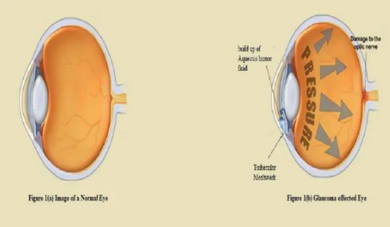
Classification is done according to the mechanism of intraocular pressure elevation [6-7]. The proposed system will first compare laser fundus images of a normal person with Glaucoma effected person and determine the result as either positive or negative. Figure 1 (a) illustrates the eye of a normal person and Figure 1 (b) shows an effected person with Glaucoma.
The images are taken from high definition laser cameras which were collected from eye hospitals. The feature extraction technique is done using color segmentation technique [8-10]. Then the patient’s result is observed and necessary action is taken.
Fig 1: Source: healthvigil.blogspot.com
For the above Figure1(b):Trabecular meshwork-The trabecular meshwork is an area of tissue in the eye located around the base of the cornea and is responsible for draining the aqueous humor from the eye.
Aqueous humor- The clear, watery fluid circulating in the chamber of the eye between the cornea and the lens.
II.
M
ATERIALA
NDM
ETHODSIn establishing treatment for glaucoma, classification according to the mechanism of intraocular pressure elevation is useful [8]. There are many types of glaucoma which are discussed below:
Primary Open Angle Glaucoma: It is the most common form of glaucoma. It occurs when the trabecular meshwork of the eye gradually becomes less efficient at draining fluid. As this happens, your eye pressure, called intraocular pressure increases.
Normal Tension Glaucoma: Some people whose eye pressure is consistently below 21 mm Hg have a type of glaucoma called normal-tension, or low-tension glaucoma. Their, optic nerve damage and visual field loss still occur.
Ocular Hypertension: It is a condition where someone has higher eye pressure than normal, but does not have other signs of glaucoma, such as optic nerve damage or blank spots that show up in their peripheral (side) vision when tested.
Computer Based Diagnosis of Glaucoma using
Digital Fundus Images
Archana Nandibewoor S B Kulkarni Sridevi Byahatti Ravindra Hegadi
Proceedings of the World Congress on Engineering 2013 Vol III,
WCE 2013, July 3 - 5, 2013, London, U.K.
ISBN: 978-988-19252-9-9
ISSN: 2078-0958 (Print); ISSN: 2078-0966 (Online)
Primary Angle Closure Glaucoma: It is less common form of glaucoma. It occurs when the drainage angle of the eye becomes blocked. Unlike open-angle glaucoma, eye pressure usually goes up very fast.
Secondary Glaucoma: It is glaucoma that results from another eye condition or disease. For example, someone who has had an eye injury, someone who is on long-term steroid therapy or someone who has a tumor may develop secondary glaucoma.
Congenital Glaucoma: It is a rare type of glaucoma that develops in infants and young children and can be inherited.
Because of the important role of the visual system imbalance and maintaining posture in human beings, glaucoma patients should consider themselves at greater risk of falls, and would be advised to take the necessary precautions to help prevent any accidents. In addition, since the peripheral visual system has such a high contribution to this intrinsic balancing mechanism, the severity of glaucoma and degree of visual field obstruction should also be considered.
III.
O
VERALLD
ESCRIPTION&
A
RCHITECTURALD
ESIGNO
FT
HEP
ROPOSEDS
YSTEMThe architectural design for a Glaucoma patient in initial state is shown in Figure 2 below.
Fig 2. Architectural design for a Glaucoma patient
The above figure 2 explains the following things:
1. Clinical observations: This includes the data chart of the Patient including his age, history of any previous accident/tumor, pressure variation etc.
2. Data Analysis consist of the following records; Data loader - uploads of a person’s eye image.
Patient record -all information about the health condition of a person. Database of all the images will have all set of images of normal case and all range of people suffering from Glaucoma.
Laser image - this is the fundus image taken by high definition camera
Image comparison - person’s image is compared with the database of all the images
3. Patient result will be displayed after the end of all the iterations.
VI.
A
LGORITHMFigure 3 shows the complete algorithm of the proposed work. We are considering the following three types of glaucoma in our study.
Fig 3. Glaucoma detection algorithm
V.
R
ESULTS ANDD
ISCUSSIONThis work will be a perfect study of any patient’s laser fundus image of the eye, and gives precise information about his/her condition. The work is carried on the basis of comparing the color of various stages encountered from the period of checkup, to treatment of a patient. We keep the color image of a normal person say pink color as reference and then compare with the person’s clinical observation. This clinical observation consists of the details of the person like his age, any previous injury the person had met and fundus photograph of the eye etc. These clinical observations collected are then compared with the standard database as given in the algorithm above and finally detect the type of glaucoma. This is beneficial to any eye clinic or eye hospital. During the treatment, the doctor as well as patient can compare the condition after every checkup. At last one thing guaranteed, is that the patients may be given an improvement chart or a graphical representation about his/her reports. Figure 4 shows sample images of glaucoma patient.
If the result is positive i.e. if the patient is found to be suffering from glaucoma then the possible outcome displayed is that how much percentage of the eye is infected. Since glaucoma treatment is possible in both the cases through medication and surgery, if the outcome percentage is more than 10% than the patient can go for medication. If the patient condition is very poor then the result of eye damage was found is more than 60% then the patient has to strictly go for surgery. A detail of such study is under progress
.
Fig 4. Sample images of glaucoma patient with color segmentation (a) Original glaucoma image (b) Cropped image showing center part (c) Edge detected and pixel value
Proceedings of the World Congress on Engineering 2013 Vol III,
WCE 2013, July 3 - 5, 2013, London, U.K.
ISBN: 978-988-19252-9-9
ISSN: 2078-0958 (Print); ISSN: 2078-0966 (Online)
VI.
C
ONCLUSIONSGlaucoma is a silent disease that comes with no symptoms and warning. Initially no one can say that the patient is having any sort of problem either by looking and touching the eye. When Glaucoma increases, the pressure inside the eye (>21mm of Hg) increases which makesthe patient feel uncomfortable (symptoms) and needs to consult a doctor. An algorithm is proposed in such a way that any disorder found inside the eye with respect to color, an immediate action is taken. By keeping a standard color as reference, the patient’s eye color is matched. If this patients eye color is darker then the reference image then the result is displayed as positive. Also the percentage of glaucoma affected is given.The risk involved in losing the eye sight is decreased due to the early detection and prevents the human from virtual impairness. The early detection of glaucoma can be done in the proposed method where an increase in the formation of color detects the glaucoma.
A
CKNOWLEDGMENTSThe authors thank SDM Medical College for providing fundus images for this work. The authors express their gratitude to Dr.S.Mohankumar, Principal, and the Management, S.D.M College of Engineering and Technology, Dharwad, Karnataka, India, and also the staff of Computer Science Engineering Department, S.D.M College of Engineering and Technology, Karnataka, India.
R
EFERENCES[1] http://en.wikipedia.org/wiki/Glaucoma#Medication
Google: search engine Google helped in various ways like getting image showing the difference between the normal condition and eye with Glaucoma
[2] Bock R, Meier J, Nyúl LG, Hornegger J, Michelson G, Glaucoma risk index: Automated glaucoma detection from color fundus images, Med Image Anal 14:471 481, 2010. [3] Automatic Detection of Glaucoma Using Fundus Image T. R. Ganesh Babu, S. Shenbagadevi European Journal of Scientific Research ISSN 1450-216X Vol.59 No.1 (2011), pp.22-32 © EuroJournals Publishing, Inc. 2011
http://www.eurojournals.com/ejsr.htm
[4] N.Anil Kumar,M.Satya Anuradha,Prakash.SSVD, Vepa,Ravuri Daniel-Active contours Techniques for Automatic Detection of Glaucoma,IJRTE,ISSN: 2277-3878, Volume-1, Issue-4,October 2012
[5] Rafael C Gonzalez, Richard E Woods, Steven L
Eddins,”Digital Image Processing using MATLAB “Pearson Education Asia Publications.
[6] Classification of Glaucoma at
http://www.webmd.com/eye-health/classification-of-glaucoma [7] Liu JH, Zhang X, Kripke DF, et al. Twenty-four-hour
intraocular pressure pattern associated with early glaucomatous changes. Invest Ophthalmol Vis Sci. 2003; 44:1586–1590. [8] Optic disk and cup segmentation from monocular color retinal images for glaucoma assessment, Joshi GD, Sivaswamy J, Krishnadas SR.
[9] Segmentation of Color Fundus Images of the Human Retina: Detection of the Optic Disc and the Vascular Tree Using Morphological Techniques Thomas Walter and Jean-Claude Klein, [email protected]
http://cmm.ensmp.fr/~walter/
[10] Comparison of Colour Spaces for Optic Disc Localisation in Retinal Images Alireza Osareh1, Majid Mirmehdi, Barry Thomas and Richard Markham, Department of Computer Science, University of Bristol, Bristol, BS8 1UB, U.K and Bristol Eye Hospital, Bristol, BS1 2LX, U.K.
(osareh, majid, [email protected])