Favorable Alteration of Tumor
Microenvironment by Immunomodulatory
Cytokines for Efficient T-Cell Therapy in Solid
Tumors
Siri Tähtinen1, Saija Kaikkonen1, Maiju Merisalo-Soikkeli1, Susanna Grönberg-Vähä-Koskela1, Anna Kanerva1,2, Suvi Parviainen1,3, Markus Vähä-Koskela1,
Akseli Hemminki1,3,4*
1Cancer Gene Therapy Group, Department of Pathology and Transplantation Laboratory, Haartman Institute, University of Helsinki, Helsinki, Finland,2Department of Obstetrics and Gynecology, Helsinki University Central Hospital, Helsinki, Finland,3TILT Biotherapeutics Ltd, Helsinki, Finland,4Department of Oncology, Helsinki University Central Hospital, Helsinki, Finland
*akseli.hemminki@helsinki.fi
Abstract
Unfavorable ratios between the number and activation status of effector and suppressor immune cells infiltrating the tumor contribute to resistance of solid tumors to T-cell based therapies. Here, we studied the capacity of FDA and EMA approved recombinant cytokines to manipulate this balance in favor of efficient anti-tumor responses in B16.OVA melanoma bearing C57BL/6 mice. Intratumoral administration of IFN-α2, IFN-γ, TNF-α, and IL-2 signifi-cantly enhanced the anti-tumor effect of ovalbumin-specific CD8+ T-cell (OT-I) therapy, whereas GM-CSF increased tumor growth in association with an increase in immunosup-pressive cell populations. None of the cytokines augmented tumor trafficking of OT-I cells significantly, but injections of IFN-α2, IFN-γand IL-2 increased intratumoral cytokine secre-tion and recruitment of endogenous immune cells capable of stimulating T-cells, such as natural killer and maturated CD11c+ antigen-presenting cells. Moreover, IFN-α2 and IL-2 increased the levels of activated tumor-infiltrating CD8+ T-cells concomitant with reduction in the CD8+ T-cell expression of anergy markers CTLA-4 and PD-1. In conclusion, intratu-moral administration of IFN-α2, IFN-γand IL-2 can lead to immune sensitization of the established tumor, whereas GM-CSF may contribute to tumor-associated immunosuppres-sion. The results described here provide rationale for including local administration of immu-nostimulatory cytokines into T-cell therapy regimens. One appealing embodiment of this would be vectored delivery which could be advantageous over direct injection of recombi-nant molecules with regard to efficacy, cost, persistence and convenience.
OPEN ACCESS
Citation:Tähtinen S, Kaikkonen S, Merisalo-Soikkeli M, Grönberg-Vähä-Koskela S, Kanerva A, Parviainen S, et al. (2015) Favorable Alteration of Tumor Microenvironment by Immunomodulatory Cytokines for Efficient T-Cell Therapy in Solid Tumors. PLoS ONE 10(6): e0131242. doi:10.1371/journal. pone.0131242
Editor:Hiroshi Shiku, Mie University Graduate School of Medicine, JAPAN
Received:March 5, 2015
Accepted:May 29, 2015
Published:June 24, 2015
Copyright:© 2015 Tähtinen et al. This is an open access article distributed under the terms of the Creative Commons Attribution License, which permits unrestricted use, distribution, and reproduction in any medium, provided the original author and source are credited.
Data Availability Statement:All relevant data are within the paper and its Supporting Information files.
Introduction
Adoptive T-cell therapies (ACT) are a potent approach for treating cancer. Immunotherapy using tumor-specific T-cells was first established by Steven Rosenberg in 1980’s and subse-quently human trials ofex vivoexpanded tumor-infiltrating lymphocytes (TILs) have shown promising results when combined to systemic high-dose interleukin-2 (IL-2) and lymphode-pletion [1]. Importantly, significant toxicities and even mortality has been associated with these concomitant treatments, while TIL therapyper sehas been considered safe [2,3]. More recently, approaches to genetically engineer peripheral blood T-cells have provided proof-of-concept data but modest response rates in advanced solid tumors [4,5]. In contrast, exceptional efficacy has been achieved in the treatment of CD19-expressing hematological malignancies using chimeric antigen receptor (CAR) T-cells [6,7], highlighting the inherent potential of the technology for any tumor type, including solid tumors, if critical obstacles such as T-cell hypo-function [8] can be overcome.
Several recombinant cytokines are routinely used in the treatment of cancer and other dis-eases [9]. Granulocyte macrophage—colony stimulating factor (GM-CSF) has been approved by FDA for the treatment of neutropenia due its capacity to stimulate the differentiation of bone marrow stem cells [10]. Interferonα2 (IFN-α2) is a type I interferon, which can activate different immune cells and has been utilized for decades in the treatment of melanoma and renal cell cancer [11]. Interferonγ(IFN-γ), a type II interferon, is FDA-approved for the ther-apy of granulomatous disease and severe osteopetrosis, and clinical studies for efficacy in oncological indications are ongoing [12]. Tumor necrosis factorα(TNF-α) is used in isolated limb perfusion of locally advanced melanoma or soft tissue sarcoma [9] due to its capacity to induce tumor cell apoptosis and subsequent immunological anti-tumor responses [13]. Lastly, interleukin-2 (IL-2) stimulates the growth, differentiation and survival of antigen-spe-cific T-cells [14] and has been used as monotherapy for several different cancer types, includ-ing melanoma [15].
As all of the aforementioned cytokines have been shown to induce innate and/or adaptive immune responses against the established tumor both in preclinical and clinical settings [9,16], we hypothesized that local administration of recombinant cytokines could manipulate the tumor microenvironment in favor of adoptive T-cell therapy. Confirming our initial hypothe-sis, more than one of the studied five cytokines proved to be able to modulate the microenvi-ronment and reduce the tumor resistance to cytotoxic CD8+ T-cells. These preclinical results support the use of intratumorally administrated, carefully selected cytokines in combination with adoptive T-cell therapy.
Materials and Methods
Cells and recombinant murine cytokines
Murine melanoma B16 cells expressing ovalbumin (OVA) [17,18] were a generous gift from Prof. Richard Vile (Mayo Clinic, MN, September 30th2010). B16.OVA were maintained in RPMI 1640, 10% FBS, 5 mg/ml G418, 20 mM L-Glutamine, 1x Pen/Strep solution and cultured at 37°C and 5% CO2. Carrier-free murine cytokines interferonα2, interferonγ(from
eBioscience, San Diego, CA), IL-2 and GM-CSF (from Invitrogen, Waltham, MA) were thawed after receipt, reconstituted in PBS at 100μg/ml and aliquots stored at -80°C until use.
Isolation and expansion of T-cells
C57BL/6-Tg(TcraTcrb)1100Mjb/J (OT-I) mice are widely used models in immunology and these mice have transgenic T-cell receptors designed to recognize OVA residues 257–264 University of Helsinki Funds and Finnish Cancer
Organizations. The funder provided support in the form of salaries for authors AH and SP, but did not have any additional role in the study design, data collection and analysis, decision to publish, or preparation of the manuscript. The specific roles of these authors are articulated in the‘author contributions’section.
(SIINFEKL) in the context of H-2Kb. Spleen and lymph nodes were collected from OT-I mice, processed into single-cell suspension and treated with ACK lysing buffer to remove red blood cells. CD8a+ T-cells were enriched by depleting non-target cells with mouse CD8 (Ly-2) Microbeads (Miltenyi Biotech, Bergisch Gladbach, Germany). Enriched T-cells were expanded for 7 days in RPMI 1640 supplemented with 10% FBS, 20 mM L-Glutamine, 1x Pen/Strep solu-tion, 15 mM HEPES, 50μM 2-mercaptoethanol, 1 mM Na pyruvate, 160 ng/ml recombinant murine IL-2 (R&D Systems, Minneapolis, MN) and 300 ng/ml soluble mouse CD3e body (clone 145-2C11, Abcam, Cambridge, UK). Last polyclonal activation with IL-2 and anti-mouse CD3 was peformed 3 days prior to adoptive transfer.
Ethics Statement
This study was carried out in strict accordance with the recommendations in the Act on the Protection of Animals Used for Scientific or Educational Purpose (497/2013) and Govern-ment Decree on the Protection of Animals Used for Scientific or Educational Purposes (564/ 2013). The protocols were approved by the National Animal Experiment Board of the Regional State Administrative Agency of Southern Finland (permit number: ESAVI/4621/ 04.10.03/2012). All injections were performed under isoflurane anesthesia and all efforts were made to minimize suffering.
Animal experiments
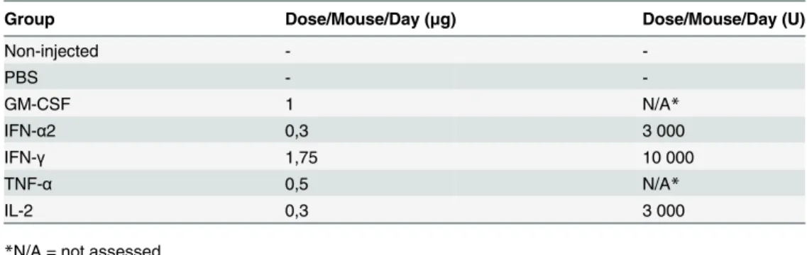
4–7-week-old C57BL/6 immunocompetent female mice were implanted subcutaneously with 2.5 x 10⁵B16.OVA cells in 50μl RPMI, 0% FBS, in the right flank. Ten days post tumor implantation, mice were divided into groups and tumors (~3 mm minimum diameter) were left non-injected or injected with either 50μl phosphate buffered saline (PBS) or carrier-free recombinant murine cytokines in 50μl PBS (Table 1). Mice received 10 doses of recombinant cytokines intratumorally in total (S1 Fig). On the first day of the intratumoral treatment, the mice were also adoptively transferred with 2 x 10⁶CD8a-enriched and expanded splenocytes from OT-I mice. The OT-I cells were administered into intraperitoneal cavity in 100μl RPMI, 0% FBS, as it has been shown that intraperitoneal injections of OT-I mimic the kinetics of intravenous delivery [19]. Tumor growth of mice was monitored every 2–3 days by using elec-tronic calipers and volume was calculated as 0.52 x length x width2. Mice were examined every day and euthanized before the designated experimental endpoint of day 14 when the tumor became ulcerated or when one of two diameters reached 18 mm.
Table 1. Doses of recombinant cytokines.
Group Dose/Mouse/Day (μg) Dose/Mouse/Day (U)
Non-injected -
-PBS -
-GM-CSF 1 N/A*
IFN-α2 0,3 3 000
IFN-γ 1,75 10 000
TNF-α 0,5 N/A*
IL-2 0,3 3 000
*N/A = not assessed
Tissue processing for Flex Set analysis
Mice were euthanized and 10–100 mg of tumor tissue was frozen in 2 ml microcentrifuge tubes on dry ice and stored at -80°C. Prior to processing ice-cold PBS supplemented with 0.1% BSA and protease inhibitor cocktail (Sigma-Aldrich, St. Louis, MO) was added and the tumor pieces were homogenized by Tissue Master 125 rotor (Omni International, Kennesaw, GA). Tumor homogenate was spun at 2000 RCF 10 min +4°C and the supernatant was analyzed with CBA Flex Set cytokine beads (BD, Franklin Lakes, NJ) on BD Accuri C6 flow cytometer with FCAP Array software (BD) per manufacturer’s instructions.
Tissue processing for flow cytometry
Mice were euthanized and tumors were processed for flow cytometric analysis by pushing the tumor tissue through a 70μm sterile strainer using a 1 ml syringe plunger. RPMI 1640 supple-mented with 10% FBS, 20 mM L-Glutamine, 1x Pen/Strep was added and the single-cell solu-tion was cultured at 37°C and 5% CO2for 24 hours, after which cells were either analyzed
directly by flow cytometry or frozen at -80°C for later analysis.
Flow cytometry
Tumor cell samples were stained according to manufacturer instructions with respective com-mercial antibodies validated by the supplier (Table 2). The labeled cells were centrifuged at 500 RCF for 5 min and the pellet was resuspended in Flow Cytometry Staining Buffer
(eBioscience). For T-cell activation assay tumor samples were treated with intracellular protein transport inhibitor brefeldin A (eBioscience) and Cell Stimulation Cocktail containing PMA and ionomycin (eBioscience) at 37°C and 5% CO2for 6 hours. After stimulation the cells were
stained for surface markers, fixed and permeabilized prior to intracellular staining. All cell sam-ples were analyzed on BD Accuri C6 flow cytometer with CFlow Sampler software (BD) count-ing at least 100000 events per sample.
Statistical analysis
Statistics was performed with GraphPad Prism 6 (GraphPad Software Inc., San Diego, CA) and SPSS version 21 (SPSS IBM, New York, NY). One-way ANOVA followed by Tukey’s post-hoc test was used for comparison of multiple groups. Log-transformed tumor volumes were ana-lyzed by repeated measures ANOVA. Differences were considered statistically significant when P values were<0.05.
Results
Anti-tumor efficacy is achieved by intratumoral administration of IFN-
α
2,
IFN-
γ
, TNF-
α
and IL-2 but not GM-CSF
In accordance with typical clinical outcomes in melanoma T-cell therapy in the absence of preconditioning [20], cells alone resulted in poor growth control of established tumors (Fig 1A; non-injected and PBS-injected groups). Instead, administration of intratumoral IFN-α2, IFN-γ
and IL-2 resulted in superior treatment efficacy over control groups (Fig 1Aand Fig A inS2 Fig). In addition, TNF-αwas found effective in curing 80% of mice by day 14 post-transfer (Fig B inS2 Fig). As an interesting side note, daily injections of phosphate buffered saline also added to the anti-tumor effect of OT-I T-cell therapy, supporting previous notions that any damage to the tumor can result in immune response [21,22]. While many of the cytokines injected improved the anti-tumor effect of OT-I therapy, GM-CSF resulted in growth-Table 2. List of antibodies used.
Antibody Monoclonal or
polyclonal
Host species Commercial supplier
Catalogue number
Concentration (per sample)
CD8b-FITC monoclonal rat eBioscience 11-0083-85 0,5μg
Foxp3-APC monoclonal rat eBioscience 17-5773-82 1μg
CD25-PE monoclonal rat eBioscience 12-0251-82 0,125μg
CD19-PE monoclonal rat eBioscience 12-0193-82 0,125μg
H-2Kb-PE monoclonal mouse eBioscience 12-5958-82 0,25μg
H-2Kb-SIINFEKL-PeCy7 monoclonal mouse eBioscience 25-5743-80 0,125μg
CD8a-APC monoclonal rat eBioscience 17-0081-82 0,125μg
CD45-APC monoclonal rat eBioscience 17-0451-82 0,125μg
CTLA-4-PE monoclonal armenian
hamster
eBioscience 12-1522-81 0,25μg
PD-1-PeCy7 monoclonal armenian
hamster
eBioscience 25-9985-80 1μg
NK1.1-FITC monoclonal mouse eBioscience 11-5941-81 0,5μg
F4/80-APC monoclonal rat eBioscience 17-4801-82 0,5μg
CD44-FITC monoclonal rat eBioscience 11-0441-81 0,5μg
CD62L-PE monoclonal rat eBioscience 12-0621-81 0,125μg
CD69-PeCy7 monoclonal armenian
hamster
eBioscience 25-0691-82 0,5μg
IFN-γ-APC monoclonal rat eBioscience 17-7311-82 0,125μg
CD4-PerCP.Cy5.5 monoclonal rat BD 550954 0,4μg
CD3-PeCy7 monoclonal rat BD 560591 0,2μg
CD11c-FITC monoclonal armenian
hamster
BD 553801 0,5μg
Gr-1-FITC monoclonal rat BD 553127 0,5μg
Ly6G-PE monoclonal rat BD 551461 0,3μg
CD11b-PerCP-Cy5.5 monoclonal rat BD 550993 0,3μg
Ly6C-APC monoclonal rat BD 560595 0,3μg
CD86-PE monoclonal rat BD 553692 0,3μg
CD3-APC monoclonal armenian
hamster
BD 553066 0,3μg
CCR7-PerCP-Cy5.5 monoclonal rat BD 560812 0,5μg
CD206-FITC monoclonal rat Biolegend 141704 0,125μg
SIINFEKL-pentamer-APC
ND* ND* Proimmune F093-4B 10μl
*ND = not determined
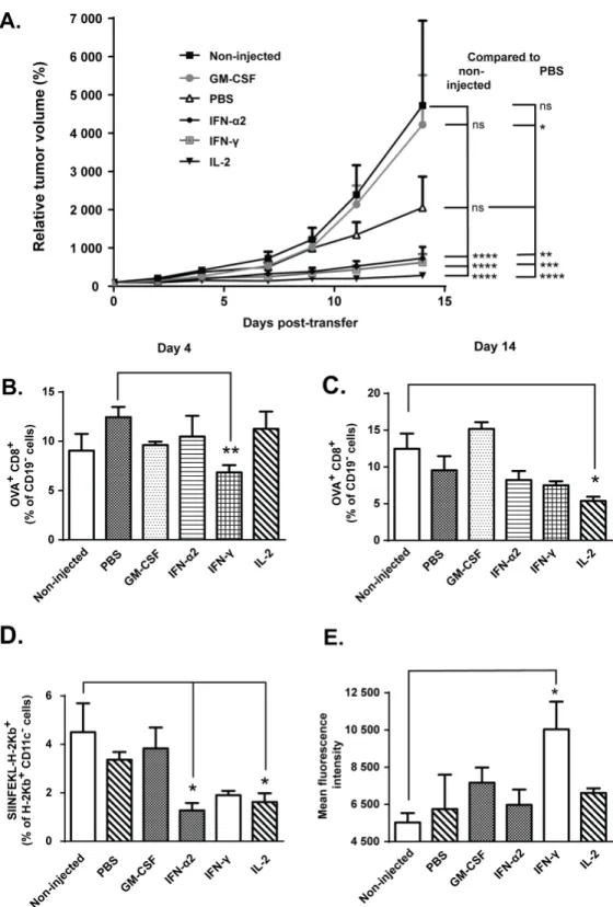
Fig 1. IFN-α2, IFN-γand IL-2 augment anti-tumor efficacy but do not increase tumor-accumulation of
transferred cells.Mice bearing syngeneic B16.OVA tumors were adoptively transferred with 2x10⁶CD8a+
OVA-stimulatory effect when compared to PBS injected control tumors (Fig 1A), as described earlier by Obermueller et al [23].
Levels of ovalbumin-specific CD8+ T-cells do not correlate with
treatment efficacy
To assess the degree of tumor-antigen specific T-cell infiltration in tumors as a possible expla-nation for the observed anti-tumor effects with the cytokine combiexpla-nations, total CD8+ T-cells specific for MHC-I-loaded chicken ovalbumin SIINFEKL peptide (endogenous + transferred OT-I cells) were quantified by flow cytometry on days 4 and 14 post-transfer. Interestingly, the levels of tumor-infiltrating pentamer-positive CD8+ T-cells were lower in IFN-γ–treated mice
compared to PBS-injected controls on day 4 post-transfer and in IL-2-injected mice compared to non-injected controls on day 14 post-transfer (Fig 1B–1C). At the same time, however, the level of putative non-DC target (tumor) cells, identified as CD11c-cells presenting the OVA-derived peptide SIINFEKL in context of MHC class I, was significantly lower in IFN-α2 and IL-2-injected tumors compared to non-injected controls on day 14 post-transfer (Fig 1D). It is possible that any target cells presenting OVA-peptides on their H-2Kb molecules, including melanoma cells, were efficiently killed by tumor-reactive cytotoxic T-cells earlier on, thus lead-ing to antigen-loss variants in the tumor cell population through immunoeditlead-ing [24]. In order to gain more insight into CD8+ T-cell-mediated tumor control, we assessed overall expression of MHC class I, depicted as H-2Kb+cells by flow cytometry on day 14 post-transfer. MHC-I expression was enhanced by intratumoral IFN-γbut not by any other cytokines (Fig 1E). Thus, MHC-I expression in the tumors did not directly predict anti-tumor efficacy with the cytokine combinations.
Combination of local cytokine injections and adoptive T-cell transfer is
associated with changes in both pro- and anti-inflammatory cytokine
levels in tumors
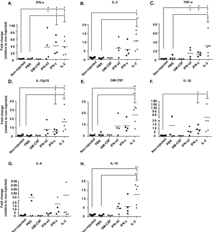
In order to gain further insight into the possible anti-tumor mechanisms of adoptive CD8+ T-cell transfer combined with intratumoral cytokine injections, which seemingly did not involve increased trafficking of CD8+ T-cells or increased intratumoral MHC-I expression (Fig 1B– 1E), we analyzed tumors at the study endpoint for several central immunomodulatory cyto-kines: T-cell growth factor IL-2, pro-inflammatory cytokines IFN-γ, TNF-αand heterodimeric IL-12p70, all of which are associated with T-cell activation and Th1 polarization; anti-inflam-matory cytokine IL-10, secreted by regulatory T-cells and Th2 cells; innate monocyte/NK immune activators GM-CSF and IL-1β; as well as the“acute phase”cytokine IL-6 [25,26]. Remarkably, intratumoral injection with IFN-γand IL2 increased the secretion of all of these cytokines compared to non-injected and PBS-injected tumors, with no clear delineation toward either pro-inflammatory (Fig 2A–2D) or anti-inflammatory profiles (Fig 2E–2H). While it was not possible to separate the relative levels of the injected recombinant cytokines from the endogenously induced cytokines, the levels of the other analyzed cytokines were still increased, confirming that local cytokine treatment modulates the overall cytokine balance in the tumor. This secretion of cytokines in the tumor may be an indicator of heightened immune
derived peptide SIINFEKL and (e) mean fluorescence intensity (MFI) of mouse MHC class I H-2kb from tumor samples was assessed by flow cytometry on day 14 post-transfer (n = 5). Data presented as mean±SEM.
*P0.05,**P0.01,***P0.001,****P0.0001 by repeated measures ANOVA (a) or one-way ANOVA followed by Tukey’s post-hoc test (b-e).
Fig 2. Recombinant cytokines induce intratumoral, endogenous secretion of cytokines associated with immune cell activation.B16.OVA-bearing mice were treated intraperitoneally with 2x10⁶CD8a+enriched OT-I lymphocytes and treated intratumorally with either PBS or recombinant cytokine (in PBS)
or left non-injected. Levels of (a) IFN-γ, (b) IL-2, (c) TNF-α, (d) IL-12p70, (e) GM-CSF, (f) IL-1β, (g) IL-6 and (h) IL-10 from tumor homogenates were measured with CBA Flex sets on day 14 post-transfer (n = 3–5). Horizontal lines represent median values.*P0.05 and**P0.01 byone-way ANOVA followed by Tukey’s post-hoc test.
detection and destruction of the established tumor [27–29], as the control groups show very low levels of endogenous cytokines and subsequently poor tumor growth control. Interestingly, daily injections of GM-CSF did not result in measurable increase of total GM-CSF over back-ground in the tumors at the sampled endpoint (Fig 2E), implying rapid turnover of the cytokine.
Local administration of IL-2 decreases the total number of CD4+ TILs but
induces CD4+ T-cell polarization into Tregs
Since we saw no clear skewing of the intratumoral cytokine balance towards tumor rejection, we proceeded by analyzing the phenotypes of immune cells present in the tumors. On day 14 post-transfer IFN-γ- and IL-2-treated tumors contained more CD45+leukocytes than control tumors (Fig A inS3 Fig), whereas the total amount of CD3+T-lymphocytes did not signifi-cantly differ between the control and treatment groups (Fig B inS3 Fig). Local administration of IL-2 decreased the levels of tumor-infiltrating CD4+T-cells compared to non-injected con-trol mice (Fig C inS3 Fig). In addition, PBS and IL-2 injections resulted in CD4+T-cell polari-zation toward regulatory phenotype (Foxp3+CD25+CD4+) within the studied T-cell
population (Fig D inS3 Fig), which for IL-2 was expected [30].
Intratumoral IFNa2 and IL-2 increase the accumulation of immune cells
capable of stimulating CD8+ T-cells
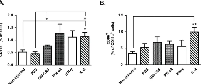
T-cell activity is regulated by antigen-presenting cells (APCs), which can either promote activa-tion of tumor-specific T-cells or induce antigen-specific peripheral tolerance in absence of co-stimulatory signals such as CD86 [31]. Following intratumoral immunomodulation with GM-CSF and IL-2, the total levels of dendritic cells were increased over control groups (Fig 3A). Analysis of the maturation status of these cells revealed that onlyin situadministration of IL-2 resulted in higher proportion of intratumoral CD11c+CD86+DCs compared to
Fig 3. Intra-tumor accumulation of antigen-presenting cells (APCs) is increased by GM-CSF and IL-2.B16.OVA bearing mice were treated with adoptive transfer of 2x10⁶CD8a+enriched OT-I lymphocytes intraperitoneally and tumors were either injected with PBS or recombinant cytokine in PBS or
left non-injected (n = 5). (a) Levels of CD11c+dendritic cells and (b) proportion of dendritic cells expressing maturation marker CD86 on cell surface were analyzed on day 14 post-transfer from tumors. Data presented as mean±SEM.*P0.05 and**P0.01 by one-way ANOVA followed by Tukey’s post-hoc test.
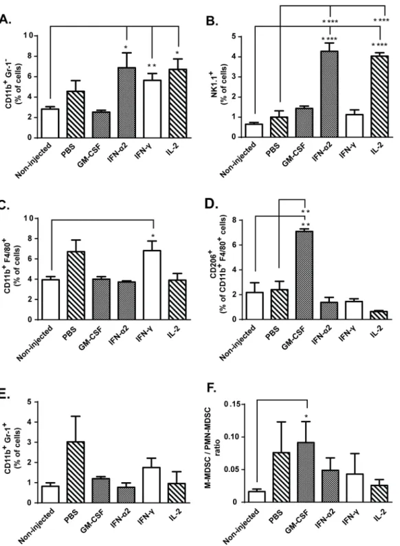
non-injected control group (Fig 3B). In addition, the number of tumor-infiltrating natural killer (NK) cells was increased in IFN-α2 and IL-2 treated mice compared to non-injected and PBS controls (Fig 4B). Thus, the greater proportion of mature DCs and NK cells following IL-2 administration compared to GM-CSF administration could have contributed to the superior anti-tumor effects of IL-2.
In situ administration of recombinant GM-CSF results in tumor-infiltrating
myeloid cells polarization into immunosuppressive phenotype
In order to assess influence of the cytokine treatments on tumor composition as a possible explanation for the observed anti-tumor effects, we characterized the myeloid cell populations in the tumors. Total number of tumor-infiltrating CD11b+Gr-1-myeloid cells was increased in IFN-α2, IFN-γand IL-2 treated groups compared to non-injected control mice (Fig 4A). As NK-cells and F4/80+ macrophages account for most of the CD11b+cells in these treatment groups (Fig 4B–4C), it is possible that the quality rather than quantity of tumor-infiltrating myeloid cells determines if tumors are rendered sensitive to killing by cytotoxic T-cells. Further analysis of macrophage polarization revealed that intratumoral GM-CSF injection skewed tumor-infiltrating macrophages towards immunosuppressive M2 phenotype, characterized by CD206 expression (Fig 4D). By contrast, as IFN-γ-treated tumors contained high levels of endogenous cytokines TNF-α, IL-12p70 and IL-1β(Fig 2C,2D, 2F and 2G) and the tumor-infiltrating macrophages did not express putative M2 marker CD206 (Fig 4D), we find it likely that IFN-γinstead skewed the macrophages towards M1 phenotype [32].
In addition to tumor-associated macrophages (TAM), intratumoral administration of exog-enous GM-CSF also resulted in increased ratio of monocytic (M-MDSC, CD11b+Gr1 +-Ly6G-Ly6Chigh) over polymorphonuclear (PMN-MDSC, CD11b+Gr1+Ly6G+Ly6Clow) myeloid-derived suppressor cells (Fig 4E–4F). Although both are part of the immune popula-tion suppressing T-cell funcpopula-tions, in some cases M-MDSCs have been considered more immu-nosuppressive than PMN-MDSCs [33].
Immunomodulation through IFN-
α
2, IFN-
γ
and IL-2 results in changes in
tumor-infiltrating T-cell phenotypes
As we did not see evidence of increased tumor-accumulation of transferred OVA-specific OT-I cells following cytokine treatments, we decided to analyze the phenotype and activation status of tumor-infiltrating CD8+ T-cells. Interestingly, tumors treated with GM-CSF,
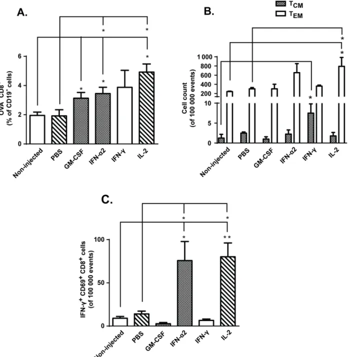
IFN-α2 and IL-2 contained more endogenous, OVA-CD8+T-cells than non-injected tumors (Fig 5A). Further analysis revealed that some of these CD8+TILs were targeting endogenous melanoma-associated antigens TRP-2 and gp100 (Figs E and F inS3 Fig), suggesting reper-toire expansion following adoptive T-cell transfer [34,35]. In addition, local immunomodula-tion with IFN-γresulted in increased levels of CD44highCD62LhighCCR7highcentral memory T-cells (TCM), whereas intratumoral IL-2 treatment led to increase in CD44high CD62L-lowCCR7loweffector memory T-cells (T
EM) (Fig 5B). As previously reported [36], IL-2
pro-motes T-cell differentiation to TEMcells, which have reduced proliferative capacity but can
Fig 4. Intratumoral myeloid cell subsets are influenced by local cytokine therapy.Mice bearing subcutaneous B16.OVA tumors received
intraperitoneal transfer of 2x10⁶CD8a+enriched OT-I lymphocytes and intratumoral injections of either PBS or recombinant cytokine in PBS (n = 5). Levels of
tumor-infiltrating (a) CD11b+myeloid cells, (b) NK1.1+natural killer cells, (c) CD11b+F4/80+ macrophages, (d) suppressive M2 macrophages (characterized by surface expression of CD206), (e) CD11b+Gr-1+myeloid-derived suppressor cells (MDSC) and (f) ratio of monocytic (M) to polymorphonuclear (PMN)
MDSCs were assessed from tumors on day 14 post-transfer by flow cytometry. Data presented as mean±SEM.*P0.05,**P0.01,***P0.001 and
****P0.0001 byone-way ANOVA followed by Tukey’s post-hoc test.
Downregulation of anergy markers on CD8+ TILs is achieved following
IFN-
α
2, IFN-
γ
and IL-2 treatment
As TIL hypofunction has been associated with upregulation of surface inhibitory receptors [8], we wanted to study whether observed changes in tumor microenvironment also affect the Fig 5. IFN-α2, IFN-γand IL-2 treatment leads to changes in CD8+ TIL phenotypes.Mice harboring subcutaneous B16.OVA tumors were treated intraperitoneally with 2x10⁶CD8a+enriched OT-I lymphocytes and injected intratumorally with either PBS or recombinant cytokine in PBS or left non-injected
(n = 5). (a) Levels of tumor-infiltrating endogenous (non-OVA) CD8+T-cells and (b) count of central memory (T
CM) and effector memory (TEM) T-cells were
assessed from tumors on day 14 post-transfer by flow cytometry. (c) Activation status of tumor-infiltrating CD8+ T-cells was evaluated on day 14 by expression of CD69 and IFN-γfollowing PMA/Ionomycin stimulationex vivo. Data presented as mean±SEM.*P0.05 and**P0.01 by one-way ANOVA followed by Tukey’s post-hoc test.
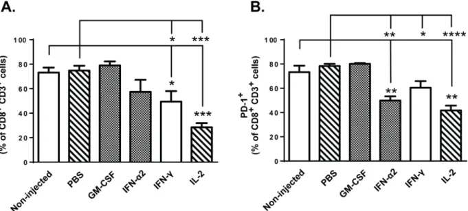
expression of anergy/exhaustion markers CTLA-4 and PD-1 on CD8+ T-cells. Flow cytometric analysis of tumor-infiltrating lymphocytes revealed that administration of IFN-γand IL-2 downregulated the expression of CTLA-4 on CD3+ CD8+ TILs compared to control groups on day 14 post-transfer (Fig 6A). Furthermore, significant reduction in expression of PD-1 on these tumor-infiltrating T-cells was observed following IFN-α2, IFN-γand IL-2 treatment on day 14 (Fig 6B). These effects seemed to develop over time, since only IL-2 could downregulate CTLA-4 expression already on day 4 post-transfer (Fig A inS4 Fig) and IFN-γtreated tumors contained high levels of PD-1+ TILs at the early time point (Fig B inS4 Fig). Notably, the high expression of both CTLA-4 and PD-1 on CD8+ T-cells infiltrating the GM-CSF—treated tumors on both time points suggested that the tumor microenvironment remained highly immunosuppressive following cytokine treatment and thus implicates that administration of recombinant GM-CSF intratumorally may not be optimal for T-cell function. Taken together, our results indicate that local tumor treatment with carefully selected immunomodulatory cytokine (such as IFN-α2, IFN-γand IL-2) can result in favorable alteration of tumor microen-vironment and thus affect T-cell activity within the tumor.
Discussion
Although immunotherapies based on gene-engineered T-cells have shown impressive clinical success in the treatment of hematological cancers such as chronic lymphocytic leukemia (CLL) and acute lymphoblastic leukemia (ALL) [6,7], the application into solid tumor types has remained difficult due to several obstacles including functional impairment of T-cell function following infiltration into tumor [4,5,8]. This T-cell hypofunction is induced by the strongly immunosuppressive tumor microenvironment, which is characterized by the lack of immune cells capable of activating anti-tumor effector cells and/or by the excess of immunosuppressive cell populations. In accord with clinical observations, our experiments showed that ACT-medi-ated anti-tumor immune responses, in the absence of preconditioning, are not potent enough Fig 6. Expression of anergy markers on CD8+ TILs are downregulated following IFN-α2, IFN-γand IL-2 treatments.Mice bearing subcutaneous B16. OVA tumors were injected with 2x10⁶CD8a+enriched OT-I lymphocytes into peritoneal cavity and beginning on the same day, tumors were injected with
either PBS or recombinant cytokine in PBS or left non-injected (n = 5). Proportion of CD3+CD8+TILs expressing surface anergy markers (a) CTLA-4 and (b) PD-1 was analyzed by flow cytometry on day 14 post-transfer. Data presented as mean±SEM.*P0.05,**P0.01,***P0.001,****P0.0001 by one-way ANOVA followed by Tukey’s post-hoc test.
to control tumor growth even in mice, as B16.OVA tumor-bearing mice treated with T-cells only displayed poor growth inhibition (Fig 1A). It has been previously suggested that resistance of solid tumors to adoptive T-cell therapy is the result of an imbalance between the number and/or activation status of tumor-infiltrating effector and suppressor immune cells [4]. Our aim was to manipulate this balance in favor of anti-tumor responses by using immunomodula-tory cytokines administered intratumorally. Notably, injections of IFN-α2, IFN-γ, TNF-αand IL-2 markedly improved the anti-tumor effect of ACT, while treatment with GM-CSF resulted in stimulation of tumor growth (Fig 1A,S2 Fig). In addition, differences in efficacy and immune cell composition of non-injected and PBS-injected control tumors also revealed that mere physical manipulation of the tumor by a needle can affect the tumor microenvironment and cause inflammation that results in minor (but not statistically significant) inhibition of rel-ative tumor growth (Fig 1A and 1B,S3 Fig). This observation presents an important detail that should be taken into consideration in the course of preclinical testing of cancer immunothera-pies even if it lacks clinical relevance.
In our hands, four of the five cytokines studied showed anti-tumor efficacy and one of the cytokine candidates, TNF-α, was so potent when given in combination with T-cells that some tumors disappeared completely. This prevented us from analyzing the immune cell content of the treated tumors, but we propose that the overall anti-tumor effect seen in this group was due to the direct anti-tumor effects of TNF-αon one hand [13] and immunological synergy with T-cell therapy on the other. A likely scenario was that TNF-αaffected the anti-tumor immune response indirectly via inducing killing of tumor cells and promoting destruction of tumor-associated vasculature [13]. However, we could not investigate this further since advanced necrosis of tumors (day 4 post-transfer) and the cures (day 14 post-transfer) resulted in com-plete lack of viable tumor material for flow cytometric analysis.
IL-2 is frequently used concomitantly with adoptive T-cell therapies and intratumoral administration of IL-2 has previously been shown to induce infiltration of CD4+, CD8+T cells and APCs in preclinical models [37,38]. Our results suggest that in addition to enhanced tumor-infiltration of these immune cells, intratumoral rIL-2 treatment also augments the acti-vation of CD11c+dendritic cells and CD8+TILs (Figs3Band5C). This may indicate that tumor-induced tolerance was partly lifted as tumor-induced immunosuppression usually pre-vents APC-mediated T-cell activation [31]. This conclusion is also supported by the observa-tion that intratumoral administraobserva-tion of IL-2 had the most prominent impact on the
downregulation of T-cell anergy markers CTLA-4 and PD-1 (Fig 6A and 6B, Fig A inS4 Fig), both of which contribute strongly to T-cell hypofunction [39]. On the other hand, IL-2 increased overall tumor cytokine secretion with no clear bias toward pro-inflammatory or anti-tumor phenotype (Fig 2A–2H), and IL-2 injections increased the relative amount of Foxp3+ CD25+CD4+regulatory T-cells in the tumors (Fig D inS3 Fig), suggesting simultaneous induction of both anti-tumor and immunosuppressive pathways. Specifically, stimulation of anti-tumor CD8+ T-cells, including the OT-I graft and endogenous TILs, is clearly a desirable effect, while untoward effects include the aforementioned stimulation of Tregs. This problem could be overcome by using variants of IL-2 which display reduced stimulation of CD25 while retaining the features sought [40].
concentrations induces immunosuppression [43]. This may implicate that the dose, timing and exposure time of immunomodulatory cytokines on the tumor microenvironment is of impor-tance in context of adoptive T-cell therapy, especially in the case of GM-CSF which has shown promising signs of anti-tumor immune stimulation when used in optimal doses [44]. Our results with recombinant murine GM-CSF also revealed that while the total level of tumor-infiltrating DCs and CD8+T-cells was increased, neither DC maturation nor T-cell activation was enhanced (Figs3A, 3B,5A and 5C). GM-CSF has been successfully used to augment anti-tumor immune responses [44,45], but we found that it can induce immune tolerance rather than activation, as reported by Bronte et al [43]. Since GM-CSF employed by several immunotherapeutic
approaches such as cancer vaccines and oncolytic viruses [46–48], the aforementioned observa-tions provide important insights into immunobiology of GM-CSF when used as a bolus dose; as opposed to protracted lower-level production locally in a tumor by a viral vector [48].
One factor that might explain the lack of complete eradication of tumors even after combi-nation therapy is the low level of MHC class I molecules expressed on B16 tumor cells, which might prevent tumor cell killing by TILs (Fig 1E). MHC molecules in mice, known as HLA (Human Leukocyte Antigen) in humans, are required for presentation of tumor epitopes to the T-cell receptor [49]. HLA has been reported to be downregulated in several cancer types [50], which results in immune escape through the inability of anti-tumor T-cells to recognize their target. This is of special concern in cell therapies based on TILs or tumor-antigen specific TCR. However, these issues can be circumvented by the use of CAR T-cell therapies [51] which do not rely of MHC/HLA. In our approach, tumor-treatment with interferon-γresulted in increased expression of MHC class I on tumor cells over non-injected tumors (Fig 1E). Inter-estingly, we also observed lower levels of activated CD8+ TILs in the IFN-γ-treated mice com-pared to IFN-α2 mice (Fig 5C), whereas treatment efficacy remained identical in these groups (Fig 1A), suggesting that enhanced tumor cell recognition can compensate for the scarcity of functional T-cells. Finally, IFN-α2 and IL-2 also induced tumor accumulation of NK cells (Fig 4B), which are capable of killing tumor cells expressing low levels of MHC class I [52] and thus compensate for the poor tumor recognition of T-cells. Thus, MHC-I levels are not automati-cally predictive of therapeutic efficacy even with approaches relying on it for efficacy, when immunostimulatory cytokines are employed. It is, however, possible that qualitative differences in immune responses between different cytokines masked the cytokine-specific role of MHC-I expression in anti-tumor efficacy, i.e. whereas therapeutic efficacy with IFN-γmay depend on MHC-I, efficacy with IFN-α2 and IL-2 might not.
Many established solid tumors are infiltrated by diverse leukocyte subsets including both myeloid- and lymphoid-lineage cells, and the tumor microenvironment plays a major role in delineation of the phenotypic profile and activation status of these cells [53,54]. More impor-tantly, the balance between anti-tumor and pro-tumor immune cells may determine the out-come of cancer immunotherapy [54] and thus encourages closer scrutiny. In our hands,
IFN-α2 and IL-2 yielded the best results in terms of T-cell activation versus anergy (Figs5Cand6A– 6B), while IFN-γtreatment decreased expression of exhaustion markers on TILs and increased the number of intratumoral central memory T-cells (Figs5Band6A–6B). The majority of tumor-infiltrating myeloid cells identified in these three treatment groups were NK cells and possibly M1 macrophages (Fig 4B–4C), both of which may facilitate T-cell functions [55,56]. Thus it can be argued that local administration of IFN-α2, IFN-γor IL-2 favorably alters the myeloid-lymphocyte balance and makes the tumor less resistant to immune cell attack.
Moreover, especially viral vectors such as adenoviruses can yield high concentrations locally for protracted periods, which low concomitant concentrations systemically [57]. A further improvement of this approach would be the use of adenoviral vectors that are replication-com-petent only in tumor cells; one of the first phases of replication of the virus is replication of the genome, resulting in 10000-fold amplification of the transgene expression cassette. If the virus is designed in a way linking transgene expression to virus replication, viral expression of immu-nomodulatory cytokinesin situcould potentially offer a safer and more tumor-selective option than recombinant cytokines, as transgene expression would not occur in non-transcomple-menting (= non-tumor) cells [57].
In summary, incorporation of immunomodulatory cytokines IFN-α2, IFN-γ, TNF-αand IL-2 into treatment regimen can alter the tumor microenvironment in favor of T-cell function, whereas in situ injections of GM-CSF can induce and sustain highly immunosuppressive immune cell populations within the tumor, thus leading to poor tumor growth control. These results have important implications in several experimental immunotherapies, and provide a strong rationale for adaptation of direct or vectored cytokine administration into T-cell therapy regimens.
Supporting Information
S1 Fig. Treatment schedule.Female C57BL/6 mice were implanted with 2,5x10⁵B16.OVA cells subcutaneously into the right flank (1 tumor/mouse). 10 days post-implantation mice were divided into groups and injected intraperitoneally with 2x10⁶polyclonally activated CD8a+-enriched OT-I lymphocytes. Beginning on the same day, tumors were injected with PBS or with one of the recombinant murine cytokines diluted in PBS. One control group of mice received only adoptive transfer of OT-I cells and the tumors were left non-injected to avoid immune responses generated by physical (needle) manipulation of the tumor microenvironment. Intra-tumoral injections were continued for 5 consecutive days per week. A set of mice were sacrificed (SAC) and organs were harvested for analysis on days 4 and 14 post-transfer.
(TIF)
S2 Fig. Intratumoral administration of TNF-αcombined with adoptive transfer of OT-I
cells results in anti-tumor efficacy.Mice bearing B16.OVA flank tumors were adoptively transferred with 2x10⁶CD8a+enriched OT-I lymphocytes intraperitoneally and tumors were either not injected or injected with PBS or recombinant cytokines in PBS (n = 10). Tumor growth was monitored every 2–3 days with an electronic caliper. (Fig A) Absolute tumor vol-umes (mm3) of all groups and (Fig B) relative tumor volumes (% of day 0 volume) of TNF-α
treatment group. Data presented as mean ± SEM.
P0.0001 by repeated measures ANOVA.
(TIF)
S3 Fig. Lymphocyte subsets in the tumors following cytokine treatment.Mice with B16. OVA flank tumors were treated with adoptive transfer of 2x10⁶CD8a+enriched OT-I lympho-cytes intraperitoneally and with 50μl PBS or recombinant cytokine in PBS intratumorally (n =-5). Levels of tumor-infiltrating (Fig A) CD45+leukocytes, (Fig B) CD3+T-lymphocytes, (Fig C) CD4+T-lymphocytes and (Fig D) proportion of regulatory T-cells of CD4+T-cells were assessed by flow cytometry on day 14 post-transfer. (Figs E–F) Amounts of endogenous CD8 + TILs targeting melanoma-associated antigens TRP-2 and gp100 were quantified on day 14 post-transfer by pentamer staining and flow cytometry. Data presented as mean ± SEM. P
0.05,P
S4 Fig. Expression of anergy markers on CD8+ TILs on day 4 post-transfer. B16.OVA-bear-ing mice were injected with 2x10⁶CD8a+enriched OT-I lymphocytes intraperitoneally and beginning on the same day, tumors were injected with either PBS or recombinant cytokine in PBS or left non-injected (n = 5). Proportion of CD3+CD8+TILs expressing surface anergy markers (Fig A) CTLA-4 and (Fig B) PD-1 was analyzed by flow cytometry on day 4 post-transfer. Data presented as mean ± SEM.
P0.05,
P0.01 and
P0.001 by one-way ANOVA followed by Tukey’s post-hoc test.
(TIF)
S5 Fig. Heat map summarizing the differenct aspects of immunostimulatory cytokines in the modulation of tumor microenvironment.Decrease (red), increase (green) or no change (gray) in activation status or proportion of different cell populations following cytokine treat-ment compared to non-injected tumors.
(TIF)
Acknowledgments
We thank Saila Pesonen, Simona Bramante, Mikko Siurala, Noora Rouvinen-Lagerström and Minna Oksanen (all from University of Helsinki, Finland) for excellent expert assistance.
Author Contributions
Conceived and designed the experiments: ST MV AH. Performed the experiments: ST SK MM SG SP. Analyzed the data: ST MV. Contributed reagents/materials/analysis tools: AK AH. Wrote the paper: ST MV AH.
References
1. Rosenberg SA, Yang JC, Sherry RM, Kammula US, Hughes MS, Phan GQ, et al. Durable complete responses in heavily pretreated patients with metastatic melanoma using T-cell transfer immunother-apy. Clin Cancer Res 2011 Jul 1; 17(13):4550–4557. doi:10.1158/1078-0432.CCR-11-0116PMID: 21498393
2. Dudley ME, Yang JC, Sherry R, Hughes MS, Royal R, Kammula U, et al. Adoptive cell therapy for patients with metastatic melanoma: evaluation of intensive myeloablative chemoradiation preparative regimens. J Clin Oncol 2008 Nov 10; 26(32):5233–5239. doi:10.1200/JCO.2008.16.5449PMID: 18809613
3. Dudley ME, Wunderlich JR, Yang JC, Sherry RM, Topalian SL, Restifo NP, et al. Adoptive cell transfer therapy following non-myeloablative but lymphodepleting chemotherapy for the treatment of patients with refractory metastatic melanoma. J Clin Oncol 2005 Apr 1; 23(10):2346–2357. PMID:15800326
4. Kunert A, Straetemans T, Govers C, Lamers C, Mathijssen R, Sleijfer S, et al. TCR-Engineered T Cells Meet New Challenges to Treat Solid Tumors: Choice of Antigen, T Cell Fitness, and Sensitization of Tumor Milieu. Front Immunol 2013 Nov 8; 4:363. doi:10.3389/fimmu.2013.00363PMID:24265631
5. Gilham DE, Debets R, Pule M, Hawkins RE, Abken H. CAR-T cells and solid tumors: tuning T cells to challenge an inveterate foe. Trends Mol Med 2012 Jul; 18(7):377–384. doi:10.1016/j.molmed.2012.04. 009PMID:22613370
6. Kalos M, Levine BL, Porter DL, Katz S, Grupp SA, Bagg A, et al. T cells with chimeric antigen receptors have potent antitumor effects and can establish memory in patients with advanced leukemia. Sci Transl Med 2011 Aug 10; 3(95):95ra73. doi:10.1126/scitranslmed.3002842PMID:21832238
7. Maude SL, Frey N, Shaw PA, Aplenc R, Barrett DM, Bunin NJ, et al. Chimeric Antigen Receptor T Cells for Sustained Remissions in Leukemia. N Engl J Med 2014; 371(16):1507–17. doi:10.1056/
NEJMoa1407222PMID:25317870
9. Vacchelli E, Eggermont A, Fridman WH, Galon J, Zitvogel L, Kroemer G, et al. Trial Watch: Immunosti-mulatory cytokines. Oncoimmunology 2013 Jul 1; 2(7):e24850. PMID:24073369
10. Metcalf D. The colony-stimulating factors and cancer. Nat Rev Cancer 2010 Jun; 10(6):425–434. doi: 10.1038/nrc2843PMID:20495576
11. Biron CA. Interferons alpha and beta as immune regulators—a new look Immunity. 2001 Jun; 14 (6):661–664.
12. Vacchelli E, Galluzzi L, Eggermont A, Galon J, Tartour E, Zitvogel L, et al. Trial Watch: Immunostimula-tory cytokines. Oncoimmunology 2012 Jul 1; 1(4):493–506. PMID:22754768
13. van Horssen R, Ten Hagen TL, Eggermont AM. TNF-alpha in cancer treatment: molecular insights, antitumor effects, and clinical utility. Oncologist 2006 Apr; 11(4):397–408. PMID:16614236
14. Lotze MT, Grimm EA, Mazumder A, Strausser JL, Rosenberg SA. Lysis of fresh and cultured autolo-gous tumor by human lymphocytes cultured in T-cell growth factor. Cancer Res 1981 Nov; 41(11 Pt 1):4420–4425. PMID:6975652
15. Dillman RO, Barth NM, VanderMolen LA, Mahdavi K, McClure SE. Should high-dose interleukin-2 still be the preferred treatment for patients with metastatic melanoma? Cancer Biother Radiopharm 2012 Aug; 27(6):337–343. doi:10.1089/cbr.2012.1220PMID:22804456
16. Lee S, Margolin K. Cytokines in cancer immunotherapy. Cancers (Basel) 2011 Oct 13; 3(4):3856–
3893.
17. Linardakis E, Bateman A, Phan V, Ahmed A, Gough M, Olivier K, et al. Enhancing the efficacy of a weak allogeneic melanoma vaccine by viral fusogenic membrane glycoprotein-mediated tumor cell-tumor cell fusion. Cancer Res 2002 Oct 1; 62(19):5495–5504. PMID:12359759
18. Fidler IJ, Gersten DM, Budmen MB. Characterization in vivo and in vitro of tumor cells selected for resistance to syngeneic lymphocyte-mediated cytotoxicity. Cancer Res 1976 Sep; 36(9 pt.1):3160–
3165. PMID:975082
19. Petersen CC, Petersen MS, Agger R, Hokland ME. Accumulation in tumor tissue of adoptively trans-ferred T cells: A comparison between intravenous and intraperitoneal injection. J Immunother 2006 May-Jun; 29(3):241–249. PMID:16699367
20. Rosenberg SA. IL-2: the first effective immunotherapy for human cancer. J Immunol 2014 Jun 15; 192 (12):5451–5458. doi:10.4049/jimmunol.1490019PMID:24907378
21. Galluzzi L, Senovilla L, Zitvogel L, Kroemer G. The secret ally: immunostimulation by anticancer drugs. Nat Rev Drug Discov 2012 Feb 3; 11(3):215–233. doi:10.1038/nrd3626PMID:22301798
22. Demaria S, Formenti SC. Radiotherapy effects on anti-tumor immunity: implications for cancer treat-ment. Front Oncol 2013 May 22; 3:128. doi:10.3389/fonc.2013.00128PMID:23734344
23. Obermueller E, Vosseler S, Fusenig NE, Mueller MM. Cooperative autocrine and paracrine functions of granulocyte colony-stimulating factor and granulocyte-macrophage colony-stimulating factor in the pro-gression of skin carcinoma cells. Cancer Res 2004 Nov 1; 64(21):7801–7812. PMID:15520186
24. Kaluza KM, Thompson JM, Kottke TJ, Flynn Gilmer HC, Knutson DL, Vile RG. Adoptive T cell therapy promotes the emergence of genomically altered tumor escape variants. Int J Cancer 2012 Aug 15; 131 (4):844–854. doi:10.1002/ijc.26447PMID:21935923
25. Burkholder B, Huang RY, Burgess R, Luo S, Jones VS, Zhang W, et al. Tumor-induced perturbations of cytokines and immune cell networks. Biochim Biophys Acta 2014 Apr; 1845(2):182–201. doi:10.1016/ j.bbcan.2014.01.004PMID:24440852
26. Henry CJ, Ornelles DA, Mitchell LM, Brzoza-Lewis KL, Hiltbold EM. IL-12 produced by dendritic cells augments CD8+ T cell activation through the production of the chemokines CCL1 and CCL17. J Immu-nol 2008 Dec 15; 181(12):8576–8584. PMID:19050277
27. Domschke C, Schuetz F, Ge Y, Seibel T, Falk C, Brors B, et al. Intratumoral cytokines and tumor cell biology determine spontaneous breast cancer-specific immune responses and their correlation to prog-nosis. Cancer Res 2009 Nov 1; 69(21):8420–8428. doi:10.1158/0008-5472.CAN-09-1627PMID: 19843863
28. Reissfelder C, Stamova S, Gossmann C, Braun M, Bonertz A, Walliczek U, et al. Tumor-specific cyto-toxic T lymphocyte activity determines colorectal cancer patient prognosis. J Clin Invest 2015 Feb; 125 (2):739–751. doi:10.1172/JCI74894PMID:25562322
29. Tartour E, Gey A, Sastre-Garau X, Lombard Surin I, Mosseri V, Fridman WH. Prognostic value of intra-tumoral interferon gamma messenger RNA expression in invasive cervical carcinomas. J Natl Cancer Inst 1998 Feb 18; 90(4):287–294. PMID:9486814
31. Mapara MY, Sykes M. Tolerance and cancer: mechanisms of tumor evasion and strategies for breaking tolerance. J Clin Oncol 2004 Mar 15; 22(6):1136–1151. PMID:15020616
32. Sica A, Mantovani A. Macrophage plasticity and polarization: in vivo veritas. J Clin Invest 2012; 122 (3):787–795. doi:10.1172/JCI59643PMID:22378047
33. Dietlin TA, Hofman FM, Lund BT, Gilmore W, Stohlman SA, van der Veen RC. Mycobacteria-induced Gr-1+ subsets from distinct myeloid lineages have opposite effects on T cell expansion. J Leukoc Biol 2007 May; 81(5):1205–1212. PMID:17307863
34. Spear P, Barber A, Sentman CL. Collaboration of chimeric antigen receptor (CAR)-expressing T cells and host T cells for optimal elimination of established ovarian tumors. Oncoimmunology 2013 Apr 1; 2 (4):e23564. PMID:23734311
35. Vignard V, Lemercier B, Lim A, Pandolfino MC, Guilloux Y, Khammari A, et al. Adoptive transfer of tumor-reactive Melan-A-specific CTL clones in melanoma patients is followed by increased frequencies of additional Melan-A-specific T cells. J Immunol 2005 Oct 1; 175(7):4797–4805. PMID:16177129
36. Klebanoff CA, Gattinoni L, Torabi-Parizi P, Kerstann K, Cardones AR, Finkelstein SE, et al. Central memory self/tumor-reactive CD8+ T cells confer superior antitumor immunity compared with effector memory T cells. Proc Natl Acad Sci U S A 2005 Jul 5; 102(27):9571–9576. PMID:15980149
37. Jackaman C, Bundell CS, Kinnear BF, Smith AM, Filion P, van Hagen D, et al. IL-2 intratumoral immu-notherapy enhances CD8+ T cells that mediate destruction of tumor cells and tumor-associated vascu-lature: a novel mechanism for IL-2. J Immunol 2003 Nov 15; 171(10):5051–5063. PMID:14607902
38. Peter I, Nawrath M, Kamarashev J, Odermatt B, Mezzacasa A, Hemmi S. Immunotherapy for murine K1735 melanoma: combinatorial use of recombinant adenovirus expressing CD40L and other immuno-modulators. Cancer Gene Ther 2002 Jul; 9(7):597–605. PMID:12082460
39. Ott PA, Hodi FS, Robert C. CTLA-4 and PD-1/PD-L1 blockade: new immunotherapeutic modalities with durable clinical benefit in melanoma patients. Clin Cancer Res 2013 Oct 1; 19(19):5300–5309. doi:10. 1158/1078-0432.CCR-13-0143PMID:24089443
40. Levin AM, Bates DL, Ring AM, Krieg C, Lin JT, Su L, et al. Exploiting a natural conformational switch to engineer an interleukin-2 'superkine'. Nature 2012 Mar 25; 484(7395):529–533. doi:10.1038/ nature10975PMID:22446627
41. Wang L, Chang EW, Wong SC, Ong SM, Chong DQ, Ling KL. Increased myeloid-derived suppressor cells in gastric cancer correlate with cancer stage and plasma S100A8/A9 proinflammatory proteins. J Immunol 2013 Jan 15; 190(2):794–804. doi:10.4049/jimmunol.1202088PMID:23248262
42. Hoque MO, Zhang Q, Liu L, Gong C, Shi H, Zeng Y, et al. Prognostic Significance of Tumor-Associated Macrophages in Solid Tumor: A Meta-Analysis of the Literature. PLoS ONE 2012; 7(12):e50946. doi: 10.1371/journal.pone.0050946PMID:23284651
43. Bronte V, Chappell DB, Apolloni E, Cabrelle A, Wang M, Hwu P, et al. Unopposed production of granu-locyte-macrophage colony-stimulating factor by tumors inhibits CD8+ T cell responses by dysregulating antigen-presenting cell maturation. J Immunol 1999 May 15; 162(10):5728–5737. PMID:10229805
44. Geynisman DM, Zha Y, Kunnavakkam R, Aklilu M, Catenacci DV, Polite BN, et al. A randomized pilot phase I study of modified carcinoembryonic antigen (CEA) peptide (CAP1-6D)/montanide/GM-CSF-vaccine in patients with pancreatic adenocarcinoma. J Immunother Cancer 2013 Jun 27; 1:8–1426-1-8. eCollection 2013. doi:10.1186/2051-1426-1-8PMID:24829746
45. Spitler LE, Grossbard ML, Ernstoff MS, Silver G, Jacobs M, Hayes FA, et al. Adjuvant therapy of stage III and IV malignant melanoma using granulocyte-macrophage colony-stimulating factor. J Clin Oncol 2000 Apr; 18(8):1614–1621. PMID:10764421
46. Dranoff G. GM-CSF-secreting melanoma vaccines. Oncogene 2003 May 19; 22(20):3188–3192. PMID:12789295
47. Hu JC, Coffin RS, Davis CJ, Graham NJ, Groves N, Guest PJ, et al. A phase I study of OncoVEXGM-CSF, a second-generation oncolytic herpes simplex virus expressing granulocyte macrophage colony-stimulating factor. Clin Cancer Res 2006 Nov 15; 12(22):6737–6747. PMID:17121894
48. Cerullo V, Pesonen S, Diaconu I, Escutenaire S, Arstila PT, Ugolini M, et al. Oncolytic Adenovirus Cod-ing for Granulocyte Macrophage Colony-StimulatCod-ing Factor Induces Antitumoral Immunity in Cancer Patients. Cancer Res 2010; 2010; 70(11):4297–4309.
49. Lenschow DJ, Walunas TL, Bluestone JA. CD28/B7 system of T cell costimulation. Annu Rev Immunol 1996; 14:233–258. PMID:8717514
50. Garrido F, Ruiz-Cabello F, Cabrera T, Perez-Villar JJ, Lopez-Botet M, Duggan-Keen M, et al. Implica-tions for immunosurveillance of altered HLA class I phenotypes in human tumours. Immunol Today 1997 Feb; 18(2):89–95. PMID:9057360
52. Karre K, Ljunggren HG, Piontek G, Kiessling R. Selective rejection of H-2-deficient lymphoma variants suggests alternative immune defence strategy. Nature 1986 Feb 20–26; 319(6055):675–678. PMID: 3951539
53. Quail DF, Joyce JA. Microenvironmental regulation of tumor progression and metastasis. Nat Med 2013 Nov; 19(11):1423–1437. doi:10.1038/nm.3394PMID:24202395
54. Gajewski TF, Schreiber H, Fu YX. Innate and adaptive immune cells in the tumor microenvironment. Nat Immunol 2013 Oct; 14(10):1014–1022. doi:10.1038/ni.2703PMID:24048123
55. Mocikat R, Braumüller H, Gumy A, Egeter O, Ziegler H, Reusch U, et al. Natural Killer Cells Activated by MHC Class ILow Targets Prime Dendritic Cells to Induce Protective CD8 T Cell Responses Immu-nity. 2003; 19(4):561–569.
56. Pozzi LA, Maciaszek JW, Rock KL. Both dendritic cells and macrophages can stimulate naive CD8 T cells in vivo to proliferate, develop effector function, and differentiate into memory cells. J Immunol 2005 Aug 15; 175(4):2071–2081. PMID:16081773