in T Cells Accelerates Chronic Rejection by Influencing
the Differentiation of CD4
+
T Cells and Alternatively
Activated Macrophages
Xiaofan Huang1., Lingyun Ren1., Ping Ye2
, Chao Cheng1, Jie Wu1, Sihua Wang3, Yuan Sun1, Zheng Liu1, Aini Xie4, Jiahong Xia1,5*
1Department of Cardiovascular Surgery, Union Hospital, Tongji Medical College, Huazhong University of Science and Technology, Wuhan, People’s Republic of China,
2Department of Cardiology, Central Hospital of Wuhan, Wuhan, People’s Republic of China,3Department of Thoracic Surgery, Union Hospital, Tongji Medical College, Huazhong University of Science and Technology, Wuhan, People’s Republic of China,4Division of Diabetes, Endocrinology and Metabolism, Department of Medicine, Baylor College of Medicine, Houston, Texas, United States of America,5Department of Cardiovascular Surgery, Central Hospital of Wuhan, Wuhan, People’s Republic of China
Abstract
Background:In a previous study, activation of the peroxisome proliferator–activated receptorc(PPARc) inhibited chronic cardiac rejection. However, because of the complexity of chronic rejection and the fact that PPARcis widely expressed in immune cells, the mechanism of the PPARc- induced protective effect was unclear.
Materials and Methods:A chronic rejection model was established using B6.C-H-2bm12KhEg (H-2bm12) mice as donors, and MHC II-mismatched T-cell-specific PPARcknockout mice or wild type (WT) littermates as recipients. The allograft lesion was assessed by histology and immunohistochemistry. T cells infiltrates in the allograft were isolated, and cytokines and subpopulations were detected using cytokine arrays and flow cytometry. Transcription levels in the allograft were measured by RT-PCR. In vitro, the T cell subset differentiation was investigated after culture in various polarizing conditions. PPARc -deficient regularory T cells (Treg) were cocultured with monocytes to test their ability to induce alternatively activated macrophages (AAM).
Results:T cell-specific PPARcknockout recipients displayed reduced cardiac allograft survival and an increased degree of pathology compared with WT littermates. T cell-specific PPARcknockout resulted in more CD4+T cells infiltrating into the allograft and altered the Th1/Th2 and Th17/Treg ratios. The polarization of AAM was also reduced by PPARcdeficiency in T cells through the action of Th2 and Treg. PPARc-deficient T cells eliminated the pioglitazone-induced polarization of AAM and reduced allograft survival.
Conclusions:PPARc-deficient T cells influenced the T cell subset and AAM polarization in chronic allograft rejection. The mechanism of PPARcactivation in transplantation tolerance could yield a novel treatment without side effects.
Citation:Huang X, Ren L, Ye P, Cheng C, Wu J, et al. (2014) Peroxisome Proliferator-Activated ReceptorcDeficiency in T Cells Accelerates Chronic Rejection by Influencing the Differentiation of CD4+T Cells and Alternatively Activated Macrophages. PLoS ONE 9(11): e112953. doi:10.1371/journal.pone.0112953
Editor:Josep Bassaganya-Riera, Virginia Tech, United States of America
ReceivedFebruary 27, 2014;AcceptedOctober 22, 2014;PublishedNovember 10, 2014
Copyright:ß2014 Huang et al. This is an open-access article distributed under the terms of the Creative Commons Attribution License, which permits unrestricted use, distribution, and reproduction in any medium, provided the original author and source are credited.
Funding:This study was funded by the National Nature Science Foundation of China (Program No: 81070205, 81130056, 81100176 and 81373170). The funders had no role in study design, data collection and analysis, decision to publish, or preparation of the manuscript.
Competing Interests:The authors have declared that no competing interests exist. * Email: [email protected]
.These authors contributed equally to this work.
Introduction
In end-stage heart disease, heart transplantation is becoming the most important clinical treatment [1]. However, even with efficient immunosuppressive therapy to prevent acute rejection in the early stage after transplantation, chronic allograft rejection is the major obstacle to the long-term survival of heart transplant recipients [2]. The principal phenomena causing chronic allograft rejection are coronary allograft vasculopathy (CAV) [3] and leukocytes from recipients infiltrating into allografts [4]. Previous
function and polarization of macrophages [9]. The classically activated macrophage (CAM)/alternatively activated macrophage (AAM) ratio in allografts has been considered to play a key role in the immune response to transplantation [10]. Within the complex mechanism of chronic allograft rejection, both T cells and macrophages participate in the lesion of allografts [3]. These cells are immunotherapy targets in chronic allograft rejection.
Peroxisome proliferator-activated receptor-c (PPARc) is a member of a nuclear receptor family that regulates glucose metabolism and lipogenesis. Recently, PPARc and its agonists were found to have immunoregulatory functions in T cells and macrophages [11–13]. Given the anti-inflammatory effects of PPARcagonists, we and other researchers have used them to treat both acute and chronic allograft rejection and have observed clear protective effects [14–16]. However, due to the complex pathological process of chronic rejection and the broad effects of PPARcon multiple immune cells, details regarding the effects of PPARcon immune cells in chronic allograft rejection are unclear. To understand the mechanism of PPARc and its agonists in chronic allograft rejection, we used B6.C-H-2bm12KhEg (H-2bm12) mice as donors and T cell specific PPARcknockout (PPARcfl/fl; Lck-Cre+
, T-cell-PPARcko
) mice or wild type (WT) littermates as recipients to establish a single major histocompatibility complex (MHC) class II-mismatched cardiac chronic allograft rejection model. We found that T cell-specific PPARcdeficiency impacted the differentiation of CD4+ T cell subsets and AAM bias in cardiac allografts. The protective effect of PPARc agonists was eliminated in PPARcdeficiency in T cells.
Materials and Methods
Animals
B6.129-Ppargtm2Rev/J (H-2b, in short PPARc fl/fl) mice, B6.Cg-Tg (Lck-cre)548Jxm/J (H-2b, in short Lck-Cre+
) mice and B6.C-H-2bm12KhEg (H-2bm12, in short bm12) mice were pur-chased from Jackson Laboratories Inc (Bar Harbor, ME, USA).
C57BL/6 (H-2b) mice were purchased from Tongji Medical
College of Huazhong University of Science and Technology (HUST) (Wuhan, China). T cell-PPARcko
mice were generated by crossbreeding and verified by the standard PCR procedure recommended by Jackson Labs. PPARc fl/fl; Lck-Cre+ T
cell-PPARcko
mice and PPARcfl/fl; Lck-Cre2WT littermates were regarded as controls for each other. All of the experimental mice were male and 6 to 8 weeks old (18–25 g body weight). The animals were bred and maintained in a specific pathogen-free (SPF) barrier facility at Tongji Medical College (Wuhan, China). All animal experiments were approved by the Institutional Animal Care and Use Committee of Tongji Medical College. The protocol was approved by the Ethics Committee of Tongji Medical College, Huazhong University of Science and Technol-ogy (IORG No: IORG0003571). All surgery was performed under sodium pentobarbital anesthesia, and all efforts were made to minimize suffering.
Cardiac transplantation model and treatment
Heterotopic vascularized cardiac transplantation models were established as described previously [17]. Donor and recipient mice were anesthetized by intraperitoneal injection with ketamine at 1 mg/kg body weight. The donor hearts were transplanted into the abdomens of the recipients; the ascending aorta was connected to the recipient abdominal aorta and the pulmonary artery was sutured with the recipient inferior vena cava. The recipient mice were maintained on a thermal place until recovery from operation. Allograft function was monitored by daily palpation. At the
endpoint of experiments, mice were sacrificed by an over dose injection of ketamine and heart grafts were harvested for further detection. Pioglitazone (Ji’nan Zhongke Yitong Chemical Co., Ltd, Ji’nan, China) was provided in the chow (3 mg/kg*d) [15] starting at 1 day prior to the operation.
Histological analyses of allografts
Allografts from recipients were harvested 40 days after the operation. The allograft was embedded in paraffin for hematox-ylin and eosin (H&E) and Masson trichrome staining. The severity of each allograft rejection was scored according to graft coronary artery disease (GAD) and parenchymal rejection (PR) grade [18,19]. A portion of the allograft was embedded in OCT (Sakura, Torrance, CA, USA) and subjected to immunohistochemical
staining using anti-mouse CD4, CD8, and cdTCR antibodies
(eBioscience, San Diego, CA).
Isolation of infiltrated lymphocytes from allografts and flow cytometry
The infiltrated lymphocytes were isolated as described previ-ously [20]. The allografts were cut into pieces and digested with 2 mg/ml collagenase D (Worthington Bio, Lakewood, NJ) combined with 10% fecal calf serum in RPMI 1640 media for 2 h at 37uC. The suspensions were filtered with cell strainers (40mm, BD Biosciences, San Diego, CA). The isolated cells were stained with FITC-anti-CD4, APC-Cy7-anti-CD11b, PerCP-Cy5.5-anti-IFN-c, eFlour660-anti-IL-13, PE-anti-IL-17A, APC-anti-Foxp3, and 7-AAD (eBioscience, San Diego, CA) according to eBioscience’s Best Protocols. The cells were assessed with a fluorescence activated cell sorter (FACS) Aria II (BD Biosciences, San Diego, CA) and analyzed using FCS Express 4 Plus (De Novo Software, Los Angeles, CA).
Immunoblots and enzyme-linked immunosorbent assay (ELISA)
To measure the cytokine secretion of the infiltrated CD4+ T cells, lymphocytes were sterilely isolated from allografts using a CD4+ T cell isolation kit (Miltenyi Biotec, Bergisch Gladbach, Germany) and cultured in neutral stimulate with plate-bound
anti-CD3 (5mm/ml, eBioscience) and anti-CD28 (10mm/ml,
eBioscience) antibodies. The supernatants were removed after 48 h, and cytokine levels were analyzed using a mouse cytokine antibody array (RayBiotech Inc., Norcross, GA).
Quantitative real-time PCR
Total RNA was extracted from allografts or cultured cells using TRIzol Reagent (Invitrogen, Carlsbad, CA) following the manu-facturer’s instructions. cDNA was prepared using the PrimeScript RT Master Mix reverse transcription kit (TaKaRa, Shiga, Japan). The PCR mixture was prepared using SYBR Premix EX Taq (TaKaRa, Shiga, Japan), and transcription levels were detected using a BioRad CFX96 (Bio-Rad Laboratories Inc., Hercules, CA). The relative expression levels were normalized to the expression of GAPDH using theDDCt method. The primers used
were as follows: T-bet, forward: 59
-CAACAACCCCTTTGC-GAAAG-39, reverse: 59-TCCCCCAAGCAGTTGACAGT-39;
GATA-3, forward: 59-AGCCACATCTCTCCCTTCAG-39,
re-verse: 59-AGGGCTCTGCCTCTCTAACC-39; RORct, forward:
59-TGCAAGACTCATCGACAAGG-39, reverse: 59
-AGGGGA-TTCAACATCAGTGC-39; Foxp3, forward: 59
-ACTGGG-GTCTTCTCCCTCAA-39, reverse: 59
-GTGGGAAGGTGCAG-AGTAG-39; iNOS, forward: 59
forward: 59-CTCCAAGCCAAAGTCCTTAGAG-39, reverse: 59
-GGAGCTGTCATTAGGGACATC-39; Mrc1, forward: 59
-AAT-GAAGATCACAAGCGCTGC-39, reverse: 59
-TGACACCCAG-CGGAATTTCT-39, GAPDH, forward: 59
-TTCACCACCATG-GAGAAGGC-39, reverse: 59
-GGCATGGACTGTGGTCATG-A-39.
Cell culture and monocyte/Treg coculture
Mouse splenic CD4+T cells were purified with a CD4+T cell isolation kit (Miltenyi Biotec, Bergisch Gladbach, Germany) and stimulated with plate-bound anti-CD3 (5mm/ml, eBioscience) and anti-CD28 (10mm/ml, eBioscience) antibodies together with IL-12 (10 ng/ml) for Th1 differentiation, IL-4 (10 ng/ml) for Th2 differentiation, TGF-b (5 ng/ml) and IL-6 (20 ng/ml) for Th17 differentiation, and TGF-b (5 ng/ml) alone for Treg differentia-tion. All cytokines were obtained from eBioscience. Monocytes and Treg were cocultured at a ratio of 2:1 according to a previously reported method [21]. Treg and monocytes were isolated from spleen using a CD4+CD25+isolation kit or CD11b+ isolation kit (Miltenyi Biotec, Bergisch Gladbach, Germany). The purity of CD11b+isolated monocytes is greater than 88% by flow cytometry. Cells were cocultured with RPMI 1640 and stimulated by anti-CD3 (5mm/ml, eBioscience) and LPS (50 ng/ml, Sigma, St Louis, USA) for 36 h.
Statistical analysis
The data are expressed as the mean6SD, and comparisons between groups were conducted using unpaired Student’sttests. The allograft survival curve was plotted using the Kaplan-Meier method, and differences were determined using a log-rank test. All data were analyzed using Prism 5.0 (GraphPad Software, La Jolla, CA). P values less than 0.05 were considered statistically significant.
Results
PPARc-deficient T cells exhibit significant decreases in cardiac allografts survival and aggravate chronic allograft rejection
We established a model of heterotrophic cardiac transplantation using an MHC class II - mismatched model. To investigate the contribution of PPARc expression in T cells to chronic allograft rejection, we used T cell-PPARcko
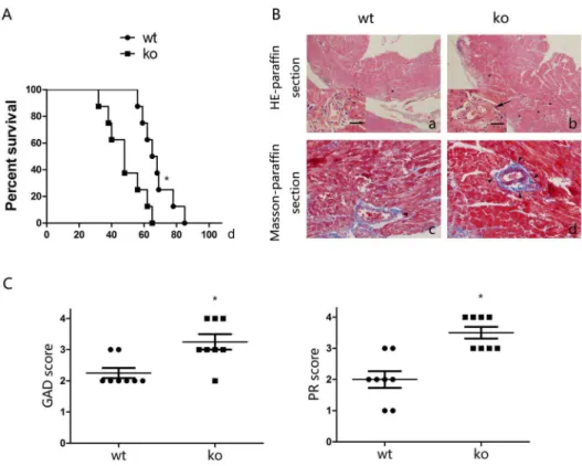
mice as recipients. We evaluated allografts daily by palpation to detect their function. In WT littermates, a median allograft survival (MST) of 67 days was observed after transplantation; however, the MST in T cell-PPARcko mice was only 48 days (Figure 1A). To detect lesions of chronic allograft rejection, HE and Masson staining were performed on the allografts 40 days after transplantation. In WT littermates, perivas-cular leukocytic infiltration, CAV and destruction of the cardiac muscle structure were observed, however, in T cell-PPARcko
mice, more intensive chronic allograft rejection signs were noted compared with the WT littermates. (Figure 1B). Consistent with these observations, the GAD and PR scores of T cell-PPARckomice were increased compared with WT littermates 40 days after transplanta-tion (Figure 1C). These results suggested that recipients with PPARc -deficient T cells exhibited decreased allograft survival and aggravated lesions of chronic allograft rejection.
T cell PPARcdeficiency increases CD4+T cell infiltration in cardiac allografts
Leukocyte infiltration into allografts is a hallmark and pathogenic factor of chronic allograft rejection. Given that we
identified more severe lesions in T cell-PPARckomice, we next detected the infiltration of T cell subsets into the allograft 40 days
after transplantation. The CD4+, CD8+, and cdTCR+ T cell
subsets in allografts were counted per high-power field (HPF) using immunohistochemical staining. Compared with the T cell subsets in the cardiac allografts of WT littermate recipients, a remarkable increase in infiltrated CD4+ T cells was observed in T cell-PPARcko
mice. In contrast, the numbers of CD8+andcdTCR+T cells did not differ between the groups (Figure 2A, B). As T cell-PPARckomice found to have CD4+, CD8+,cdTCR+T cells and
CD11b+ monocytes ratio comparable to those of C57BL/6 and
WT littermate initiate before operation (Figure S1).
PPARc-deficient T cells alter cytokine secretion and the polarization of CD4+T cells in cardiac allografts
Most studies have focused on Th1 (secreting IFN-c), Th2 (secreting IL-4/13) and Th17 (secreting IL-17) cells, which were considered the predominant contributors to chronic allograft rejection. Because we found increased CD4+T cell infiltration in PPARc-deficient T cell recipients in a previous study, we used a cytokine antibody array to detect cytokine secretion by infiltrated CD4+ T cells in vitro under the neutral stimulation conditions with anti-CD3/28 40 days after transplantation. We confirmed that in PPARc-deficient T cell recipients, CD4+T cells secreted higher levels of Th1-related IFN-c and Th17-related IL-17. In contrast, Th2-related IL-5, IL-10 and IL-13 cytokine secretion decreased compared with WT littermates (Figure 3A). We also used RT-PCR to measure the mRNA levels of transcription factors specific for CD4+ T cells in the cardiac allografts. The cardiac allografts from PPARc-deficient T cell recipients expressed higher levels of Th1- and Th17- related T-bet and RORct and reduced levels of Th2- and Treg- related GATA-3 and Foxp3 (Figure 3B). The flow cytometry data on the infiltrated CD4+T cells in the allograft revealed a variation trend consistent with the RT-PCR data (Figure 3C, D).
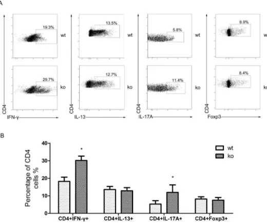
PPARc-deficient CD4+T cells enhance Th1 and Th17 polarizationin vitro
Previous experiments indicated that CD4+T cell subsets varied in PPARc-deficient T cell allografts. We tested whether the PPARc-deficient T cells exhibited altered polarization into Th1, Th2, Th17, and Treg cells. We isolated CD4+ T cells from the spleens of T cell-PPARckomice or WT littermates and cultured them under Th1/Th2/Th17/Treg-polarizing conditions for 4 days. Cells were then harvested, and intracellular cytokine levels were detected using flow cytometry with intracellular staining for IFN-c, IL-13, IL-17A, and Foxp3. A remarkable increase in IFN-c
and IL-17A production was observed in the PPARc-deficient
CD4+ T cells. However, the percentage of CD4+IL-13+ and
CD4+Foxp3+ T cells did not change (Figure 4A, B). These
findings suggested increased differentiation into the Th1 and Th17 subsets in PPARc-deficient CD4+T cells. The decreased Th2 and Treg subsets in the allografts were likely attributed to the increase in Th1 and Th17 cells and not to a failure to differentiate.
The effect of T cell PPARcdeficiency on macrophage polarizationin vivoandin vitro
infiltration at 7, 14, and 40 days after transplantation. In the cardiac allografts from T cell-PPARcko
mice, the number of macrophages was significantly increased compared with the WT littermates allografts (Figure 5A, B). To identify macrophage polarization into CAM and AAM, we measured the expression of CAM-related-iNOS, AAM-related-Arg1, and Mrc1 using RT-PCR at 7, 14, and 40 days after transplantation. Arg1 and Mrc1 mRNA levels did not increase in T cell-PPARcko
mice, whereas the levels were significantly elevated in WT littermates on days 14 and 40 post-grafting. In addition, no significant differences in the
mRNA levels of CAM-related-iNOS were noted in the two groups. Therefore, in T cell-PPARcko
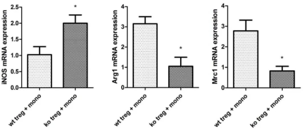
recipients mice, macro-phages failed to differentiate into AAM in local allografts. Because some studies have demonstrated that Treg can induce AAM polarization, we hypothesized that the PPARc-deficient Treg failed to induce AAM polarization. We isolated autologous CD4+ CD25+ Treg from the spleens of T cell-PPARcko
mice or their WT littermates and cocultured these cells with CD11b monocytes from C57BL/6 mice. After 2 days, cells were collected for RT-PCR detection. Arg1 and Mrc1 transcription levels were reduced Figure 1. PPARc-deficient T cells reduce survival and augment lesions associated with chronic rejection.A. The survival time of cardiac allografts in T cell-PPARckomice was significantly shorter than that of reduced compared with WT littermates. B. Cardiac allograft (bm12) sections of lesions 40 days after transplantation from T cell-PPARckomice (ko) or WT littermates. (a, b) HE staining, major view magnification 406, left bottom
view magnification 4006. Scale bars = 100mm. (c, d) Masson trichrome staining, magnification 4006. T cell-PPARckomice exhibited severe lesions.
Arrows indicate obvious intimal hyperplasia, asterisks indicate leukocytic infiltration, and filled arrowheads indicate collagen deposition. C. Graft coronary artery disease (GAD) and parenchymal rejection (PR) scores in T cell-PPARcko mice were considerably increased compared with WT littermates. The data are presented as the mean6SD for each group (n = 8), *p,0.05.
doi:10.1371/journal.pone.0112953.g001
Figure 2. Infiltration CD4+, CD8+, and cdTCR+ cells in donor hearts from T cell-PPARcko mice or WT littermates. A. Immunohistochemical staining of CD4+, CD8+, andcdTCR+cells in allografts 40 days after transplantation. Top, allograft from T cell-PPARcko mice (ko); bottom, allograft from WT littermates (wt). B. Quantitative analysis of CD4+, CD8+, andcdTCR+cells in allografts counted per high power
in PPARc-deficient Treg cocultured with monocytes (Figure 6). All of the above results demonstrated that PPARc-deficient T cells impact AAM polarizationin vivoandin vitro.
PPARc-deficient T cells abrogate the protective effect of a PPARc agonist
In our prior study, we demonstrated that PPARc-deficient T cells reduce the polarization of AAM. Other researchers have observed that PPARcagonists enhance AAM differentiation [42]. We examined whether PPARc-deficient T cells neutralize the protective effect of PPARcagonists in chronic allograft rejection. After treatment with the PPARcagonist pioglitazone, the median allograft survival significantly decreased in T cell-PPARckomice (Figure 7A). Moreover, Arg1 and Mrc1 transcription levels in allografts significantly decreased in T cell-PPARcko groups compared with WT littermates administered pioglitazone 40 days after transplantation (Figure 7B).
Discussion
Several studies have demonstrated the protective role of PPARc
agonists in chronic allograft rejection [15,22,23]. In these studies, the allogeneic reactions of cell infiltration and proinflammatory cytokine secretion are suppressed by PPARc agonists. However, the details regarding how PPARcinfluences the immune response in chronic allograft rejection are unknown. Recent studies have
reported that PPARc plays an important immunoregulatory
function in autoimmune diseases, such as experimental
autoim-mune encephalomyelitis (EAE) [24], allergic airway inflammation [25], trinitrobenzene sulfonic acid-induced colitis [26], and insulin resistance [27]. In these disease models, PPARcis the key factor promoting the differentiation of CD4+T cells and macrophages. Our previous study found that PPARcactivation with eicosapen-taenoic acid can impact the balance between Th17/Treg cells and protect cardiac allografts [14], consistent with its effect on autoimmune diseases. Therefore, we hypothesize that PPARc
controls T cell subset polarization in chronic allograft rejection and influences tolerance to allografts after transplantation.
In this study, we established a MHC class II-mismatched chronic allograft rejection model. To eliminate confusion due to non-selective PPARc agonists in the entire immune system, we used B6 background PPARcfl/fl; Lck-Cre+
T cell-specific PPARc
knockout mice and WT littermates as recipients. In the T cell-PPARcko
group, leukocyte infiltration and CAV were aggravated, and allograft survival decreased compared with WT littermates. These results confirmed that although PPARcinfluences both T cells and macrophages [28], in chronic allograft rejection, T cells expressing PPARc may participate in the primary function of transplant tolerance. Previous studies have suggested that CD4+, CD8+, and cdTCR+ T cells infiltrated into the allografts and contributed to allograft rejection [19,29]. We used immunohisto-chemical staining to detect infiltrated T cells in cardiac allografts 40 days after transplantation. The number of infiltrated CD4+T cells was significantly increased in allografts in T cell-PPARcko recipients. This result suggested that the infiltration of CD4+ T Figure 3. Cytokines and transcription factor expression in infiltrating CD4+T cells in cardiac allografts.A. Analysis of cytokine secretion
by infiltrating CD4+T cells from T cell-PPARckomice (ko) and WT littermates (wt) using a cytokine antibody array 40 days after transplantation. B. The transcription levels of the transcription factors T-bet, GATA-3, RORct, and Foxp3 in allografts were measured by RT-PCR 40 days after transplantation. C. Graft-infiltrating CD4+T cells were isolated 40 days after transplantation. These cells were stained with CD45, CD4, and 7-AAD then intracellularly
stained with IFN-c, IL-13, IL-17A, and Foxp3 for assessment by flow cytometry. D. The percentage of CD4+IFN-c+/CD4+IL-13+/CD4+IL-17A/CD4+
Foxp3+cells in infiltrating CD4+T cells was determined by flow cytometry. The data are presented as the mean6SD for each group (In panels A and B n = 10; in panels C and D n = 5), *p,0.05.
Figure 4. T cell-specific-PPARcknockout changes CD4+T cell subsets differentiation.A. CD4+T cells were isolated from T cell-PPARcko mice (ko) and WT littermates (wt) spleens and then cultured in protocols that induce T cell polarization into Th1, Th2, Th17, and Treg. After 4 days of culture, these cells were assessed by flow cytometry. B. The percentage of CD4+IFN-c+/CD4+IL-13+/CD4+IL-17A/CD4+Foxp3+cells in each cultured group of total CD4+cells. The data are presented as the mean6SD for each group (n = 5), *p,0.05.
doi:10.1371/journal.pone.0112953.g004
Figure 5. Analysis of macrophage infiltration and polarization in cardiac allografts.A. Flow cytometry analyses of the proportion of CD11b+F4/80+ macrophages infiltrating cardiac allografts 40 days after transplantation. B. Dynamic analyses of the proportion of infiltrated
macrophages in the total mononuclear cells in allografts at 7, 14, and 40 days after transplantation. C. iNOS, Arg1 and Mrc1 transcription levels in allografts were measured by RT-PCR at 7, 14 and 40 days after transplantation. The data are presented as the mean6SD for each group (n = 5), *p, 0.05.
cells in allografts increased with PPARcknockout and caused the intensive lesion.
CD4+T cells and their secretion of various cytokines participate in allogeneic rejection [30]. We isolated infiltrated CD4+T cells in allografts and examined their secreted cytokines. Based on RT-PCR and flow cytometry, the percentage of Th1 and Th17 increased, whereas Th2 and Treg decreased in the T cell-PPARcko
group. These results implied that PPARc knockout altered the subsets of CD4+ T cells. We next examined T cell differentiationin vitro. In contrast to the allografts, Th1 and Th17 differentiated more efficiently when PPARc was knocked out, whereas Th2 and Treg differentiation remained unchanged. These results are consistent with those of previous studies that also used conditional PPARcknockout mice cells [24,26]. Other laboratories using PPARc agonists reached similar conclusions. Augstein et al. used the PPARcagonist troglitazone to reduce T cell production of IFN-cin an autoimmune diabetes model [31]. In an allergic airway inflammation model, a PPARc agonist decreased NF-kB activity and reduced IL-17 release [25]. We concluded that the number of Th1 and Th17 cells infiltrated into allografts increased remarkably and led to a relative decrease in Th2 and Treg cells. Th1 is a classic proinflammatory cell and has been shown to be principally responsible for chronic allograft rejection [32]. Yuan et al. demonstrated that Th17 secretion of the proinflammatory cytokine IL-17A resulted in CAV when T-bet
was knocked out [6]. Th2 and Treg cells have always been recognized as elements that prolong allograft survival [33,34] and were present at a lower ratio in allografts than in WT littermates. Their protective effect was limited with PPARcdeficiency.
Recent studies have reported that macrophages are a major component in the immune response involved in allograft rejection [35,36]. CAM accelerate the immune response, whereas AAM have repair and anti-inflammatory abilities [37]. Moreover,
PPARc agonists skewed monocytes toward AAM polarization
[13]. We have observed that without PPARcin T cells, PPARc
agonists cannot exert protective function on macrophages. In additional studies, we investigated macrophage polarization in T cell-PPARckomice. Previous studies have indicated that IL-4 and IL-13 binding to IL-4Ra and IL-13Ra1 resulted in downstream phosphorylation of STAT6, thereby causing macrophage polar-ization [38–40]. More recently, Szanto et al. proposed that PPARc plays a critical role in these path ways because IL-4 signaling activates PPARc through STAT6 interactions with PPARcand promotes their binding to PPRE [41]. Indeed, the
IL-4/13-STAT6-KLF4-PPARc axis is regarded as an essential
regulator of CAM/AAM polarization and function [42]. In our experiments, the ratio of Th2 decreased, and Th2-related cytokine production was reduced. The AAM markers Arg1 and Mrc1 were
down-regulated after T cell specific knockout of PPARc.
Additionally, Tiemessen et al indicated that Treg have the ability Figure 6. PPARckoTreg fail to induce AAM in a monocyte and T cell coculture.CD4+CD25+T cells were isolated from the spleens of T cell-PPARckomice (ko) and WT littermates (wt) and cocultured with CD11b
+monocytes from PBMC C57BL/6 mice. The transcription level of iNOS, Arg1,
and Mrc1 was assessed in monocytes after 48 h of coculture by RT-PCR. The data are presented as the mean6SD for each group (n = 5), *p,0.05. doi:10.1371/journal.pone.0112953.g006
Figure 7. The beneficial effects of pioglitazone on the survival of cardiac allografts were counteracted in T cell-PPARcko.A. T
cell-PPARckomice (ko) abrogate the protective effect of pioglitazone on the survival curve. B. iNOS, Arg1, and Mrc1 transcription levels in allografts were measured by RT-PCR 40 days after transplantation. The data are presented as the mean6SD for each group (n = 5), *p,0.05.
to induce AAM in vitro [21]. Thus, we employed the same method to demonstrate that PPARc-deficient Treg failed to induce AAM polarization. The reduced Th2-related cytokine release together with the elimination of the ability of Treg to induce AAMs indicated that the decline in number of AAMs in PPARc-deficient allografts might be the cause of the severity of chronic rejection. To confirm that T cells influenced by PPARc
were the primary cause of AAM polarization in transplantation tolerance, we administered a PPARc agonist to T-cell-PPARcko
mice and WT littermate recipients. Indeed, PPARc-deficient T cells significantly reduced the survival of allografts and suppressed AAM polarization in the pioglitazone-treated groups.
In conclusion, the present study demonstrated that PPARc
expression is important for maintaining subsets of CD4+ T cells and macrophages in transplantation tolerance. The protective effect of a PPARc agonist in allograft rejection potentially functions through these immune processes. However, one of the remarkable features in chronic rejection is CAV, whose pathologic lesions occasionally appear similar to atherosclerosis but occur through a unique mechanism [43]. Vladimir et al. reported that macrophage-specific PPARc knockout increased atherosclerosis [44]. PPARcexpression in macrophages requires further investi-gation. Although PPARc agonists have effective immunoregula-tory capacity in many autoimmune diseases and transplant rejection animal models, the side effects of conventional
thiazolidinedione drugs (TZD) have been reported in recent clinical studies [12]. Because the mechanism of the protective effect of PPARcin chronic rejection has been demonstrated, we expect that PPARc-targeted therapeutics might serve as a novel method to promote transplant tolerance.
Supporting Information
Figure S1 T cell-PPARckomice have normal T cell and monocyte subpopulations. Flow cytometry analyzing the proportion of T cell and monocyte subpopulations from T cell-PPARcko
mice are normally comparable to those of C57BL/6 mice and WT littermate before operation in the spleens. The data are presented as the mean6SD for each group (n = 5).
(TIF)
Acknowledgments
The authors of this manuscript have certified that they comply with the Principles of Ethical Publishing in thePLOS ONE.
Author Contributions
Conceived and designed the experiments: XH JW SW AX JX. Performed the experiments: XH LR PY CC. Analyzed the data: XH. Wrote the paper: XH. Revised the manuscript: YS ZL.
References
1. Stehlik J, Edwards LB, Kucheryavaya AY, Benden C, Christie JD, et al. (2012) The registry of the international society for heart and lung transplantation: 29th official adult heart transplant report-2012. J Heart Lung Transplant 31: 1052– 1064.
2. Schmauss D, Weis M (2008) Cardiac allograft vasculopathy: recent develop-ments. Circulation 117: 2131–2141.
3. Suzuki J-i, Isobe M, Morishita R, Nagai R (2010) Characteristics of Chronic Rejection in Heart Transplantation. Circ J 74: 233–239.
4. El-Sawy T, Fahmy NM, Fairchild RL (2002) Chemokines: directing leukocyte infiltration into allografts. Curr Opin Immunol 14: 562–568.
5. Piccotti JR, Li K, Chan SY, Eichwald EJ, Bishop DK (1999) Cytokine Regulation of Chronic Cardiac Allograft Rejection: Evidence Against A Role for Th1 in the Disease Process1. Transplantation 67: 1548–1555.
6. Yuan X, Paez-Cortez J, Schmitt-Knosalla I, D’Addio F, Mfarrej B, et al. (2008) A novel role of CD4 Th17 cells in mediating cardiac allograft rejection and vasculopathy. J Exp Med 205: 3133–3144.
7. Tay SS, Plain KM, Bishop GA (2009) Role of IL-4 and Th2 responses in allograft rejection and tolerance. Curr Opin Organ Transplant 14: 16–22. 8. Moraes-Vieira PM, Bassi EJ, Larocca RA, Castoldi A, Burghos M, et al. (2013)
Leptin deficiency modulates allograft survival by favoring a Th2 and a regulatory immune profile. [corrected]. Am J Transplant 13: 36–44. 9. Martinez FO, Helming L, Gordon S (2009) Alternative activation of
macrophages: an immunologic functional perspective. Annu Rev Immunol 27: 451–483.
10. Mannon RB (2012) Macrophages: contributors to allograft dysfunction, repair, or innocent bystanders? Curr Opin Organ Transplant 17: 20–25.
11. da Rocha Junior LF, Dantas AT, Duarte AL, de Melo Rego MJ, Pitta Ida R, et al. (2013) PPARgamma Agonists in Adaptive Immunity: What Do Immune Disorders and Their Models Have to Tell Us? PPAR Res 2013: 519724. 12. Ahmadian M, Suh JM, Hah N, Liddle C, Atkins AR, et al. (2013) PPARgamma
signaling and metabolism: the good, the bad and the future. Nat Med 19: 557– 566.
13. Bouhlel MA, Derudas B, Rigamonti E, Dievart R, Brozek J, et al. (2007) PPARgamma activation primes human monocytes into alternative M2 macrophages with anti-inflammatory properties. Cell Metab 6: 137–143. 14. Ye P, Li J, Wang S, Xie A, Sun W, et al. (2012) Eicosapentaenoic acid disrupts
the balance between Tregs and IL-17+T cells through PPARgamma nuclear receptor activation and protects cardiac allografts. J Surg Res 173: 161–170. 15. Kosuge H, Haraguchi G, Koga N, Maejima Y, Suzuki J, et al. (2006)
Pioglitazone prevents acute and chronic cardiac allograft rejection. Circulation 113: 2613–2622.
16. Tobiasova Z, Zhang L, Yi T, Qin L, Manes TD, et al. (2011) Peroxisome proliferator-activated receptor-gamma agonists prevent in vivo remodeling of human artery induced by alloreactive T cells. Circulation 124: 196–205. 17. Corry RJ, Winn HJ, Russell PS (1973) Primarily vascularized allografts of hearts
in mice. The role of H-2D, H-2 K, and non-H-2 antigens in rejection. Transplantation 16: 343–350.
18. Furukawa Y, Libby P, Stinn JL, Becker G, Mitchell RN (2002) Cold ischemia induces isograft arteriopathy, but does not augment allograft arteriopathy in non-immunosuppressed hosts. The American journal of pathology 160: 1077– 1087.
19. Wang S, Xu X, Xie A, Li J, Ye P, et al. (2012) Anti-interleukin-12/23p40 antibody attenuates chronic rejection of cardiac allografts partly via inhibition gammadeltaT cells. Clin Exp Immunol 169: 320–329.
20. Itoh S, Kimura N, Axtell RC, Velotta JB, Gong Y, et al. (2011) Interleukin-17 accelerates allograft rejection by suppressing regulatory T cell expansion. Circulation 124: S187–196.
21. Tiemessen MM, Jagger AL, Evans HG, van Herwijnen MJ, John S, et al. (2007) CD4+CD25+Foxp3+regulatory T cells induce alternative activation of human monocytes/macrophages. Proc Natl Acad Sci U S A 104: 19446–19451. 22. Chen Y, Liu Y, Yuan Z, Tian L, Dallman MJ, et al. (2007) Rosiglitazone
suppresses cyclosporin-induced chronic transplant dysfunction and prolongs survival of rat cardiac allografts. Transplantation 83: 1602–1610.
23. Chen Y, Li D, Tsang JY, Niu N, Peng J, et al. (2011) PPAR-gamma signaling and IL-5 inhibition together prevent chronic rejection of MHC Class II-mismatched cardiac grafts. J Heart Lung Transplant 30: 698–706.
24. Klotz L, Burgdorf S, Dani I, Saijo K, Flossdorf J, et al. (2009) The nuclear receptor PPAR gamma selectively inhibits Th17 differentiation in a T cell-intrinsic fashion and suppresses CNS autoimmunity. J Exp Med 206: 2079– 2089.
25. Park SJ, Lee KS, Kim SR, Min KH, Choe YH, et al. (2009) Peroxisome proliferator-activated receptor gamma agonist down-regulates IL-17 expression in a murine model of allergic airway inflammation. J Immunol 183: 3259–3267. 26. Hontecillas R, Bassaganya-Riera J (2007) Peroxisome proliferator-activated receptorcis required for regulatory CD4+T cell-mediated protection against colitis. The Journal of Immunology 178: 2940–2949.
27. Odegaard JI, Ricardo-Gonzalez RR, Goforth MH, Morel CR, Subramanian V, et al. (2007) Macrophage-specific PPARgamma controls alternative activation and improves insulin resistance. Nature 447: 1116–1120.
28. Glass CK, Saijo K (2010) Nuclear receptor transrepression pathways that regulate inflammation in macrophages and T cells. Nat Rev Immunol 10: 365– 376.
29. Fischbein MP, Yun J, Laks H, Irie Y, Fishbein MC, et al. (2001) CD8+ lymphocytes augment chronic rejection in a MHC class II mismatched model. Transplantation 71: 1146–1153.
30. Liu Z, Fan H, Jiang S (2013) CD4(+) T-cell subsets in transplantation. Immunol Rev 252: 183–191.
31. Augstein P, Dunger A, Heinke P, Wachlin G, Berg S, et al. (2003) Prevention of autoimmune diabetes in NOD mice by troglitazone is associated with modulation of ICAM-1 expression on pancreatic islet cells and IFN-cexpression in splenic T cells. Biochem Biophys Res Commun 304: 378–384.
33. He XY, Chen J, Verma N, Plain K, Tran G, et al. (1998) Treatment with interleukin-4 prolongs allogeneic neonatal heart graft survival by inducing T helper 2 responses. Transplantation 65: 1145–1152.
34. Ge W, Jiang J, Liu W, Lian D, Saito A, et al. (2010) Regulatory T Cells Are Critical to Tolerance Induction in Presensitized Mouse Transplant Recipients Through Targeting Memory T Cells. Am J Transplant 10: 1760–1773. 35. Wyburn KR, Jose MD, Wu H, Atkins RC, Chadban SJ (2005) The role of
macrophages in allograft rejection. Transplantation 80: 1641–1647. 36. Magil AB (2009) Monocytes/macrophages in renal allograft rejection.
Transplant Rev (Orlando) 23: 199–208.
37. Gordon S, Martinez FO (2010) Alternative Activation of Macrophages: Mechanism and Functions. Immunity 32: 593–604.
38. Heller NM, Qi X, Junttila IS, Shirey KA, Vogel SN, et al. (2008) Type I IL-4Rs selectively activate IRS-2 to induce target gene expression in macrophages. Sci Signal 1: ra17.
39. Martinez-Nunez RT, Louafi F, Sanchez-Elsner T (2011) The interleukin 13 (IL-13) pathway in human macrophages is modulated by microRNA-155 via direct targeting of interleukin 13 receptora1 (IL13Ra1). J Biol Chem 286: 1786–1794.
40. Dhakal M, Hardaway JC, Guloglu FB, Miller MM, Hoeman CM, et al. (2013) IL-13Ralpha1 is a surface marker for M2 macrophages influencing their differentiation and function. Eur J Immunol.
41. Szanto A, Balint BL, Nagy ZS, Barta E, Dezso B, et al. (2010) STAT6 transcription factor is a facilitator of the nuclear receptor PPARgamma-regulated gene expression in macrophages and dendritic cells. Immunity 33: 699–712.
42. Liu G, Yang H (2013) Modulation of macrophage activation and programming in immunity. J Cell Physiol 228: 502–512.
43. Rahmani M, Cruz RP, Granville DJ, McManus BM (2006) Allograft vasculopathy versus atherosclerosis. Circ Res 99: 801–815.