Received 08 Apr., 2014 Accepted 09 July, 2014 (006342)
1Federal University of São Paulo – UNIFESP, Botucatu, SP, Brazil, e-mail: ericalmeida2000@yahoo.com.br *Corresponding author
Prions: the danger of biochemical weapons
Eric Almeida XAVIER
1*
1 Introduction
People have had a passion for weapons of mass destruction
since the government military agencies search for chemical
weapons culminating with the use of poisonous gases in the First
War. During the Cold War, there were many rumors of research
on biochemical and biological weapons since intelligence
agencies of North America and Russian, used diferent types
of weapons, such as radioactive, poison and contagious disease
weapons, agaist their political enemies because these weapons
cause diseases that could be above suspicion. It is important to
emphasize that Prions also have this characteristic. herefore
it is plausible that Prion weapons can be used not only by
governments but also by terrorists.
Furthermore, hypothetically, if Prions were used as a
biochemical weapon, they could damage not only humans and
animals, but the worldwide economies; therefore, even if Prions
do not kill instantly a target, they can be a very persuasive object
for those who have access to it.Prion diseases include a group
of fatal neurodegenerative and infectious disorders in humans
such as Creutzfeldt-Jacob disease (CJD), a variant form of CJD
(vCJD), Gerstmann-Sträussler-Scheinker syndrome (GSS), and
kuru, fatal familial insomnia (FFI); in animals, they include:
scrapie of sheep and goats, chronic wasting disease (CWD)
of mule deer and elk, and bovine spongiform encephalopathy
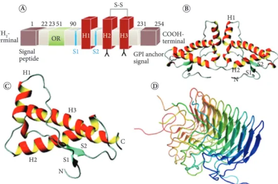
(BSE) of cattle (Prusiner, 1996). he central feature of these
disorders is the conformational change of the host encoded,
cellular Prion protein (PrPc), see figures (Figure 1A and
Figure 2B, C) to an abnormal, partially proteinase K resistant
and infectious isoform (PrPSc) with an aggregation propensity
accumulating in the brain of diseased individuals (Figure 1B
and Figure 2D) (Gambetti et al., 2003; Tatzelt & Schätzl, 2007).
Cases of vCJD in Great Britain and France raised the possibility
that BSE has been transmitted to humans (Bateman et al., 1995;
Cousens et al., 1997). Macaque monkeys and marmosets both
developed neurologic disease several years ater inoculation
with bovine Prions (Baker et al., 1993; Ono et al., 2011). hese
experiments clearly show that the inoculation of Prions is
potentially fatal, and the fact that Prion strains are transmitted
between species raised the possibility that a large section of
the population is at high risk as a result of exposure to BSE
Prions (Anderson et al., 1996; Sweeting et al., 2010; Bruce et al.,
1997; Collinge et al., 1996; Griin, 1997; Lasmézas et al., 1996;
Scott et al., 1993).
Prions have key features which help them to survive in the
environment for long periods, such as the resistance to protein
degradation, “partially resistant to proteinase K” (Bolton et al.,
1982; Prusiner, 1991), high resistant when exposed to
irradiation, heat, and harsh chemical treatments (Plum, 1997),
plus the fact that they can be attached to soil (Johnson et al.,
2006; Saunders et al., 2011a) and spread through air, and
therefore they are extremely hazardous (Denkers et al., 2010).
Although Prions are just only a polypeptide sequence, they are
resistant to heat since people who consumed contaminated
meat ater preparation got sick; this raised the possibility that
a particular conformation of bovine PrPSc was selected for
heat resistance during the manufacture of meat and bone meal
(MBM). herefore, it is believed that MBM is the source of
Prions responsible for BSE (Prusiner, 1997).
herefore, hypothetically, Prions can be manufactured
based on the molecular characteristics of the Prion protein
to make deadly biochemical weapons. he study on how the
conversion of PrPC (the normal cellular protein) into PrPSc
(the abnormal disease-causing isoform) (Supattapone, 2010)
can generate polypeptide sequences designed exclusively to kill.
Basically, an ideal Prion polypeptide sequence to be used as a
biochemical weapon must be easily recombined and produced
in large scale (Supattapone, 2010), be resistant to cold and heat,
be fatal in very small quantities, and be transmitted through
air, as demonstrated by (Denkers et al., 2010; Haybaeck et al.,
2011), and through water, food and soil, as demonstrated
Abstract
he knowledge of biotechnology increases the risk of using biochemical weapons for mass destruction. Prions are unprecedented
infectious pathogens that cause a group of fatal neurodegenerative diseases by a novel mechanism. hey are transmissible
particles that are devoid of nucleic acid. Due to their singular characteristics, Prions emerge as potential danger since they
can be used in the development of such weapons. Prions cause fatal infectious diseases, and to date there is no therapeutic or
prophylactic approach against these diseases. Furthermore, Prions are resistant to food-preparation treatments such as high
heat and can ind their way from the digestive system into the nervous system; recombinant Prions are infectious either bound
to soil particles or in aerosols. herefore, lethal Prions can be developed by malicious researchers who could use it to attack
political enemies since such weapons cause diseases that could be above suspicion.
Keywords:
Prions; biochemical weapons; Prion diseases; Prions danger to the environment; Prions risk alert.
OI:
is a possible mechanism for triggering the conversion of
PrPc into PrPSc where α-helices appear to be converted into
β-sheets (Zhang et al., 1995). However, no atomic-resolution
structure of the ibrillar state, which is likely to be infectious,
has been reported to date because characterizing the structure
of PrPSc has been challenging due to the diiculty in studying
it through Nuclear magnetic resonance (NMR) or X-ray
crystallography methods (Wasmer et al., 2008). However, there
is a structural model based on solid-state nuclear magnetic
resonance restraints for amyloid ibrils from the Prion-forming
domain (residues 218 to 289) of the HET-s (het-s/S locus)
that occur naturally in the filamentous fungus Podospora
anserine (Figure 2D) (Wasmer et al., 2008; Dalstra et al., 2005).
Nevertheless, the basis of PrPSc conversion has been elucidated.
Some models with the earliest conversion events involving
PrPSc have been established suggesting that the formation of
the disease-causing isoform involves refolding in two of the PrPc
NH2-terminal (N-terminal) α-helix (H1 and H2) into β-sheets
or the two β-strands (S1 and S2) and proposing to “seed” β-sheet
elongation as the short α-helix H1 (Figure 1) (Huang et al.,
1996; Muramoto et al., 1996); the single disulide bond joining
COOH-terminal helices (C-terminal) would remain intact
because the disulide is required for PrPSc formation (Figure 1)
(Pan et al., 1993; Muramoto et al., 1996).
by (Saunders et al., 2009; Saunders et al., 2011a), Gough &
Maddison (2010) and (Smith et al., 2011). If Prions do not meet
all speciications necessary of a biochemical weapon to kill,
they at least have key features such as surviving through the
digestive system, as demonstrated by Sales (2006), resistance to
high temperatures during food preparation processes (Prusiner,
1997), recombinant Prions are infectious, as demonstrated by
(Wang et al., 2010; Legname et al., 2004; Makarava et al., 2010),
and lastly, a few molecules can produce a chain reaction in the
conversion of PrPc into PrPSc causing a fatal neurodegenerative
disease (Rigter et al. 2009).
2 Prion molecular factors involved in the conversion
of the native form PrPc into the infective form PrPSc,
which could be explored for evil plans
The basic mechanisms involved in the conversion
PrPc into PrPSc are mainly achieved in four routes: the
first includes the conformational mechanisms; the second
includes the structural mechanisms; the third is related to the
environmental pH; and fourth is the pathologic route.
he irst route is associated to the lethality of the peptides,
which involves a conformational change, through which the
alfa (α)-helical content diminishes and the amount of beta
(β)-sheet increases (Pan et al., 1993). PrPSc formation is a
posttranslational process involving only a conformational
change in PrPC. A comparison of the secondary structures
shows that PrPc is 42% α-helical with a very low (3%) β-sheet
content, whereas PrPSc consists of 30% α-helices and
43% β-sheets (Figure 1). Although the precise physiological
role of PrPSc and the chemical diferences between PrPSc and
normal Prion protein (PrP) remain unknown, it appears that
the diferences are conformational (Prusiner, 1998). he Prions
conformational transition from PrPc to PrPSc is accompanied
by profound changes in the properties of the protein: PrPc is
soluble in nondenaturing detergents, whereas PrPSc is not
(Meyer et al., 1986) and PrPC is readily digested by proteases,
whereas PrPSc is partially resistant (Kocisko et al., 1994;
Hsiao et al., 1989) (Figure 1B). However, it is not fully known
how disease-causing Prions arise in patients with sporadic
forms; the main hypothesis is the horizontal transmission of
Prions from humans or animals (Haley et al., 2011). herefore it
seems that the diseases caused by Prions are diseases of dietary
origin, raising a great possibility that the PrPSc conformation
is highly resistant to the mechanisms of animal polypeptides
digestion, which is a key feature for a biochemical weapon (Sales,
2006). he second route is the structural mechanisms, which
are associated with the basic structural transition from PrPc
to PrPSc. Peptide fragments corresponding to Syrian hamster
PrP residues 90 to 145 and 109 to 141, which contain the most
conserved residues of the Prion protein and the irst two putative
α-helical regions, were studied using infrared spectroscopy
and circular dichroism. he peptides could be induced to form
α-helical structures in aqueous solutions and in the presence
of organic solvents such as triluoroethano or detergents such
as sodium dodecyl. On the other hand, NaCl at physiological
concentration or acetonitrile induced the peptides to acquire
substantial β-sheet. he results suggest that perturbation of
the packing environment of the highly conserved residues
pH 4.1 can convert into the β-sheet conformation at pH 3.6
but not vice versa a loss of α-helix since the gain of β-sheets
and a number of PrPSc-like conformations can be generated
by incubating recombinant PrPc at low pH, indicating that
the protonation of key residues is likely to destabilize PrPc
facilitating its conversion to PrPSc. Fold stability of human PrPc
as a function of pH is signiicantly reduced by the protonation
of two histidine residues, His187 and His155. Mutation of
His187 to an arginine imposes a permanently positively charged
residue in this region of the protein and has a dramatic efect
on the folding of PrPc resulting in a molecule that displays a
markedly increased propensity to oligomerize. he oligomeric
form is characterized by an increased β-sheet content, loss of
ixed side chain interactions, and partial proteinase resistance.
herefore, the protonation state of H187 appears to be crucial
in determining the conformation of PrP; the unprotonated
form favors native PrPc, while the protonated form favors
PrPSc-like conformations (Hosszu et al., 2010; Gerber et al.,
2008; Hosszu et al., 2009). If these conditions are relevant, as
there is considerable evidence that endosome-like organelles or
Theoretically, the high β-sheet content of PrPSc was
predicted based on its ability to polymerize into amyloid ibrils
(Prusiner et al., 1983; Caughey et al., 1991). Studies have found
that the deletion of each of the four predicted helices prevented
PrPSc formation, as did the deletion of the stop transfer efector
region and the C178A mutation. he removal of a 36-residue
loop between helices 2 and 3 did not prevent formation of
protease-resistant PrP; the resulting scrapie-like protein,
designated PrPSc106, contained 106 residues ater cleavage
of an N-terminal signal peptide and a C-terminal sequence
for glycolipid anchor addition.he addition of the detergent
Sarkosyl to cell lysates solubilized PrPSc106, which retained
resistance to digestion by proteinase K. hese results suggest
that the regions of a proposed secondary structure in PrP
and the disulide bond stabilizing helices 2 and 3 are required
for PrPSc formation (Rogers et al., 1993; Fischer et al., 1996;
Muramoto et al., 1996). he third route is related to the pH
environment. he pH seems to be important for the change
of conformation because human PrPc has a pH-dependent
conformational change. he α-helical intermediate formed at
for all of the transport of Prions, and other cells, including
tingible-body macrophages (phagocytic cells in lymphoid
germinal centers) are plausible locations for PrPSc propagation
(Arnold et al., 1995; Aguzzi & Sigurdson, 2004) since PrP is
captured by phagocytes of the immune system.herefore, the
conformational convertion of PrPc into PrPSc can be triggered
by endocytosis of a Prion particle, and a phagocytic cell may
trigger the disease with a particle reaching the brain by the
sympathetic nervous system from the lymphatic tissues (Harris
& True, 2006; Aguzzi et al., 2008; Aguzzi et al., 2001; Venneti,
2010). Therefore, the immune system is a target of lethal
Prions, which could be added to adjuvants that are intended to
perform phagocytosis by DCs to increase their eiciency and
thus be transmitted through small wounds or scratches on the
victim’s skin.
3 Discussion
Recombinant Prions with fatal features could be developed
in relatively simple laboratories using animals such as rats,
mice, and monkeys (Supattapone, 2010; Wang et al., 2010;
Legname et al., 2004; Makarava et al., 2010). Ricin has already
been used as a weapon (Augerson, 2000; National Security Notes,
2004), for example in the case that caught the full attention of
international media and was described by Papaloucas et al
(2008) and which was about a political dissident that was killed
by a supposed KGB agent using a single ricin-tipped umbrella
as a weapon. Consequently, the same mechanism can be used to
deliver Prions using simple objects without giving the victim a
chance to receive a vaccine, treatment, or a speciic anti-serum
injection. Some political enemies must be eliminated and Prions
can be a possible alternative to the use of venoms, precisely
because Prions do not kill instantly and make the investigation
process very diicult to trace the assassin agent. Another class
of venom that have been used before and can be substituted by
Prions are the radioactive venom (Jordan & Finn, 2006) because
Prions can cause the same efect. One advantage is leaving no
traces detectable by anti-gama radiation equipment, such as
Geiger counter, and another advantage is being less dangerous
to the assassin agent willing to use it.
he psychological efect caused by the use of Prions to
extinguish rivals could be as powerful as the radioactive efects,
and it can send a strong message such as “Do not play with the
government interests”. he use of such weapons seems to have
strong personal issues involved because it would be easier to
kill someone simply using a gun, but with Prions, the victim
agonizes for months before dying (Papaloucas et al., 2008). he
most frightening possibility would be the use of Prions to get
rid of enemies in large regions of ongoing conlicts or political
separatist wars. In theory, it would not raise any suspicion by
the international community because of its silence mechanisms,
but ater years a lot of people would start dying presenting the
same symptoms and the alert would have come too late. If
Prions are made in laboratories with the purpose to be spread
in the air, it could kill a large number of people since it has
been demonstrated that CWD can be dispersed as aerosol
(Denkers et al., 2010; Haybaeck et al., 2011; Ford et al., 2002).
In addition, the decontamination of the environment can be a
lysosomes, with their locally acidic environments are plausible
locations for PrPSc propagation (Arnold et al., 1995), these
models of Prions for these molecular regions are crucial for the
development of disease via PrPSc, and theerefore the structure
of these regions of the molecule can be exploited as a catalytic
core for elaboration of new lethal Prions. Exploring mechanisms
that allow the Prions to be captured by the airways as an aerosol
microparticles; thus lesser amounts of Prions can be dispersed
through the air and contaminate the water, soil, plantations,
and very large regions. he fourth route is the pathologic
route. Another mechanism of infection that could be very
well explored is the pathogenesis. Prions could be related to
microspheres directed to speciic target cells and be embraced
and activated by the endosomes pathway of many types of cells
(Arnold et al., 1995). Pathogenesis can be divided into natural or
congenital transmission and external transmission. he native or
natural pathogenesis of Prion disease varies including mutation
in human Prion protein gene (PRNP) and external transmitted
causes (Tranulis et al., 2011). Figure 2A shows a normal structure
of PRNP gene (Manson & Tuzi, 2001). Missense mutations and
expansions in the octapeptide region (OR) result in familial
forms of Creutzfeldt-Jakob disease (fCJD) and GSS (Beck et al.,
2010; Jansen et al., 2011; Kovács et al., 2002). On the other hand,
FFI is caused by the D178N mutation, the disease progresses
quickly, and the patient dies within a few months ater the onset
of symptoms sleep disorders with agitation, fractionated sleep,
snoring, and daytime sleepiness (Ayuso Blanco et al., 2006;
Montagna et al., 2003). he inheritable familial forms of all
Prion diseases (fCJD, GSS, and FFI) are inherited as
autosomal-dominant disorders (Mastrianni, 2003). he polymorphism
coding for methionine (M) or valine (V) at codon 129 of the
Prion protein gene (PRNP M129V) plays a pivotal role in
the susceptibility to CJD, inluencing familial, transmitted,
and sporadic forms of the disease (Alperovitch et al., 1999),
homozygosity for methionine at position 129 (met/met at codon
129) predisposes susceptibility and earlier age of onset of disease
(Kretzschmar & Illig, 2009; Mead et al., 2009). herefore, the
PRNP polymorphisms is related to speciic clinical forms of
Prion diseases, and polymorphism in the regulatory region
of PRNP is associated with increased risk of sporadic CJD
(Sanchez-Juan et al., 2011).
epidemiology of BSE in British cattle. Nature, 382(6594), 779-788. Retrieved from http://www.ncbi.nlm.nih.gov/pubmed/8752271 Arnold, J. E., Tipler, C., Laszlo, L., Hope, J., Landon, M., & Mayer, R.
J. (1995). he abnormal isoform of the prion protein accumulates in late-endosome-like organelles in scrapie-infected mouse brain.
he Journal of Pathology, 176(4), 403-411. Retrieved from http:// www.ncbi.nlm.nih.gov/pubmed/7562256 PMid:7562256. http:// dx.doi.org/10.1002/path.1711760412
Augerson, W. S. (2000). A review of the scientiic literature as it pertains to Gulf War Illnesses (Vol. 5). Santa Monica: Rand Corporation. Ayuso Blanco, T., Urriza Mena, J., Caballero Martínez, C., Iriarte
Franco, J., Munoz, R., & García-Bragado, F. (2006). [Fatal familiar insomnia: clinical, neurophysiological and histopathological study of two cases]. Neurologia, 21(8), 414-420. Retrieved from http:// www.ncbi.nlm.nih.gov/pubmed/17013786 PMid:17013786. Baker, H., Ridley, R., & Wells, G. (1993). Experimental transmission
of BSE and scrapie to the common marmoset. he Veterinary Record, 132(16), 403-406. Retrieved from http://www.ncbi.nlm. nih.gov/entrez/query.fcgi?cmd=Retrieve&db=PubMed&dopt=Cita tion&list_uids=8488658 PMid:8488658. http://dx.doi.org/10.1136/ vr.132.16.403
Bateman, D., Hilton, D., Love, S., Zeidler, M., Beck, J., & Collinge, J. (1995). Sporadic Creutzfeldt-Jakob disease in a 18-year-old in the UK. Lancet, 346(8983), 1155-1156. Retrieved from http://www. ncbi.nlm.nih.gov/entrez/query.fcgi?cmd=Retrieve&db=PubMe d&dopt=Citation&list_uids=7475612 http://dx.doi.org/10.1016/ S0140-6736(95)91828-0
Beck, J. A., Poulter, M., Campbell, T. A., Adamson, G., Uphill, J. B., Guerreiro, R., Jackson, G. S., Stevens, J. C., Manji, H., Collinge, J., & Mead, S. (2010). PRNP allelic series from 19 years of prion protein gene sequencing at the MRC Prion Unit. Human Mutation,
31(7|), E1551-63. Retrieved from http://www.ncbi.nlm.nih.gov/ pubmed/20583301 http://dx.doi.org/10.1002/humu.21281 Bolton, D. C., McKinley, M. P., & Prusiner, S. B. (1982). Identiication
of a protein that purifies with the scrapie prion. Science,
218(4579), 1309-1311. Retrieved from http://www.ncbi.nlm.nih. gov/pubmed/6815801 PMid:6815801. http://dx.doi.org/10.1126/ science.6815801
Bruce, M. E., Will, R. G., Ironside, J. W., McConnell, I., Drummond, D., Suttie, A., McCardle, L., Chree, A., Hope, J., Birkett, C., Cousens, S., Fraser, H., & Bostock, C. J. (1997). Transmissions to mice indicate that ‘new variant’ CJD is caused by the BSE agent. Nature,
389(6650), 498-501. Retrieved from http://www.ncbi.nlm.nih.gov/ pubmed/9333239 PMid:9333239. http://dx.doi.org/10.1038/39057 Caughey, B. W., Dong, A., Bhat, K. S., Ernst, D., Hayes, S. F., & Caughey,
W. S. (1991). Secondary structure analysis of the scrapie-associated protein PrP 27-30 in water by infrared spectroscopy. Biochemistry,
30(31), 7672-7680. Retrieved from http://www.ncbi.nlm.nih.gov/ entrez/query.fcgi?cmd=Retrieve&db=PubMed&dopt=Citation &list_uids=1678278 PMid:1678278. http://dx.doi.org/10.1021/ bi00245a003
Collinge, J., Sidle, K. C., Meads, J., Ironside, J., & Hill, A. F. (1996). Molecular analysis of prion strain variation and the aetiology of ‘new variant’ CJD. Nature, 383(6602), 685-690. Retrieved from http://www.ncbi.nlm.nih.gov/pubmed/8878476 PMid:8878476. http://dx.doi.org/10.1038/383685a0
Cousens, S. N., Vynnycky, E., Zeidler, M., Will, R. G., & Smith, P. G. (1997). Predicting the CJD epidemic in humans. Nature, 385(6613), 197-198. Retrieved from http://www.ncbi.nlm.nih.gov/entrez/ query.fcgi?cmd=Retrieve&db=PubMed&dopt=Citation&list_ uids=9000063 PMid:9000063. http://dx.doi.org/10.1038/385197a0
huge problem if Prions are not rapidly degraded in the soil by
microorganisms; some studies have demonstrated that the soil
is as possible reservoir of scrapie and CWD agents, which can
persist in the environment for years. Attachment to soil particles
likely inluences the persistence and infectivity of Prions in the
environment (Smith et al., 2011; Gough & Maddison, 2010;
Saunders et al., 2009).
he evidence that soil and other environmental surfaces
can play a role as reservoir of Prions dissemination contributes
to the imminent threat these particles can represent if they
are released into the environment (Maddison et al., 2010;
Saunders et al., 2011b). Finally, the impact that Prions could
cause to wildlife, especially mammals, is terrifying; people
who had been in contact to contaminated environments or had
ingested inoculated animals could die in days, months, or years.
he long efects of Prion contamination can be terrible
and are similar to radioactive efects. Concluding, it is utmost
important to alert the scientiic community scientiic community,
agencies, and governments worldwide to discourage, inhibit,
and investigate those who have this evil intention.
4 Final conclusions
Bioterrorism is as emerging threat that is growing with
the development of biotechnology. he risk of biochemical
weapons falling into wrong hands can be devastating; it could
contaminate cattle, humans and many other animal species
leading to thousands of deaths and would lead to a global
pandemic and economic crisis too.
Acknowledgements
he authors are grateful to Cayman Chemical Company,
Ann Arbor, Michigan. USA.
References
Aguzzi, A., Montrasio, F., & Kaeser, P. S. (2001). Prions: health scare and biological challenge. Nature Reviews: Molecular Cell Biology, 2(2), 118-126. Retrieved from http://www.ncbi.nlm. nih.gov/pubmed/11252953 PMid:11252953. http://dx.doi. org/10.1038/35052063
Aguzzi, A., & Sigurdson, C. J. (2004). Antiprion immunotherapy: to suppress or to stimulate? Nature Reviews: Immunology, 4(9), 725-736. Retrieved from http://www.ncbi.nlm.nih.gov/pubmed/15343371 PMid:15343371. http://dx.doi.org/10.1038/nri1437
Aguzzi, A., Baumann, F., & Bremer, J. (2008). he prion’s elusive reason for being. Annual Review of Neuroscience, 31, 439-477. Retrieved from http://www.ncbi.nlm.nih.gov/pubmed/18558863 PMid:18558863. http://dx.doi.org/10.1146/annurev.neuro.31.060407.125620 Alperovitch, A., Zerr, I., Pocchiari, M., Mitrova, E., Pedro Cuesta, J.,
Hegyi, I. Collins, S., Kretzschmar, H., Van Duijn, C., & Will, R. G. (1999). Codon 129 prion protein genotype and sporadic Creutzfeldt-Jakob disease. Lancet, 353(9165), 1673-1674. Retrieved from http:// www.ncbi.nlm.nih.gov/pubmed/10335789
PMid:21249178 PMCid:PMC3020930. http://dx.doi.org/10.1371/ journal.ppat.1001257
Hosszu, L. L., Trevitt, C. R., Jones, S., Batchelor, M., Scott, D. J., Jackson, G. S., Collinge, J., Waltho, J. P., & Clarke, A. R. (2009). Conformational properties of beta-PrP. Journal of Biological Chemistry, 284(33), 21981-21990. Retrieved from http:// www.ncbi.nlm.nih.gov/pubmed/19369250 PMid:19369250 PMCid:PMC2755922. http://dx.doi.org/10.1074/jbc.M809173200 Hosszu, L. L., Tattum, M. H., Jones, S., Trevitt, C. R., Wells, M. A.,
Waltho, J. P., Collinge, J., Jackson, G. S., & Clarke, A. R. (2010). he H187R mutation of the human prion protein induces conversion of recombinant prion protein to the PrP(Sc)-like form. Biochemistry,
49(40), 8729-38. Retrieved from http://www.ncbi.nlm.nih.gov/ pubmed/20718410 PMid:20718410. http://dx.doi.org/10.1021/ bi100572j
Hsiao, K., Baker, H. F., Crow, T. J., Poulter, M., Owen, F., Terwilliger, J. D., Westaway, D., Ott, J., & Prusiner, S. B. (1989). Linkage of a prion protein missense variant to Gerstmann-Sträussler syndrome. Nature, 338(6213), 342-345. Retrieved from http://www.ncbi. nlm.nih.gov/entrez/query.fcgi?cmd=Retrieve&db=PubMed&d opt=Citation&list_uids=2564168 PMid:2564168. http://dx.doi. org/10.1038/338342a0
Huang, Z., Prusiner, S., & Cohen, F. (1996). Scrapie Prions: a three-dimensional model of an infectious fragment. Folding & Design,
1(1), 13-19. Retrieved from http://www.ncbi.nlm.nih.gov/entrez/ query.fcgi?cmd=Retrieve&db=PubMed&dopt=Citation&li st_uids=9079359 http://dx.doi.org/10.1016/S1359-0278(96)00007-7 Jansen, C., Voet, W., Head, M. W., Parchi, P., Yull, H., Verrips, A.,
Wesseling, P., Meulstee, J., Baas, F., Van Gool, W. A., Ironside, J. W., & Rozemuller, A. J. (2011). A novel seven-octapeptide repeat insertion in the prion protein gene (PRNP) in a Dutch pedigree with Gerstmann-Sträussler-Scheinker disease phenotype: comparison with similar cases from the literature. Acta Neuropathologica,
121(1), 59-68. Retrieved from http://www.ncbi.nlm.nih.gov/ pubmed/20198483 PMid:20198483 PMCid:PMC3015204. http:// dx.doi.org/10.1007/s00401-010-0656-3
Johnson, C. J., Phillips, K. E., Schramm, P. T., McKenzie, D., Aiken, J. M., & Pedersen, J. A. (2006). Prions adhere to soil minerals and remain infectious. PLoS Pathog, 2(4), e32. Retrieved from http://www.ncbi.nlm.nih.gov/pubmed/16617377 PMid:16617377 PMCid:PMC1435987. http://dx.doi.org/10.1371/journal. ppat.0020032
Jordan, M., & Finn, P. (2006, November 25). Radioactive poison killed ex-spy. he Washington Post. Retrieved from http://www. washingtonpost.com/wp-dyn/content/article/2006/11/24/ AR2006112400410.html
Kocisko, D. A., Come, J. H., Priola, S. A., Chesebro, B., Raymond, G. J., Lansbury, P. T., & Caughey, B. (1994). Cell-free formation of protease-resistant prion protein. Nature, 370(6489), 471-474. Retrieved from http://www.ncbi.nlm.nih.gov/pubmed/7913989 PMid:7913989. http://dx.doi.org/10.1038/370471a0
Kovács, G. G., Trabattoni, G., Hainfellner, J. A., Ironside, J. W., Knight, R. S., & Budka, H. (2002). Mutations of the prion protein gene phenotypic spectrum. Journal of Neurology, 249(11), 1567-1582. Retrieved from http://www.ncbi.nlm.nih.gov/pubmed/12420099 PMid:12420099. http://dx.doi.org/10.1007/s00415-002-0896-9 Kretzschmar, H., & Illig, T. (2009). Are further genetic factors associated
with the risk of developing variant Creutzfeldt-Jakob disease? he Lancet: Neurology, 8(1), 25-26. Retrieved from http://www.ncbi. nlm.nih.gov/pubmed/19081509 http://dx.doi.org/10.1016/S1474-4422(08)70266-7
Dalstra, H. J., Van der Zee, R., Swart, K., Hoekstra, R. F., Saupe, S. J., & Debets, A. J. (2005). Non-mendelian inheritance of the HET-s prion or HET-s prion domains determines the het-S spore killing system in Podospora anserina. Fungal Genetics and Biology: FG & B, 42(10), 836-847. Retrieved from http://www.ncbi.nlm.nih.gov/ pubmed/16043372 PMid:16043372. http://dx.doi.org/10.1016/j. fgb.2005.05.004
Denkers, N. D., Seelig, D. M., Telling, G. C., & Hoover, E. A. (2010). Aerosol and nasal transmission of chronic wasting disease in cervidized mice. he Journal of General Virology, 91(Pt 6), 1651-1658. Retrieved from http://www.ncbi.nlm.nih.gov/ pubmed/20164261 PMid:20164261 PMCid:PMC2888164. http:// dx.doi.org/10.1099/vir.0.017335-0
Fischer, M., Rülicke, T., Raeber, A., Sailer, A., Moser, M., Oesch, B., Brandner, S., Aguzzi, A., & Weissmann, C. (1996). Prion protein (PrP) with amino-proximal deletions restoring susceptibility of PrP knockout mice to scrapie. he EMBO Journal, 15(6), 1255-1264. Retrieved from http://www.ncbi.nlm.nih.gov/entrez/query.fcgi?c md=Retrieve&db=PubMed&dopt=Citation&list_uids=8635458 PMid:8635458 PMCid:PMC450028.
Ford, M. J., Burton, L. J., Morris, R. J., & Hall, S. M. (2002). Selective expression of prion protein in peripheral tissues of the adult mouse.
Neuroscience, 113(1), 177-192. Retrieved from http://www.ncbi. nlm.nih.gov/pubmed/12123696 http://dx.doi.org/10.1016/S0306-4522(02)00155-0
Gambetti, P., Kong, Q., Zou, W., Parchi, P., & Chen, S. G. (2003). Sporadic and familial CJD: classiication and characterisation.
British Medical Bulletin, 66(1), 213-239. Retrieved from http://www. ncbi.nlm.nih.gov/pubmed/14522861 PMid:14522861. http://dx.doi. org/10.1093/bmb/66.1.213
Gerber, R., Tahiri-Alaoui, A., Hore, P. J., & James, W. (2008). Conformational pH dependence of intermediate states during oligomerization of the human prion protein. Protein Science,
17(3), 537-544. Retrieved from http://www.ncbi.nlm.nih.gov/ pubmed/18218718 PMid:18218718 PMCid:PMC2248315. http:// dx.doi.org/10.1110/ps.073163308
Gough, K. C., & Maddison, B. C. (2010). Prion transmission: prion excretion and occurrence in the environment. Prion, 4(4), 275-282. Retrieved from http://www.ncbi.nlm.nih.gov/pubmed/20948292 PMid:20948292 PMCid:PMC3268960. http://dx.doi.org/10.4161/ pri.4.4.13678
Griin, J. M. (1997). BSE and British cattle exports. he Veterinary Record, 141(11), 286-287. Retrieved from http://www.ncbi.nlm. nih.gov/pubmed/9316247 PMid:9316247.
Haley, N. J., Mathiason, C. K., Carver, S., Zabel, M., Telling, G. C., & Hoover, E. A. (2011). Detection of CWD Prions in salivary, urinary, and intestinal tissues of deer: potential mechanisms of prion shedding and transmission. Journal of Virology, 85(13), 6309-6318. Retrieved from http://www.ncbi.nlm.nih.gov/pubmed/21525361 PMid:21525361 PMCid:PMC3126547. http://dx.doi.org/10.1128/ JVI.00425-11
Harris, D. A., & True, H. L. (2006). New insights into prion structure and toxicity. Neuron, 50(3), 353-357. Retrieved from http://www. ncbi.nlm.nih.gov/pubmed/16675391 PMid:16675391. http://dx.doi. org/10.1016/j.neuron.2006.04.020
& Sata, T. (2011). Experimental transmission of bovine spongiform encephalopathy (BSE) to cynomolgus macaques, a non-human primate. Japanese Journal of Infectious Diseases, 64(1), 50-54. Retrieved from http://www.ncbi.nlm.nih.gov/pubmed/21266755 PMid:21266755.
Pan, K. M., Baldwin, M., Nguyen, J., Gasset, M., Serban, A., Groth, D., Mehlhorn, I., Huang, Z., Fletterick, R. J., & Cohen, F. E. (1993). Conversion of alpha-helices into beta-sheets features in the formation of the scrapie prion proteins. Proceedings of the National Academy of Sciences of the United States of America,
90(23), 10962-10966. Retrieved from http://www.ncbi.nlm.nih.gov/ entrez/query.fcgi?cmd=Retrieve&db=PubMed&dopt=Citation&li st_uids=7902575 PMid:7902575 PMCid:PMC47901. http://dx.doi. org/10.1073/pnas.90.23.10962
Papaloucas, M., Papaloucas, C., & Stergioulas, A. (2008). Ricin and the assassination of Georgi Markov. Pakistan Journal of Biological Sciences, 11(19), 2370-2371. Retrieved from http://www.ncbi. nlm.nih.gov/pubmed/19137875 PMid:19137875. http://dx.doi. org/10.3923/pjbs.2008.2370.2371
Plum, J. (1997). BSE: can we predict the future? Bulletin et Mémoires de l’Académie Royale de Médecine de Belgique, 152(6), 264-273. Retrieved from http://www.ncbi.nlm.nih.gov/pubmed/9581370 PMid:9581370.
Prusiner, S. B. (1991). Molecular biology of prion diseases. Science,
252(5012), 1515-1522. Retrieved from http://www.ncbi.nlm.nih. gov/pubmed/1675487 PMid:1675487. http://dx.doi.org/10.1126/ science.1675487
Prusiner, S. B. (1996). Prion biology and diseases--laughing cannibals, mad cows, and scientiic heresy. Medicinal Research Reviews, 16(5), 487-505. Retrieved from http://www.ncbi.nlm. nih.gov/pubmed/8865151 http://dx.doi.org/10.1002/(SICI)1098-1128(199609)16:5<487::AID-MED4>3.0.CO;2-R
Prusiner, S. B. (1997). Prion diseases and the BSE crisis. Science,
278(5336), 245-251. Retrieved from http://www.ncbi.nlm.nih. gov/entrez/query.fcgi?cmd=Retrieve&db=PubMed&dopt=Citati on&list_uids=9323196 PMid:9323196. http://dx.doi.org/10.1126/ science.278.5336.245
Prusiner, S. B. (1998). Prions. Proceedings of the National Academy of Sciences of the United States of America, 95(23), 13363-13383. Retrieved from http://www.ncbi.nlm.nih.gov/pubmed/9811807 PMid:9811807 PMCid:PMC33918. http://dx.doi.org/10.1073/ pnas.95.23.13363
Prusiner, S. B., McKinley, M. P., Bowman, K. A., Bolton, D. C., Bendheim, P. E., Groth, D. F., & Glenner, G. G. (1983). Scrapie Prions aggregate to form amyloid-like birefringent rods. Cell,
35(2 Pt 1), 349-358. Retrieved from http://www.ncbi.nlm.nih.gov/ entrez/query.fcgi?cmd=Retrieve&db=PubMed&dopt=Citation&li st_uids=6418385 http://dx.doi.org/10.1016/0092-8674(83)90168-X Rigter, A., Priem, J., Timmers-Parohi, D., Langeveld, J. P., Van
Zijderveld, F. G., & Bossers, A. (2009). Prion protein self-peptides modulate prion interactions and conversion. BioMed Central Biochemistry, 10, 29. Retrieved from http://www.ncbi.nlm.nih.gov/ pubmed/19943977
Rogers, M., Yehiely, F., Scott, M., & Prusiner, S. B. (1993). Conversion of truncated and elongated prion proteins into the scrapie isoform in cultured cells. Proceedings of the National Academy of Sciences of the United States of America, 90(8), 3182-3186. Retrieved from http://www.ncbi.nlm.nih.gov/entrez/query.fcgi?cmd=Retrieve& db=PubMed&dopt=Citation&list_uids=8475059 PMid:8475059 PMCid:PMC46263. http://dx.doi.org/10.1073/pnas.90.8.3182 Lasmézas, C. I., Deslys, J. P., Demaimay, R., Adjou, K. T., Lamoury,
F., Dormont, D., Robain, O., Ironside, J., & Hauw, J. J. (1996). BSE transmission to macaques. Nature, 381(6585), 743-744. Retrieved from http://www.ncbi.nlm.nih.gov/pubmed/8657276 PMid:8657276. http://dx.doi.org/10.1038/381743a0
Legname, G., Baskakov, I. V., Nguyen, H. O., Riesner, D., Cohen, F. E., DeArmond, S. J., & Prusiner, S. B. (2004). Synthetic mammalian Prions. Science, 305(5684), 673-676. Retrieved from http://www. ncbi.nlm.nih.gov/pubmed/15286374 PMid:15286374. http://dx.doi. org/10.1126/science.1100195
Maddison, B. C., Baker, C. A., Terry, L. A., Bellworthy, S. J., horne, L., Rees, H. C., & Gough, K. C. (2010). Environmental sources of scrapie Prions. Journal of Virology, 84(21), 11560-11562. Retrieved from http://www.ncbi.nlm.nih.gov/pubmed/20739536 http:// dx.doi.org/10.1128/JVI.01133-10
Makarava, N., Kovacs, G. G., Bocharova, O., Savtchenko, R., Alexeeva, I., Budka, H., Rohwer, R. G., & Baskakov, I. V. (2010). Recombinant prion protein induces a new transmissible prion disease in wild-type animals. Acta Neuropathologica, 119(2), 177-187. Retrieved from http://www.ncbi.nlm.nih.gov/pubmed/20052481 PMid:20052481 PMCid:PMC2808531. http://dx.doi.org/10.1007/s00401-009-0633-x
Manson, J. C., & Tuzi, N. L. (2001). Transgenic models of the transmissible spongiform encephalopathies. Expert Reviews In Molecular Medicine, 2001, 1-15. Retrieved from http://www.ncbi. nlm.nih.gov/pubmed/14987360 PMid:14987360.
Mastrianni, J. A. (2003). Genetic prion diseases. In R. A. Pagon , M. P. Adam, H. H. Ardinger, T. D. Bird, C. R. Dolan, C. T. Fong, R. J. H. Smith, K. Stephens, editors. Gene Reviews. Seattle: University of Washington. 1993-2004. PMID:20301407
Mead, S., Poulter, M., Uphill, J., Beck, J., Whitield, J., Webb, T. E., Campbell, T., Adamson, G., Deriziotis, P., Tabrizi, S. J., Hummerich, H., Verzilli, C., Alpers, M. P., Whittaker, J. C., & Collinge, J. (2009). Genetic risk factors for variant Creutzfeldt-Jakob disease: a genome-wide association study. he Lancet: Neurology, 8(1), 57-66. Retrieved from http://www.ncbi.nlm.nih.gov/pubmed/19081515 http:// dx.doi.org/10.1016/S1474-4422(08)70265-5
Meyer, R. K., McKinley, M. P., Bowman, K. A., Braunfeld, M. B., Barry, R. A., & Prusiner, S. B. (1986). Separation and properties of cellular and scrapie prion proteins. Proceedings of the National Academy of Sciences of the United States of America, 83(8), 2310-2314. Retrieved from http://www.ncbi.nlm.nih.gov/entrez/query.fcgi?cmd=Retriev e&db=PubMed&dopt=Citation&list_uids=3085093 PMid:3085093 PMCid:PMC323286. http://dx.doi.org/10.1073/pnas.83.8.2310 Montagna, P., Gambetti, P., Cortelli, P., & Lugaresi, E. (2003). Familial
and sporadic fatal insomnia. he Lancet: Neurology, 2(3), 167-176. Retrieved from http://www.ncbi.nlm.nih.gov/pubmed/12849238 http://dx.doi.org/10.1016/S1474-4422(03)00323-5
Muramoto, T., Scott, M., Cohen, F. E., & Prusiner, S. B. (1996). Recombinant scrapie-like prion protein of 106 amino acids is soluble. Proceedings of the National Academy of Sciences of the United States of America, 93(26), 15457-15462. Retrieved from http://www.ncbi.nlm.nih.gov/entrez/query.fcgi?cmd=Retrieve& db=PubMed&dopt=Citation&list_uids=8986833 PMid:8986833 PMCid:PMC26426. http://dx.doi.org/10.1073/pnas.93.26.15457 National Security Notes. (July, 2004). he recipe for ricin, Part III: US
patent, “the production of toxic ricin”, intellectual property of the US Army. Pasadena. Retrieved from http://www.globalsecurity.org/ org/nsn/nsn-040723.htm
nlm.nih.gov/pubmed/20185716 PMid:20185716. http://dx.doi. org/10.1126/science.1187790
Sweeting, B., Khan, M. Q., Chakrabartty, A., & Pai, E. F. (2010). Structural factors underlying the species barrier and susceptibility to infection in prion disease. Biochemistry and Cell Biology,
88(2), 195-202. Retrieved from http://www.ncbi.nlm.nih.gov/ pubmed/20453922 PMid:20453922. http://dx.doi.org/10.1139/ O09-172
Tatzelt, J., & Schätzl, H. (2007). Molecular basis of cerebral neurodegeneration in prion diseases. he FEBS Journal, 274(3), 606-611. Retrieved from http://www.ncbi.nlm.nih.gov/entrez/ query.fcgi?cmd=Retrieve&db=PubMed&dopt=Citation&list_ uids=17288549 PMid:17288549. http://dx.doi.org/10.1111/j.1742-4658.2007.05633.x
Tranulis, M. A., Benestad, S. L., Baron, T., & Kretzschmar, H. (2011). Atypical prion diseases in humans and animals. Topics in Current Chemistry, 305, 23-50. Retrieved from http://www.ncbi. nlm.nih.gov/pubmed/21598097 PMid:21598097. http://dx.doi. org/10.1007/128_2011_161
Venneti, S. (2010). Prion diseases. Clinics in Laboratory Medicine,
30(1), 293-309. Retrieved from http://www.ncbi.nlm.nih.gov/ pubmed/20513552 PMid:20513552. http://dx.doi.org/10.1016/j. cll.2009.11.002
Wang, F., Wang, X., Yuan, C. G., & Ma, J. (2010). Generating a prion with bacterially expressed recombinant prion protein. Science,
327(5969), 1132-1135. Retrieved from http://www.ncbi.nlm.nih. gov/pubmed/20110469 PMid:20110469 PMCid:PMC2893558. http://dx.doi.org/10.1126/science.1183748
Wasmer, C., Lange, A., Van Melckebeke, H., Siemer, A. B., Riek, R., & Meier, B. H. (2008). Amyloid ibrils of the HET-s(218-289) prion form a beta solenoid with a triangular hydrophobic core. Science, 319(5869), 1523-1526. Retrieved from http://www.ncbi.nlm.nih. gov/pubmed/18339938 PMid:18339938. http://dx.doi.org/10.1126/ science.1151839
Zhang, H., Kaneko, K., Nguyen, J. T., Livshits, T. L., Baldwin, M. A., Cohen, F. E., James, T. L., & Prusiner, S. B. (1995). Conformational transitions in peptides containing two putative alpha-helices of the prion protein. Journal of Molecular Biology. 250(4), 514-526. Retrieved from http://www.ncbi.nlm.nih.gov/entrez/query.fcgi?c md=Retrieve&db=PubMed&dopt=Citation&list_uids=7542350 PMid:7542350. http://dx.doi.org/10.1006/jmbi.1995.0395 Sales, N. (2006). What can we learn from the oral intake of Prions by
sheep? Journal of Pathology, 209(1), 1-3. Retrieved from http://www. ncbi.nlm.nih.gov/pubmed/16575798 PMid:16575798. http://dx.doi. org/10.1002/path.1977
Sanchez-Juan, P., Bishop, M. T., Croes, E. A., Knight, R. S., Will, R. G., Van Duijn, C. M., & Manson, J. C. (2011). A polymorphism in the regulatory region of PRNP is associated with increased risk of sporadic Creutzfeldt-Jakob disease. BMC Medical Genetics, 12(1), 73. Retrieved from http://www.ncbi.nlm.nih.gov/ pubmed/21600043 PMid:21600043 PMCid:PMC3114709. http:// dx.doi.org/10.1186/1471-2350-12-73
Saunders, S. E., Bartz, J. C., & Bartelt-Hunt, S. L. (2009). Inluence of prion strain on prion protein adsorption to soil in a competitive matrix. Environmental Science & Technology, 43(14), 5242-5248. Retrieved from http://www.ncbi.nlm.nih.gov/pubmed/19708348 http://dx.doi.org/10.1021/es900502f
Saunders, S. E., Shikiya, R. A., Langenfeld, K., Bartelt-Hunt, S. L., & Bartz, J. C. (2011a). Replication eiciency of soil-bound Prions varies with soil type. Journal of Virology, 85(11), 5476-5482. Retrieved from http://www.ncbi.nlm.nih.gov/pubmed/21430062 PMid:21430062 PMCid:PMC3094980. http://dx.doi.org/10.1128/ JVI.00282-11
Saunders, S. E., Yuan, Q., Bartz, J. C., & Bartelt-Hunt, S. (2011b). Efects of solution chemistry and aging time on prion protein adsorption and replication of soil-bound Prions. PLoS One, 6(4), e18752. Retrieved from http://www.ncbi.nlm.nih.gov/pubmed/21526178 PMid:21526178 PMCid:PMC3079715. http://dx.doi.org/10.1371/ journal.pone.0018752
Scott, M., Groth, D., Foster, D., Torchia, M., Yang, S. L., DeArmond, S. J., & Prusiner, S. B. (1993). Propagation of Prions with artiicial properties in transgenic mice expressing chimeric PrP genes.
Cell, 73(5), 979-88. Retrieved from http://www.ncbi.nlm.nih.gov/ entrez/query.fcgi?cmd=Retrieve&db=PubMed&dopt=Citation&li st_uids=8098995 http://dx.doi.org/10.1016/0092-8674(93)90275-U Smith, C. B., Booth, C. J., & Pedersen, J. A. (2011). Fate of Prions in
soil: a review. Journal of environmental quality, 40(2), 449-461. Retrieved from http://www.ncbi.nlm.nih.gov/pubmed/21520752 PMid:21520752 PMCid:PMC3160281. http://dx.doi.org/10.2134/ jeq2010.0412
Supattapone, S. (2010). Biochemistry: what makes a prion infectious?