RESEARCH ARTICLE
Structural Determinants of Phenotypic
Diversity and Replication Rate of Human
Prions
Jiri G. Safar1,2,3, Xiangzhu Xiao4, Mohammad E. Kabir1, Shugui Chen4, Chae Kim1, Tracy Haldiman1, Yvonne Cohen3, Wei Chen3, Mark L. Cohen1,3, Witold K. Surewicz4*
1Department of Pathology, Case Western Reserve University, Cleveland, Ohio, United States of America,
2Department of Neurology, Case Western Reserve University, Cleveland, Ohio, United States of America,
3National Prion Disease Pathology Surveillance Center, Case Western Reserve University, Cleveland, Ohio, United States of America,4Department of Physiology and Biophysics, Case Western Reserve University, Cleveland, Ohio, United States of America
Abstract
The infectious pathogen responsible for prion diseases is the misfolded, aggregated form of the prion protein, PrPSc. In contrast to recent progress in studies of laboratory
rodent-adapted prions, current understanding of the molecular basis of human prion diseases and, especially, their vast phenotypic diversity is very limited. Here, we have purified proteinase resistant PrPScaggregates from two major phenotypes of sporadic Creutzfeldt-Jakob
dis-ease (sCJD), determined their conformational stability and replication tempoin vitro, as well
as characterized structural organization using recently emerged approaches based on hydrogen/deuterium (H/D) exchange coupled with mass spectrometry. Our data clearly demonstrate that these phenotypically distant prions differ in a major way with regard to their structural organization, both at the level of the polypeptide backbone (as indicated by backbone amide H/D exchange data) as well as the quaternary packing arrangements (as indicated by H/D exchange kinetics for histidine side chains). Furthermore, these data indi-cate that, in contrast to previous observations on yeast and some murine prion strains, the replication rate of sCJD prions is primarily determined not by conformational stability but by specific structural features that control the growth rate of prion protein aggregates.
Author Summary
Sporadic Creutzfeldt-Jakob disease (sCJD) represents ~90% of all human prion diseases worldwide. This neurodegenerative disease, which is transmissible and invariably fatal, is characterized by variable progression rates and remarkable diversity of clinical and patho-logical traits. The infectious sCJD prions propagating the pathology mainly in the brain are assemblies of abnormally folded isoform (PrPSc) of a host-encoded prion protein (PrPC). The structure and replication mechanisms of human prions are unknown, and whether the PrPScsubtypes identified by proteolytic fragmentation represent distinct
OPEN ACCESS
Citation:Safar JG, Xiao X, Kabir ME, Chen S, Kim C, Haldiman T, et al. (2015) Structural Determinants of Phenotypic Diversity and Replication Rate of Human Prions. PLoS Pathog 11(4): e1004832. doi:10.1371/journal.ppat.1004832
Editor:Surachai Supattapone, Dartmouth Medical School, UNITED STATES
Received:February 3, 2015
Accepted:March 24, 2015
Published:April 14, 2015
Copyright:© 2015 Safar et al. This is an open access article distributed under the terms of the
Creative Commons Attribution License, which permits unrestricted use, distribution, and reproduction in any medium, provided the original author and source are credited.
Data Availability Statement:All relevant data are within the paper and its Supporting Information files.
Funding:This work was supported by National Institutes of Health (grant NS074317 to WKS and JGS), Center for Disease Control and Prevention (grant UR8/CCU515004 to JGS) and Charles S. Britton Fund (to JGS). The funders had no role in study design, data collection and analysis, decision to publish, or preparation of the manuscript.
strains of sCJD prions has been debated. Here, we isolated sCJD prions from patients with two very distant phenotypes of the disease, compared their structural organization using recently developed biophysical techniques, and investigated their replicationin vitro. Our data indicate that these sCJD prions are characterized by different secondary structure or-ganization and quaternary packing arrangements, and that these structural differences are responsible for distinct prion replication rates and unique phenotypic characteristics of the disease. Furthermore, our analysis reveals that, contrary to previous observations for yeast prions, the replication tempo of sCJD prions is determined not so much by their con-formational stability but by specific structural features that control the growth speed of prion particles.
Introduction
Prions are a novel class of infectious agents that are composed solely of self-replicating mis-folded protein aggregates [1]. In mammals, prions cause a group of invariably fatal and rapidly progressive neurodegenerative diseases, originally described as transmissible spongiform en-cephalopathies (TSEs) [1,2]. The most common of the human prion diseases is sporadic Creutzfeldt-Jakob disease (sCJD) [3], accounting for ~90% of all CJD cases worldwide [4]. One of the most intriguing features of these diseases is their vast phenotypic heterogeneity [1,4]. In patients homozygous for methionine in thePRNPgene, there are two major subtypes of sCJD: MM1 and MM2. These types differ with regard to the progression rate of the disease, pattern of proteinase K (PK)-resistant fragments of infectious prion protein aggregates PrPSc, (Fig 1a), neuropathological characteristics of brain lesions, and transmissibility properties in transgenic mice [4–10].
A substantial progress has been made in recent years in prion research using laboratory rodent-adapted, cloned prion strains. These studies revealed, among others, that phenotypic variability of these model prions is directly linked to (and likely encoded in) structural differ-ences of PrPSc, and suggested that prion replication rates are inversely proportional to confor-mational stability of rodent PrPSc(as defined by the concentration of denaturant needed to dissociate/unfold PrPScaggregates) [11,12]. By contrast, our understanding of the molecular basis of human prions such as those causing sCJD is far less advanced. These prions are present in human brain at a very low concentration (approximately 100-fold lower compared to that in a prion-infected rodent brain) and, thus, are much more difficult to purify and characterize. In fact, no direct structural data are available for PrPScpresent in sCJD brains beyond the evidence that the N-terminus is variably resistant to denaturation and proteolytic digestion (Fig 1a) [5,7,13–17]. Even though earlier studies suggest that phenotypic diversity in human prion dis-ease is somehow related to distinct PrPScisoforms, conformational spectrum of these isoforms and the issue of strains of human prions are poorly understood, hindering efforts to develop generally accepted international classification of human prion disease. Moreover, the classical approach—isolation and definition of a full repertoire of sCJD prion strains in transgenic mice models with uniform genetic background—had not been successful due to the constrains im-posed by the extensive phenotypic and genetic diversity of sCJD [4] and very long incubation time and/or limited transmissibility to transgenic mice [6,8–10]. The characterization of human prions is further complicated by the frequent co-existence of diverse prion particles [18,19] and prion adaptation and evolution in a new host [9,19].
organization using recently emerged approaches based on mass spectrometry-detected hydrogen/deuterium exchange. Our data provide direct experimental evidence that different phenotypes of sCJD are associated with structurally distinct PrPScaggregates. Furthermore, these data suggest that, in contrast to the observations for murine prion strains [11,12], the rep-lication rate of sCJD prions is not a pure function of conformational stability but is rather dictat-ed by specific structural features of PrPSc.
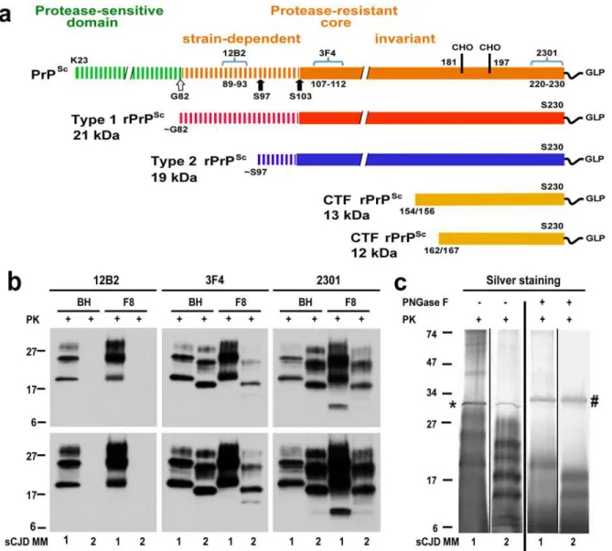
Fig 1. Schematic representation of PK-resistant fragments in rPrPSccorresponding to Type 1 (MM1) and Type 2 (MM2) sCJD prions and molecular characteristics of purified human rPrPSc used in structural studies.(a) Outline of classification of Type 1 and Type 2 human prions based on proteolytic fragmentation of PrPSc[5,52]. Major cleavage sites by PK are indicated by arrows; GLP—glycolipid; CHO- complex N-glycosylation chains. The codes above light blue brackets represent monoclonal antibodies used in differentiation of various domains of human prions, and the numbers below these brackets indicate linear epitopes recognized by these antibodies. (b) Distinct glycosylation patterns and electrophoretic mobilities of purified human Type 1 (MM1) and Type 2 (MM2) sCJD rPrPSc(homozygous for methionine (M) in codon 129) used in structural studies. To differentiate Type 1, Type 2 prions, and their C-terminal fragments, Western blots of purified rPrPSc(fraction 8; F8) from the brain homogenate (BH) of type MM1 and MM2 sCJD were developed with mAb 12B2 (epitope residues 89–93) [53], mAb 3F4 (epitope residues 107–112) [54], and rabbit polyclonal antibody 2301 (epitope residues 220–231) [55]. The lower panels correspond to prolonged exposure of the same WB to detect less abundant low molecular weight fragments of rPrPSc. (c) Distinct fragmentation patterns of purified MM1 and MM2 sCJD prions in silver stained SDS-PAGE before and after deglycosylation with PNGase F. The symbols (*) and (#) indicate bands corresponding to PK and PNGase F, respectively. The molecular weights of marker proteins are in kDa.
doi:10.1371/journal.ppat.1004832.g001
Results and Discussion
From the collection of samples obtained from 340 patients with an unequivocal diagnosis of Type 1 (MM1) and Type 2 (MM2) sCJD, we selected one case that is representative of each neuropathology group (S1 Fig) and displayed99% pure Type 1 or Type 2 proteinase K-resistant PrPSc(rPrPSc), as detected by both conformation dependent immunoassay (CDI) and Western blots [13,14,20]. The disease duration in these representative cases, as well as bio-chemical characteristics of brain PrPScassociated with them (levels of total PrPScand rPrPSc, size of PrPScparticles, conformational stability of PrPSc) correspond to the respective median values reported previously for each group [13,14] (Table 1).
The native prion particles containing rPrPScfrom these two cases were purified for structural studies with a scaled up protocol we developed previously for purification of infectious and structurally intact Sc237 prions from Syrian hamster brains [21]. The Western blot patterns of purified MM1 and MM2 rPrPScin the final fraction 8 (F8) and in the original brain homogenates (BH) were superimposable, documenting complete qualitative recovery of rPrPScfrom brain ho-mogenates (Fig 1b). As expected [22], the mass of unglycosylated fragments was ~21 kDa in Type 1 and ~19 kDa in Type 2 rPrPSc, and Type 2 rPrPScwas not detectable with mAb 12B2 due to the missing N-terminal epitope (Fig 1bandS2b Fig). The 12–13 kDa C-terminal fragments were more abundant in Type 1 rPrPScand detectable in Type 2 after longer exposure (Fig 1b). The silver-stained gels demonstrated the pattern of rPrPSccorresponding to the major bands on Western blots, and the isolated rPrPScwas ~90% pure (Fig 1c). These patterns were highly repro-ducible upon purification of rPrPScfrom different cortical areas of the same brain (S2 Fig).
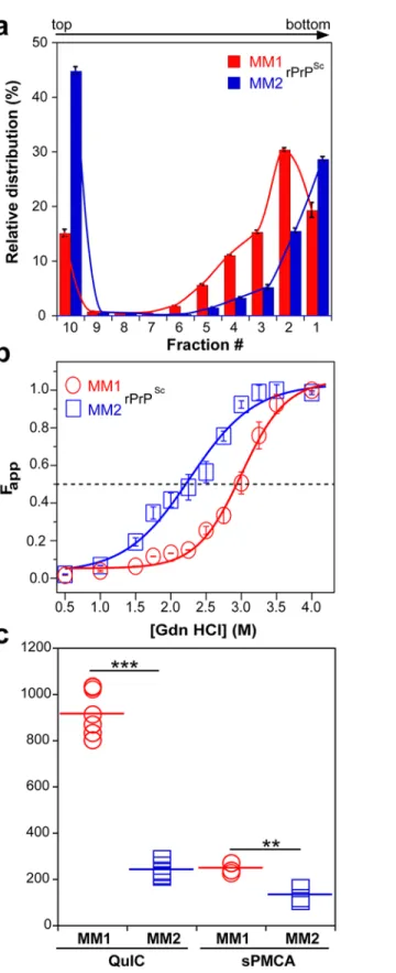
To investigate the prion size, we separated sCJD prion particles according to sedimentation velocity in sucrose gradient [13]. Consistent with previous data, the peak sedimentation veloci-ty of MM1 rPrPScwas found to be substantially slower than that of MM2 rPrPSc[13] (Fig 2a). Based on calibration with standard proteins [13], we estimate that the majority of MM1 rPrPSc particles have a molecular mass of 9-11x106Da (~380–460 monomers), whereas the respective value for MM2 rPrPScparticles is14x106Da (600 monomers) (Fig 2aandTable 1).
Using CDI, we compared the conformational stability of rPrPScobtained from different brain cortex areas in four independent purification rounds from each sCJD case (Fig 2b). The average stability of MM1 rPrPScagainst denaturation by GdnHCl was significantly higher than that of MM2 rPrPSc, with GdnHCl concentration corresponding to midpoint denaturation of 3.0 and 2.3 M, respectively (Fig 2bandTable 1).
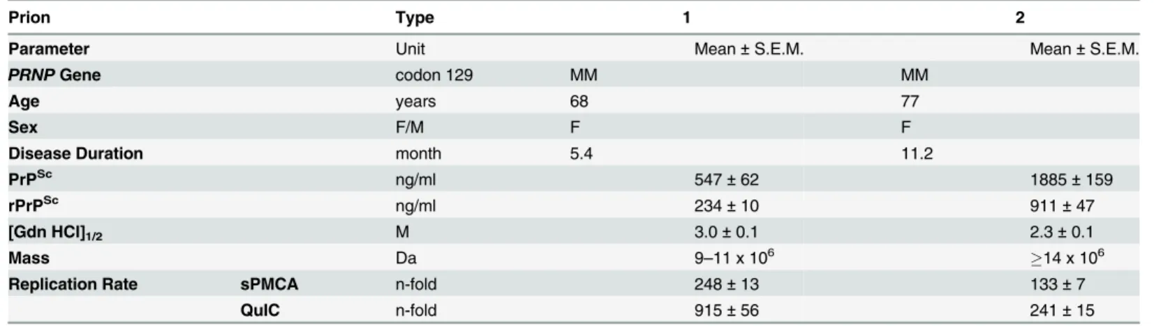
Table 1. Source, biophysical characteristics, and replication rate of Type 1 and Type 2 sCJD prions.
Prion Type 1 2
Parameter Unit Mean±S.E.M. Mean±S.E.M.
PRNPGene codon 129 MM MM
Age years 68 77
Sex F/M F F
Disease Duration month 5.4 11.2
PrPSc ng/ml 547±62 1885±159
rPrPSc ng/ml 234±10 911±47
[Gdn HCl]1/2 M 3.0±0.1 2.3±0.1
Mass Da 9–11 x 106 14 x 106
Replication Rate sPMCA n-fold 248±13 133±7
QuIC n-fold 915±56 241±15
Fig 2. Sedimentation velocity, conformational stability, and seeding potency of isolated sCJD prions.
(a) Distinct sedimentation velocity profiles of MM1 and MM2 prions. The samples were fractionated by ultracentrifugation in sucrose gradient and fractions were collected from the bottom of the tubes and analyzed for rPrPScby CDI. The bars represent average±SEM; CDI was performed on each sCJD sample in triplicate. (b) The conformational stability of MM1 and MM2 rPrPSc. The curves represent best fit to a sigmoidal
Next, we assessed the seeding efficacy (amplification index) of MM1 rPrPScand MM2 rPrPScin vitrousing two different methods, QuIC and sPMCA. The amplification index (po-tency) of different seeds is expressed as a ratio between the concentration of the PrPSc conform-ers produced with PMCA or QuIC divided by the concentration of PrPScin the seed after subtracting the background obtained in control unseeded samples. Detailed protocols of these methods and control experiments showing lack of spontaneous prion protein conversion in the unseeded reactions have been described previously [13]. In both assays, the seeding efficacy of MM1 rPrPScwas markedly higher compared to that of MM2 rPrPSc(Fig 2candTable 1). This higher replication efficacy of MM1 prions from the case selected for the present structural studies is consistent with ~4-fold higher median replication potency of MM1 prions compared to MM2 prions we previously observed (using both QuIC and sPMCA techniques) for prions from ten MM1 and ten MM2 sCJD cases (SupplementalS2 Figin [13]). These data in vitro are also in accord with available bioassay data that demonstrate higher transmission rates and sig-nificantly shorter incubation times of MM1 sCJD prions in transgenic mice expressing human PrPC(129M) or human/mouse PrPCchimeras [8,9]. The higher replication efficiency of the conformationally more stable MM1 rPrPScis both intriguing and unexpected, as some previous experiments with mouse prion strains suggest that there is an inverse correlation between prion replication rate and conformational stability of total PrPSc(i.e., the less stable conformers should replicate faster) [11,12]. Our present data indicate that this previously suggested rela-tionship does not apply to sCJD rPrPSc.
Clearly, understanding the molecular basis of phenotypic variability in sCJD requires struc-tural characterization of PrPScthat goes beyond relatively crude assays such as proteolytic frag-mentation followed by Western blotting or conformational stability measurements. This is a challenging task because most of the methods developed for structural studies of protein aggre-gates are not applicable to brain-derived PrPSc, as they require isotopic labeling or introduction of other spectroscopic probes. However, new opportunities in this regard are offered by two mass spectrometry based methods: backbone amide hydrogen/deuterium exchange coupled with mass spectrometry (HXMS) [23] and histidine hydrogen/deuterium exchange mass spec-trometry (His-HXMS) [24]. Here, we used these two methods for structural comparison of MM1 rPrPScand MM2 rPrPSc.
The HXMS method measures the rate of H/D exchange of protein backbone amide hydro-gen atoms. Since the exchange rates are much faster for protein segments that are unstructured as compared to those that are involved in H-bonded structures such asα-helices orβ-sheets, these measurements provide a sensitive tool for conformational analysis. This approach, which we recently successfully used for structural analysis of strain-specific differences in murine pri-ons [23], is especially useful for studying amyloids and related protein aggregates, as the ex-change rates within theβ-sheet cores of these aggregates are exceptionally slow [25–30].
The first step in HXMS analysis is the generation of peptic fragments that can be separated by ultrahigh performance liquid chromatography (UHPLC) and identified by MS. Both for MM1 and MM2 rPrPSc, we were able to identify 27 peptic fragments that give rise to MS spectra with a signal-to-noise ratio sufficient for reliable calculation of deuterium incorporation. These
function. The values of apparent fractional change (Fapp) are mean±SEM obtained from four batches of purified MM1 and MM2 prions, each determined in triplicate measurements. (c) Amplification of MM1 and MM2 sCJD prions by QuIC using recombinant human PrP(23–231, 129M) substrate and by sPMCA using brain homogenate of Tg mice expressing human PrPC(129M). The amplification index is the ratio between the concentration of PrPScbefore and after PMCA measured with CDI. The data points represent results of six QuIC and three sPMCA experiments, each measured in triplicate with CDI. The mean values are indicated by horizontal lines.***P<0.001,**P<0.005 determined by ANOVA.
fragments (some of them partially overlapping) cover ~85% of the C-terminal region 117–224, with the only significant gap for the segment 169–181 that contains one of the glycosylation sites (likely due to a very low concentration of peptic fragment(s) derived from the nonglycosy-lated component of rPrPSc). No peptic fragments could be analyzed from the N-terminal region up to residue 116, presumably due to the ragged N-terminus of human rPrPSc. The extent of deuterium incorporation for MM1 rPrPScand MM2 rPrPScafter 5 min and 240 h incubation in D2O is shown inFig 3. For both rPrPSctypes, the region of relatively little protection against
deuterium incorporation maps to residues ~145–160. This is in striking contrast to murine prion strains studied to date, in which case this central region is characterized by high degree of
Fig 3. Deuterium incorporation for peptic fragments derived from MM1 rPrPSc(red) and MM2 rPrPSc(blue).(a) 5 min incubation in D2O. (b) 240 h incubation in D2O. Error bars indicate standard deviation (3 independent experiments).*, P<0.05;**, P<0.02.
doi:10.1371/journal.ppat.1004832.g003
protection against H/D exchange [23]. Thus, it appears that the ~145–160 region of sCJD prions is structurally less ordered than the same region in cloned murine prion strains.
In contrast to similar protection against deuterium incorporation in the ~145–160 regions of MM1 and MM2 rPrPSc, there are substantial differences in other parts of rPrPSc correspond-ing to distinct sCJD phenotypes. This is especially evident for the 117–144 region, as peptic fragments derived from this part of MM1 rPrPScconsistently show higher degree of H/D ex-change as compared to those corresponding to the same region in MM2 rPrPSc. The difference is particularly striking for the 117–133 region, in which case the degree of deuterium incorpo-ration after 240 h exchange is 2.5–3 fold lower for MM1 rPrPSc, indicating markedly higher level of structural order in this part of MM1 rPrPScas compared to MM2 rPrPSc. An opposite trend is observed for the C-terminal region ~161–224, where higher protection against H/D ex-change is observed for MM2 rPrPSc(Fig 3). Altogether, these data clearly demonstrate substan-tial structural differences between rPrPSccorresponding to two different phenotypes of sCJD. The resolution of HXMS alone is not sufficient to propose any specific structural model that could account for these differences. However, within the context of the frequently considered model based on the parallel in-registerβ-structure motif [31,32], region-specific differences in resistance to H/D exchange observed between MM1 PrPScand MM2 PrPSccould likely reflect factors such as different proportions in these regions of residues involved inβ-strands and loops between them and/or packing differences between individualβ-strands. As in the case of murine prions [23], high level of protection against H/D exchange in the C-terminal region of sCJD PrPScis not compatible with the structural model proposing that residues ~89–175 form left-handedβ-helices, with the C-terminal region retaining the native-likeα-helical conforma-tion of PrPC[33]. However, the present data alone do not exclude the possibility that the entire PK-resistant region of PrPSccould form aβ-helix-like structure.
Structural properties of sCJD prions were further probed using the recently developed ap-proach of His-HXMS which measures the rate of H/D exchange of C2 protons in histidine side chains [34–36]. Information provided by this method is complementary to that obtained from amide HXMS measurements: while amide HXMS probes protein structural organization and dynamics at the level of the polypeptide backbone, His-HXMS probes the microenvironment (water accessibility) of specific His side chains [34–36]. As shown in a recent study with recom-binant prion protein amyloid fibrils, the latter approach can be particularly useful in probing quaternary structure of ordered protein aggregates, providing information about the packing arrangement and interfaces betweenβ-sheets [31].
There are six His residues in the PK-resistant region of human PrPSc(His99, His111, His140, His150, His177 and His187). MS signal for the peptide fragment containing His99 was too weak to allow reliable measurements. However, high quality H/D exchange data could be obtained for five other His residues. In the native structure of the PrPCmonomer, all these His side chains are fully exposed to water. Thus, as expected for unprotected histidines [34,35], the half-times of exchange are about 2–3 days. In the rPrPScstructures, these half-times are substantially longer, indicating that all His side chains are located in at least partially water-protected environment (Fig 4). However, the degree of this protection for individual His side chains varies greatly between MM1 rPrPScand MM2 rPrPSc. For example, in MM1 rPrPSc, His177 is still in a relatively water accessible environment (exchange half-time of 9 days), whereas in MM2 PrPSc, this side chain is much more protected from water (exchange half-time of 56 days). An opposite situation is observed for His111, in which the environment of the side chain is much more water-protected in the structure of MM1 rPrPScthan that of the MM2 counterpart (exchange half-times of 67 and 16 days, respectively).
interdigitation of side chains (“dry”steric zipper) and one that is more accessible to water [37,38]. Within this context, the differences in water exposure of individual His side chains ob-served between MM1 rPrPScand MM2 rPrPSccould be explained by distinct packing arrange-ments ofβ-sheets in these two structures (i.e., the same His side chain being in dry or wet interface depending on the rPrPSctype). It should be noted, however, that even for the most protected His side chains in rPrPSc, the exchange half-times are substantially shorter than those recently observed in synthetic amyloid fibrils prepared from the recombinant PrP (hun-dreds of hours), suggesting that steric zippers in brain-derived rPrPScmight be less perfect than those in synthetic amyloid fibrils. This is not entirely surprising given that rPrPScparticles con-tain glycosylated isoforms, and glycans may interfere with packing betweenβ-sheets.
Experiments with two strains of yeast prion [PSI+] demonstrated that, in this case, the criti-cal determinant of the strength of prion phenotype is the susceptibility of prion aggregates to fragmentation (that creates new ends for monomer recruitment), with the less stable structure corresponding to the stronger phenotype [39]. This fragmentation and maintenance of the yeast prion statein vivois believed to be mediated by the molecular chaperone Hsp104 [39,40]. Even though there are no known mammalian homologs of the disaggregating chaperone HsP104, the general hypothesis that less stable prions are more virulent has been adopted in the field of mammalian prions, and this model appeared to be supported by studiesin vivo
with some rodent prion strains [11,12,41], even though the results of these studies could also be explained by strain-dependent differences in prion clearance rates. Furthermore, an inverse correlation was found between the replication tempoin vitroand the conformational stability of the protease-sensitive sCJD PrPSc(but not the protease-resistant component, rPrPSc) [13]. However, data for some other rodent prion strains appear to be inconsistent with this model [42,43]. The picture is further complicated by the fact that there are no reliable direct assays to
Fig 4. Histidine H/D exchange for monomeric PrPC(black), MM1 rPrPSc(red) and MM2 rPrPSc(blue).
The parameter t1/2represents the half-time of exchange reaction for individual His residues. Error bars indicate standard deviation (3 independent experiments).**, P<0.01;***, P<0.001.
doi:10.1371/journal.ppat.1004832.g004
probe fragmentation susceptibility of PrPScaggregates, and their stability is typically assessed by measuring resistance to denaturation with SDS or chaotropes; the relationship between the latter property and fragility is not necessarily straightforward.
Our present data clearly demonstrate that, in contrast to the observations for some murine prions, lower conformational stability of sCJD rPrPScdoes not result in higher replication rate of these prions. Thus, at least in the case of human prions, conformational stability of rPrPSc (as defined by resistance to denaturation with SDS or chaotropes) is definitely not a reliable predictor of the incubation period of the disease. Importantly, our structural studies allowed us to identify substantial differences between the molecular organization of MM1 and MM2 rPrPSc, both at the level of the polypeptide backbone as well as the quaternary packing arrange-ments. As shown in our recent study with recombinant PrP amyloid fibrils[24], the differences in packing betweenβ-sheets may result in distinct conformational stabilities. However, it ap-pears that it is not the conformational stabilityper sethat controls the replication rate of rPrPSc, as the observed faster replication of MM1 sCJD prions when compared to MM2 counterparts would imply a paradoxical scenario in which higher stability of rPrPScresults in a faster replica-tion tempo. Instead, our data strongly suggest that distinct replicareplica-tion rates of MM1 and MM2 sCJD prions are dictated by specific structural features of corresponding rPrPScaggregates, features that control the intrinsic growth rate of these aggregates (i.e., the rate of templated conformational conversion of the PrPCsubstrate). Thus, the balance of factors controlling strain-specific replication tempo of sCJD prions appears to be diametrically different from that described for yeast prions [PSI+] that are associated with aggregation of Sup35 protein. In the latter case, the intrinsic elongation rate of Sup35 amyloid fibrils Sc4 corresponding to the stron-ger (faster replicating) prion phenotype is slower than that of fibrils Sc37 corresponding to the weaker phenotype, but this is more than compensated by lower stability (and thus higher effec-tive concentration of ends) for Sc4 fibrils [39]. By contrast, the faster replicating strain of sCJD prion is characterized by higher conformational stability, implying that, in this case, the domi-nant factor in controlling the replication tempo is not prion stability but the intrinsic growth rate (i.e., the rate of the conversion of PrPCmonomers).
Considerable structural differences between type 1 and type 2 PrPScin sCJD are especially intriguing given frequent coexistence of these two prion strains in affected individuals [20]. Whether this strain coexistence is the result of a primordial spontaneous misfolding or confor-mational evolution due to the template flipping during passage through cells expressing differ-ent post-translationally modified PrPCremains to be determined [19]. It should also be noted that type 1 and type 2 sCJD prions represent only a small fraction of the spectrum of human prions. It is likely that the structural variability among PrPSccorresponding to different familial forms of human prion diseases might be even larger than the extent of structural differences described herein for type 1 and type 2 sCJD prions.
Materials and Methods
Ethics statement and clinicopathologic characteristics of sCJD cases
duration; (2) methionine homozygous at codon 129 of the human prion protein (PrP) gene (PRNP); (3) unequivocal classification as pure Type 1 or Type 2 sCJD according to WB pattern; (4) unequivocal classification of pathology as definite Type 1 or 2 at the National Prion Disease Pathology Surveillance Center (NPDPSC) in Cleveland, Ohio; (5) demographic data distribu-tion within 95% confidence interval of the whole group, resulting in no difference between se-lected cases and the whole group in any of the statistically followed parameters.
Purification of prions from sCJD human brains
The purification of rPrPScfrom human brains was performed as described previously for 263K prions from Syrian hamster brains [21] with the following additional steps. The partially puri-fied samples containing ~10μg of human PrPScwere resuspended in PBS, pH7.4 containing 2
mM CaCl2and 2% Sarkosyl, sonicated in a sonication bath (3 x 5 s), and incubated with 70μg/
ml of Collagenase (Worthington Biochemical Corporation) with shaking at 600 rpm in Eppen-dorf Thermomixer for 4 h at 37°C. After adding MgCl2to a final concentration of 5 mM, the
samples were incubated with 50 IU/ml of Benzonase (Novagen/EMP) for additional 1 h at 37°C, followed by 1 h incubation with 100μg/ml of proteinase K (Amresco, Solon, OH/
Invi-trogen) at 37°C. The PK was blocked with protease inhibitor (PI) cocktail containing 0.5mM PMSF, and 5μg/ml of aprotinin and leupeptin, respectively. The pellet obtained after
centrifu-gation (30 min, 18,000 x g, 4°C) in Allegra centrifuge equipped with F2402H rotor was resus-pended in 400μl of 10% NaCl containing 1% Sarkosyl and PI cocktail, and spun again. The
final pellet was resuspended in PBS containing 2% Sarkosyl and PI cocktail (1:1000, v/v), and delipidated overnight with four volumes of Methanol/Chloroform (2:1, v/v) at -20°C. Finally, the sample was collected by centrifugation, resuspended in water containing 0.1% Sarkosyl and stored at -80°C.
Physicochemical properties and molecular characteristics of purified
sCJD prions
The purified rPrPScwas analyzed by SDS PAGE followed by silver staining and/or western blots, and by conformation-dependent immunoassay (CDI). The latter assay was performed as described previously [6,14] with the following minor modifications. First, we used white Lumi-trac 600 High Binding Plates (E&K Scientific, Santa Clara, California) coated with mAb 8H4 (epitope 175–185)[46] in 200 mM NaH2PO4containing 0.03% (w/v) NaN3, pH 7.5. Second,
al-iquots of 20μl from each fraction containing 0.007% (v/v) of Patent Blue V (Sigma) were
di-rectly loaded into wells of white strip plates prefilled with 200μl of Assay Buffer (Perkin Elmer,
Waltham, Massachusetts). Finally, the captured PrP was detected by a europium-conjugated [47] anti-PrP mAb 3F4 (epitope 107–112) and the time-resolved fluorescence (TRF) signals were measured by the multi-mode microplate reader PHERAstar Plus (BMG LabTech, Dur-ham, North Carolina). The recHuPrP(90–231,129M) and PrP(23–231,129M) used as a cali-brant in the CDI was prepared and purified as described previously [48]. The conformational stability of rPrPScwas determined with CDI as described previously [14,47] and the raw CDI signal was converted into the apparent fractional change and fitted by least square method with a sigmoidal transition model to determine GdnHCl concentration where 50% of PrPScis unfolded ([Gdn HCl]1/2) [14]. The sedimentation velocity and mass of sCJD prions was
deter-mined with calibrated sucrose gradient ultracentrifugation as described [13].
Replication rate of sCJD prions measured in vitro
The Quaking-induced Conversion (QuIC) [49] and sonication-driven serial Protein Misfolding Cyclic Amplification (sPMCA) [50] procedures were performed essentially as described
previously [13,14]. Briefly, rhuPrP(23–231,129M) used as a substrate in QuIC was expressed, purified, and refolded toα-helical conformation as described previously [48], and its initial concentration was calculated from absorbance at 280 nm using the molar extinction coefficient 56650 M-1cm-1. The stock of rhuPrP(23–231) in 10 mM sodium acetate buffer, pH 4.0, was pretreated with 12 mM HCl [rhuPrP:HCl ratio (v/v) of 1:3.9] for 7.5 min and immediately di-luted to a final concentration of 0.1 mg/ml into the reaction buffer composed of 20 mM NaH2PO4, 130 mM NaCl, pH 6.9, and containing 0.1% SDS, 0.1% Triton X-100, and 1:5000
(v/v) N2 (Invitrogen, Carlsbad, California). The QuIC was performed with final volume of 100μl per well in a sterile V-bottom, low-binding polypropylene 96-well plate (VWR,
Arling-ton Heights, Illinois) equipped with a 3 mm diameter PTFE bead (Fisher Scientific, Pittsburgh, Pennsylvania) in each well. The aliquots of sCJD brain homogenates were diluted into the com-plete QuIC reaction buffer to obtain final 10-4dilution of sCJD prions, and the plates were sealed with sterile AxyMat Silicone Sealing Mat (VWR, Arlington Heights, Illinois). The QuIC was performed in samples seeded with sCJD PrPScat 55°C for 20 hrs in an Eppendorf Thermo-mixer (Eppendorf, Hauppauge, New York) set for 1 min shaking at 1400 rpm, followed by 1 min incubation. The reaction was stopped by adding to each well 50μl of PBS (pH 6.9)
con-taining 3% (w/v) Sarkosyl and Proteinase K (PK; Amresco, Solon, Ohio) to obtain the final Sar-kosyl concentration of 1% (w/v) and PrP/PK ratio of 10:1 (w/w). The plates were incubated for 1 h at 37°C with shaking at 1200 rpm on the Eppendorf Thermomixer with 1 min intervals. The PK was blocked in each well with protease inhibitors (0.5 mM PMSF, 5 ug/ml of aprotinin and leupeptin) and the PK-resistant PrP was measured with CDI [13,14].
Sonication-driven serial Protein Misfolding Cyclic Amplification (sPMCA) of sCJD samples was performed as described [50] with the following modifications. Human PrPScwas replicated using brains of transgenic mice overexpressing human PrP with methionine at position 129 [51]. The 10% brain homogenates from sCJD patients were diluted 1000-fold into 10% normal brain homogenate and 100μl was transferred into 0.2 ml PCR tubes equipped with 2.38 mm
diameter PTFE ball (K-mac Plastics, Wyoming, Michigan). Tubes were positioned on an adap-tor placed on the plate holder of a microsonicaadap-tor (Misonix Model 3000, Farmingdale, New York) programmed to perform cycles of 60 min incubation at 32°C followed by a 30 s pulse of sonication set at 80% power. Samples were incubated, without shaking, and immersed in the water of the sonicator bath. After a round of 24 cycles, a 10μl aliquot of the amplified material
was diluted into 90μl of normal transgenic mouse brain homogenate and a new round of 24
PMCA cycles was performed. This procedure was repeated four times to reach a final 106-fold dilution of the initial sCJD brain homogenate, and the replication rate was calculated from PrPSccontent measured before and after sPMCA with CDI [13,14,19].
Backbone amide hydrogen/deuterium exchange mass spectrometry
experiments (HXMS)
To initiate deuterium labeling, 10μl aliquots of purified sCJD rPrPSc(~1.8μg) were collected
by centrifugation (21000×g, 30 min, 4°C) and added to 100μl of 10 mM phosphate buffer (pH
7.3) in D2O. After incubation at room temperature for different time periods, samples were
col-lected by centrifugation and dissociated into monomers by adding 20μl of ice cold 100 mM
column were analyzed by an LTQ Orbitrap XL mass spectrometer (ThermoElectron, San Jose, CA). To minimize back-exchange, both the trap and the analytical column were placed in a cooled chamber (~2°C) integrated with a LEAP TriValve system (LEAP Technologies, USA). The extent of deuterium incorporation in each peptic fragment was determined from mass spectra (with a correction for back-exchange) as described previously [23].
Histidine hydrogen/deuterium exchange (His-HXMS) experiments
For these measurements, samples of purified sCJD rPrPScfrom human brain (~3μg) were
sus-pended in D2O buffer (10 mM sodium phosphate, 10μM EDTA, 50μM Pefabloc, 1 ug/ml
Aprotinin, pH 9.0). After incubation for 5 days at 37°C, samples were collected by centrifugation and deglycosylated with PNGase F. To obtain fragments containing single His residues, samples were then digested with immobilized pepsin, followed by digestion with immobilized trypsin. Finally, the peptic fragments were separated on an UPLC column and analyzed by mass spec-trometry as described above for HXMS experiments. The pseudo-first-order rate constant (k) of His hydrogen exchange reaction was determined by the equation:k = -ln{1-[((R(t)—R(0))/
((1 + R(t)—R(0))] x 1/P}/t, where P is the fractional D2O content in the solvent, R is the ratio of
M+1/M isotopic peak of a given peptide before (time = 0) and after the H/X reaction (time = t). The half-life (t1/2, days) of His exchange reaction was calculated using the equation:t1/2(day) =
ln2/k/24, where k (hour-1) is the rate constant at the alkaline conditions (pH = 9) [34,35].
Supporting Information
S1 Fig. Distinct neuropathologic characteristics of the occipital neocortex in Type 1 (a, c) and Type 2 (b, d) sCJD cases homozygous for methionine in codon 129 of PRNP gene and used as a source of human prions in structural studies.(a, b) Spongiform degeneration. Typical fine vacuole-type spongiform changes with diffuse small round vacuoles in Type 1 (a) contrast with large coarse fused vacuoles in Type 2 sCJD (b). (c, d) PrPScdeposition. Dispersed punctate (synaptic-type) PrPScdeposition in occipital cortex of Type 1 sCJD (c) contrasts with large pla-que-like deposits frequently associated with vacuols in Type 2 sCJD (d). Scale bar is 50μm.
(PDF)
S2 Fig. Highly reproducible electrophoretic patterns of MM1 and MM2 sCJD prions puri-fied from different cortex areas of the same human brain.(a) The silver staining after SDS-PAGE of ~300 ng of purified rPrPScfrom different cortical areas of the same sCJD Type 1 (lanes I-III in the left panel) and Type 2 (lanes I-IV in the right panel) case before and after deglycosylation. Asterisk () and double dagger (#) point to the bands of PK and PNGase F,
re-spectively. (b) Western blot analysis of the purified human MM1 and MM2 sCJD prions before and after deglycosylation. The lower panels are from the same WB taken after longer exposure to detect less abundant low mass fragments of rPrPSc. The molecular weights of the marker proteins are in kD.
(PDF)
Acknowledgments
The authors are grateful to the patient’s families, the CJD Foundation, and all the members of the National Prion Disease Pathology Surveillance Center for invaluable technical help. We thank Dr. Glenn Telling for brains of transgenic mice expressing PrPC, Dr. Man-Sun Sy for making available hybridoma clone 8H4, and Dr. Earl Poptic for scaled up antibody production.
Author Contributions
Conceived and designed the experiments: JGS WKS. Performed the experiments: XX SC MEK CK TH. Analyzed the data: JGS WKS XX SC. Wrote the paper: JGS WKS. Sampled the brains and performed diagnostic assays: YC WC. Performed diagnostic pathology: MLC.
References
1. Prusiner SB, Scott MR, DeArmond SJ, Carlson G (2004) Transmission and replication of prions. In: Prusiner SB, editor. Prion Biology and Diseases. 2nd ed. Cold Spring Harbor: Cold Spring Harbor Laboratory Press. pp. 187–242.
2. Gajdusek DC, Gibbs CJ Jr., Alpers M (1966) Experimental transmission of a kuru-like syndrome to chimpanzees. Nature 209: 794–796. PMID:5922150
3. Gibbs CJ Jr., Gajdusek DC, Asher DM, Alpers MP, Beck E, et al. (1968) Creutzfeldt-Jakob disease (spongiform encephalopathy): transmission to the chimpanzee. Science 161: 388–389. PMID: 5661299
4. Puoti G, Bizzi A, Forloni G, Safar JG, Tagliavini F, et al. (2012) Sporadic human prion diseases: molec-ular insights and diagnosis. Lancet Neurol 11: 618–628. doi:10.1016/S1474-4422(12)70063-7PMID: 22710755
5. Gambetti P, Kong Q, Zou W, Parchi P, Chen SG (2003) Sporadic and familial CJD: classification and characterisation. Br Med Bull 66: 213–239. PMID:14522861
6. Safar JG, Geschwind MD, Deering C, Didorenko S, Sattavat M, et al. (2005) Diagnosis of human prion disease. Proc Natl Acad Sci USA 102: 3501–3506. PMID:15741275
7. Safar JG (2012) Molecular Mechanisms Encoding Quantitative and Qualitative Traits of Prion Strains. In: Gambetti P, editor. Prions and Diseases. New York: Springer Verlag.
8. Bishop MT, Will RG, Manson JC (2010) Defining sporadic Creutzfeldt-Jakob disease strains and their transmission properties. Proc Natl Acad Sci U S A 107: 12005–12010. doi:10.1073/pnas.1004688107 PMID:20547859
9. Giles K, Glidden DV, Patel S, Korth C, Groth D, et al. (2010) Human prion strain selection in transgenic mice. Ann Neurol 68: 151–161. doi:10.1002/ana.22104PMID:20695008
10. Wadsworth JD, Joiner S, Linehan JM, Desbruslais M, Fox K, et al. (2008) Kuru prions and sporadic Creutzfeldt-Jakob disease prions have equivalent transmission properties in transgenic and wild-type mice. Proc Natl Acad Sci U S A 105: 3885–3890. doi:10.1073/pnas.0800190105PMID:18316717
11. Legname G, Nguyen H-OB, Peretz D, Cohen FE, DeArmond SJ, et al. (2006) Continuum of prion pro-tein structures enciphers a multitude of prion isolate-specified phenotypes. Proc Natl Acad Sci USA 103: 19105–19110. PMID:17142317
12. Colby DW, Giles K, Legname G, Wille H, Baskakov IV, et al. (2009) Design and construction of diverse mammalian prion strains. Proc Natl Acad Sci U S A 106: 20417–20422. doi:10.1073/pnas.
0910350106PMID:19915150
13. Kim C, Haldiman T, Surewicz K, Cohen Y, Chen W, et al. (2012) Small Protease Sensitive Oligomers of PrP(Sc) in Distinct Human Prions Determine Conversion Rate of PrP(C). PLoS Pathog 8: e1002835. doi:10.1371/journal.ppat.1002835PMID:22876179
14. Kim C, Haldiman T, Cohen Y, Chen W, Blevins J, et al. (2011) Protease-Sensitive Conformers in Broad Spectrum of Distinct PrP Structures in Sporadic Creutzfeldt-Jakob Disease Are Indicator of Progression Rate. PLoS Pathog 7: e1002242. doi:10.1371/journal.ppat.1002242PMID:21931554
15. Uro-Coste E, Cassard H, Simon S, Lugan S, Bilheude JM, et al. (2008) Beyond PrP9res) type 1/type 2 dichotomy in Creutzfeldt-Jakob disease. PLoS Pathog 4: e1000029. doi:10.1371/journal.ppat. 1000029PMID:18389084
16. Wadsworth JD, Collinge J (2011) Molecular pathology of human prion disease. Acta Neuropathol 121: 69–77. doi:10.1007/s00401-010-0735-5PMID:20694796
17. Gambetti P, Cali I, Notari S, Kong Q, Zou WQ, et al. (2011) Molecular biology and pathology of prion strains in sporadic human prion diseases. Acta Neuropathol 121: 79–90. doi: 10.1007/s00401-010-0761-3PMID:21058033
18. Polymenidou M, Stoeck K, Glatzel M, Vey M, Bellon A, et al. (2005) Coexistence of multiple PrPSc types in individuals with Creutzfeldt-Jakob disease. Lancet Neurol 4: 805–814. PMID:16297838
20. Cali I, Castellani R, Alshekhlee A, Cohen Y, Blevins J, et al. (2009) Co-existence of scrapie prion pro-tein types 1 and 2 in sporadic Creutzfeldt-Jakob disease: its effect on the phenotype and prion-type characteristics. Brain 132: 2643–2658. doi:10.1093/brain/awp196PMID:19734292
21. Safar J, Roller PP, Gajdusek DC, Gibbs CJ Jr. (1993) Conformational transitions, dissociation, and un-folding of scrapie amyloid (prion) protein. J Biol Chem 268: 20276–20284. PMID:8104185
22. Parchi P, de Boni L, Saverioni D, Cohen ML, Ferrer I, et al. (2012) Consensus classification of human prion disease histotypes allows reliable identification of molecular subtypes: an inter-rater study among surveillance centres in Europe and USA. Acta Neuropathol 124: 517–529. doi: 10.1007/s00401-012-1002-8PMID:22744790
23. Smirnovas V, Baron GS, Offerdahl DK, Raymond GJ, Caughey B, et al. (2011) Structural organization of brain-derived mammalian prions examined by hydrogen-deuterium exchange. Nat Struct Mol Biol 18: 504–506. doi:10.1038/nsmb.2035PMID:21441913
24. Cobb NJ, Apostol MI, Chen S, Smirnovas V, Surewicz WK (2014) Conformational stability of mammali-an prion protein amyloid fibrils is dictated by a packing polymorphism within the core region. J Biol Chem 289: 2643–2650. doi:10.1074/jbc.M113.520718PMID:24338015
25. Del Mar C, Greenbaum EA, Mayne L, Englander SW, Woods VL Jr. (2005) Structure and properties of alpha-synuclein and other amyloids determined at the amino acid level. Proc Natl Acad Sci U S A 102: 15477–15482. PMID:16223878
26. Lu X, Wintrode PL, Surewicz WK (2007) Beta-sheet core of human prion protein amyloid fibrils as deter-mined by hydrogen/deuterium exchange. Proc Natl Acad Sci U S A 104: 1510–1515. PMID:17242357
27. Smirnovas V, Kim JI, Lu X, Atarashi R, Caughey B, et al. (2009) Distinct Structures of Scrapie Prion Protein (PrPSc)-seeded Versus Spontaneous Recombinant Prion Protein Fibrils Revealed by Hydrogen/Deuterium Exchange. J Biol Chem 284: 24233–24241. doi:10.1074/jbc.M109.036558 PMID:19596861
28. Toyama BH, Kelly MJ, Gross JD, Weissman JS (2007) The structural basis of yeast prion strain vari-ants. Nature 449: 233–237. PMID:17767153
29. Toyama BH, Weissman JS (2011) Amyloid structure: conformational diversity and consequences. Annu Rev Biochem 80: 557–585. doi:10.1146/annurev-biochem-090908-120656PMID:21456964
30. Miller MB, Wang DW, Wang F, Noble GP, Ma J, et al. (2013) Cofactor molecules induce structural transformation during infectious prion formation. Structure 21: 2061–2068. doi:10.1016/j.str.2013.08. 025PMID:24120764
31. Cobb NJ, Sonnichsen FD, McHaourab H, Surewicz WK (2007) Molecular architecture of human prion protein amyloid: a parallel, in-register beta-structure. Proc Natl Acad Sci U S A 104: 18946–18951. PMID:18025469
32. Groveman BR, Dolan MA, Taubner LM, Kraus A, Wickner RB, et al. (2014) Parallel in-register intermo-lecular beta-sheet architectures for prion-seeded prion protein (PrP) amyloids. J Biol Chem 289: 24129–24142. doi:10.1074/jbc.M114.578344PMID:25028516
33. Govaerts C, Wille H, Prusiner SB, Cohen FE (2004) Evidence for assembly of prions with left-handed beta-helices into trimers. Proc Natl Acad Sci U S A 101: 8342–8347. PMID:15155909
34. Miyagi M, Nakazawa T (2008) Determination of pK(a) values of individual histidine residues in proteins using mass spectrometry. Anal Chem 80: 6481–6487. doi:10.1021/ac8009643PMID:18665614
35. Miyagi M, Wan Q, Ahmad MF, Gokulrangan G, Tomechko SE, et al. (2011) Histidine Hydrogen-Deuterium Exchange Mass Spectrometry for Probing the Microenvironment of Histidine Residues in Dihydrofolate Reductase. PLoS One 6.
36. Tran DT, Banerjee S, Alayash AI, Crumbliss AL, Fitzgerald MC (2012) Slow histidine H/D exchange protocol for thermodynamic analysis of protein folding and stability using mass spectrometry. Anal Chem 84: 1653–1660. doi:10.1021/ac202927pPMID:22185579
37. Nelson R, Sawaya MR, Balbirnie M, Madsen AO, Riekel C, et al. (2005) Structure of the cross-beta spine of amyloid-like fibrils. Nature 435: 773–778. PMID:15944695
38. Sawaya MR, Sambashivan S, Nelson R, Ivanova MI, Sievers SA, et al. (2007) Atomic structures of am-yloid cross-beta spines reveal varied steric zippers. Nature 447: 453–457. PMID:17468747
39. Tanaka M, Collins SR, Toyama BH, Weissman JS (2006) The physical basis of how prion conforma-tions determine strain phenotypes. Nature 442: 585–589. PMID:16810177
40. Uptain SM, Lindquist S (2002) Prions as protein-based genetic elements. Annu Rev Microbiol 56: 703–741. PMID:12142498
41. Bett C, Joshi-Barr S, Lucero M, Trejo M, Liberski P, et al. (2012) Biochemical properties of highly neu-roinvasive prion strains. PLoS Pathog 8: e1002522. doi:10.1371/journal.ppat.1002522PMID: 22319450
42. Peretz D, Scott M, Groth D, Williamson A, Burton D, et al. (2001) Strain-specified relative conformation-al stability of the scrapie prion protein. Protein Sci 10: 854–863. PMID:11274476
43. Ayers JI, Schutt CR, Shikiya RA, Aguzzi A, Kincaid AE, et al. (2011) The strain-encoded relationship between PrP replication, stability and processing in neurons is predictive of the incubation period of dis-ease. PLoS Pathog 7: e1001317. doi:10.1371/journal.ppat.1001317PMID:21437239
44. World Health Organization (1999) WHO infection control guidelines for transmissible spongiform en-cephalopathies. Geneva. 38 p.
45. Geschwind MD, Shu H, Haman A, Sejvar JJ, Miller BL (2008) Rapidly progressive dementia. Ann Neu-rol 64: 97–108. doi:10.1002/ana.21430PMID:18668637
46. Zanusso G, Liu D, Ferrari S, Hegyi I, Yin X, et al. (1998) Prion protein expression in different species: Analysis with a panel of new mAbs. Proc Natl Acad Sci USA 95: 8812–8816. PMID:9671761
47. Safar J, Wille H, Itri V, Groth D, Serban H, et al. (1998) Eight prion strains have PrPScmolecules with different conformations. Nat Med 4: 1157–1165. PMID:9771749
48. Morillas M, Swietnicki W, Gambetti P, Surewicz WK (1999) Membrane environment alters the confor-mational structure of the recombinant human prion protein. J Biol Chem 274: 36859–36865. PMID: 10601237
49. Atarashi R, Wilham JM, Christensen L, Hughson AG, Moore RA, et al. (2008) Simplified ultrasensitive prion detection by recombinant PrP conversion with shaking. Nat Methods 5: 211–212. doi:10.1038/ nmeth0308-211PMID:18309304
50. Castilla J, Morales R, Saa P, Barria M, Gambetti P, et al. (2008) Cell-free propagation of prion strains. EMBO J 27: 2557–2566. doi:10.1038/emboj.2008.181PMID:18800058
51. Telling GC, Scott M, Mastrianni J, Gabizon R, Torchia M, et al. (1995) Prion propagation in mice ex-pressing human and chimeric PrP transgenes implicates the interaction of cellular PrP with another pro-tein. Cell 83: 79–90. PMID:7553876
52. Parchi P, Capellari S, Chen SG, Petersen RB, Gambetti P, et al. (1997) Typing prion isoforms. Nature 386: 232–233. PMID:9069279
53. Langeveld JP, Jacobs JG, Erkens JH, Bossers A, van Zijderveld FG, et al. (2006) Rapid and discrimina-tory diagnosis of scrapie and BSE in retro-pharyngeal lymph nodes of sheep. BMC Vet Res 2: 19. PMID:16764717
54. Kascsak RJ, Rubenstein R, Merz PA, Tonna-DeMasi M, Fersko R, et al. (1987) Mouse polyclonal and monoclonal antibody to scrapie-associated fibril proteins. J Virol 61: 3688–3693. PMID:2446004