Correlação entre o diagnóstico clínico e análise do PCR de amostras do soro, aquoso e
vítreo em pacientes com doença inflamatória ocular
Study carried out at Department of Ophthalmology, Escola Paulista de Medicina, Universidade Federal de São Paulo - UNIFESP - São Paulo (SP) - Brazil; Clinical Immunology Section, National Eye Institute NIH -Bethesda - Md - USA; Department of Immunology, University of São Paulo - USP - São Paulo (SP) - Brazil. 1Doutor e médico voluntário do Setor de Uveite e AIDS da Universidade Federal de São Paulo - UNIFESP - São Paulo (SP) - Brasil.
2Livre docente, chefe do Setor de Uveite e AIDS da UNIFESP - São Paulo (SP) - Brasil.
3Professor Titular do Departamento de Oftalmologia da UNIFESP - São Paulo (SP) - Brasil.
4Professor do Departamento de Imunologia da Universi-dade de São Paulo - USP - São Paulo (SP) - Brasil. Address to correspondence: Kimble Matos.Rua Pintassilgo, 480 - Apto. 135 - São Paulo (SP) CEP 04514-032
E-mail: kimblematos@hotmail.com Recebido para publicação em 14.09.2005 Última versão recebida em 08.06.2006 Aprovação em 25.07.2006
The author (KM) received a grant from the Ministry of Education (CAPES) - Brazil, and the paper is also part of his Doctoral thesis degree; thanks are due to Dr. Gustavo Victor for his electronical assistance. Nota Editorial: Depois de concluída a análise do ar-tigo sob sigilo editorial e com a anuência do Dr. Maria Auxiliadora M. Frazão Sibinelli sobre a divulgação de seu nome como revisor, agradecemos sua participação neste processo.
Kimble Matos1
Cristina Muccioli2
Rubens Belfort Junior3
Luiz Vicente Rizzo4
INTRODUCTION
The clinical diagnosis of some uveitic disorders can be troublesome because of the lack of detectable systemic disease(1-5). Clinical evaluation
of patients may be further complicated because of vitreous opacities and vitreous haze which may confound fundoscopic examination(3).
Immuno-compromised patients such as those with AIDS (acquired immunodefi-ciency syndrome) present additional problems for the clinical diagnosis of
of serum, aqueous, and vitreous samples in patients
with inflammatory eye disease
Keywords: Polymerase chain reaction; Uveitis; Inflammation; Eye diseases
Purpose: To study the applicability (sensitivity, specificity) of polymerase chain reaction (PCR) tests in the detection of cytomegalovirus (CMV), herpes virus (HSV) and varicella zoster (VZV), Epstein-Barr virus (EBV),
Mycobacterium sp and Toxoplasma gondii in the diagnosis of patients
with or without AIDS, with presumably infectious uveitis, using serum, aqueous humor and vitreous humor samples. Methods: Twenty individuals with uveitis of presumed infectious origin were evaluated. Sixteen of them had AIDS, four were immunocompetent individuals. We also evaluated 4 normal controls who underwent vitrectomy surgery. Clinical evaluation of the patients was performed together by three clinicians. PCR evaluations of the serum, aqueous, and vitreous humor were performed in a masked fashion by the laboratory staff. Results: Twelve patients had a clinical diagnosis of CMV retinitis. Of these 6 (50%) had a positive PCR for CMV in the vitreous, three (25%) had a positive PCR for CMV in the serum, and none were positive in the aqueous. Five patients had a clinical diagnosis of acute retinal necrosis (ARN). Three (60%) of these had positive PCR for HSV/VZV in the vitreous. One of these patients had a positive PCR reaction for both EBV and HSV/VZV in the vitreous samples. One patient with cutaneous herpes zoster had a positive PCR reaction for HSV/VZV in the serum. Four patients had a presumed diagnosis of ocular toxoplasmosis, one patient (25%) had a positive PCR for Toxoplasma gondii in the serum,
3 (75%) had positive results in the aqueous, and 2 (50%) had positive results in the vitreous. One patient with presumed ocular tuberculosis had a positive PCR reaction both in the serum and in the vitreous samples. Finally, none of the four control individuals revealed any positive PCR reaction. Conclusion: PCR is an auxiliary diagnostic procedure that should be evaluated together with ophthalmological aspects of the patient.
infectious uveitis because of the frequency of concomitant infections and the difficulty in obtaining reliable results for immunodiagnostic assays. In these patients, evaluating the presence of an infectious agent in ocular samples by mole-cular biology techniques can be a dependable alternative to conventional laboratory evaluation.
Toxoplasmosis is the most frequent cause of infectious uveitis in Brazil, occurring in approximately 30% of published series(1-5). Its typical presentation in immunocompetent hosts
is easy to diagnose because of the characteristic atrophic le-sions with hyperpigmented borders(6-7). Atypical lesions have
been reported more frequently and in immunocompromised individuals it may be especially difficult to establish a diag-nosis based solely upon on the lesion’s appearance.
Cytomegalovirus is the most frequent cause of infectious retinitis in patients with AIDS but is rare in immunocompetent individuals(4,8). Introduction of new therapies, such as HAART
(Highly Active Antiretroviral Therapy), can cause a dramatic change in the presentation of ocular CMV (cytomegalovirus) and make a clinical diagnosis more difficult. Herpetic uveitis may be caused by HSV (herpes simplex virus) types I and II and also by types IV and V(9-10). The most common presentations of
herpetic uveitis are acute retinal necrosis (ARN)(11) or
pro-gressive outer retinal necrosis (PORN), which are mainly des-cribed in AIDS patients(12). The ocular manifestations of
her-pes infection in AIDS patients may occasionally be confused with those of CMV and Toxoplasma gondii and concomitant infection by these agents cannot always be ruled out(6,9,13).
Ocular Mycobacterium infection presents a wide range of
clinical manifestations which may be confused with other infectious diseases as well as with non-infectious uveitis(2,14).
Although it is part of the differential diagnosis of uveitis in immunocompromised hosts in Brazil, despite the high incidence of pulmonary tuberculosis reports of ocular disease are rare(14-15).
Additionally, other causes of uveitis in AIDS patients must be considered, among them Mycobacterium avium, Tre-ponema pallidum, Cryptococcus neoformans, and Pneumo-cystis carinii(13,15).
Due to the rapidly progressive course that retinitis, e.g. ARN, may take in immunocompromised patients, a rapid diag-nosis is paramount to avoid loss of vision. Many complemen-tary laboratory tests are available to assist in the diagnosis of the above mentioned diseases(16-19). Aside from the inherent
problems any laboratory test may have, all the tests available for the diagnosis of infectious uveitis in individuals with AIDS are complicated by the immune status of the patient. We decided to test the efficacy of PCR for the detection of these parasites. Advantages of PCR as a diagnostic test are its ability to analyze minute samples, the rapidity with which results can be obtained and because its performance is not altered by the immune response of the patient.
The objective of this study is the applicability (sensitivity, specificity) of polymerase chain reaction (PCR) tests in the detection of cytomegalovirus (CMV), herpes virus (HSV) and
varicella zoster (VZV), Epstein-Barr virus (EBV), Mycobacte-rium sp and Toxoplasma gondii in the diagnosis of patients
with or without AIDS, with presumably infectious uveitis, using serum, aqueous humor and vitreous humor samples.
METHODS
Twenty patients were selected from the Uveitis Service at “Escola Paulista de Medicina”, Brazil, 16 with uveitis and HIV infection, 4 with uveitis without HIV infection. The mean age of the patients was 37 (range 24-57); 16 were male and 4 female in a group of patients with uveitis, and 3 male and one female in the control group. The average of onset of ocular disease was 12.5 months (range 6-48 months) prior to PCR testing. Four patients underwent intravitreal therapy with formivirsem, one with ganciclovir, one with a ganciclovir implant, 8 with IV gan-ciclovir (5 mg/kg/day), 4 with acyclovir 800 mg/day, 3 with pyrimethamine plus sulfadiazine (25 mg/day + 4 g/day respec-tively) and one with no systemic treatment. None of patients displayed associated other systemic infectious disease with the exception of the HIV infection when it was the case. Eighteen patients underwent vitrectomy due to retinal detachment, and 2 had vitreous aspiration. Tables 1, 2 and 3 summarize the compa-risons between clinical diagnosis and PCR results in the AIDS patients, immunocompetent and controls respectively.
Informed consent was obtained from each patient who received detailed explanation of all the procedures involved as part of the study. The study protocol had the approval of the review board of the Ethics Committee of the “Escola Paulista de Medicina”.
The diagnosis of uveitis was based on the clinical charac-teristics according to the criteria established by the Interna-tional Uveitis Study Group and the Research Committee of the American Uveitis Society(20-21).
Sample preparation. Serum, aqueous and vitreous samples from 20 patients with uveitis of presumed infectious etiology were collected, of whom 16 patients had AIDS and 4 did not. The control group consisted of 4 patients who underwent vitrectomy; one for treatment of a macular hole, 2 patients for retinal detachment repair, and one for dislocation of an intra-ocular lens into the vitreous. Samples were stored at -20ºC until use for nucleic acid extraction.
PCR Analysis. DNA was isolated from the samples accor-ding to the method described by Boom et al(22). Primers for
CMV, HSV/VZV (herpes simplex virus/varicella zoster virus), EBV(Epstein-Barr virus), Toxoplasma gondii and
mycobac-teria were selected from published sequences(23-25). Primers
were tested for optimal anneling temperature and Mg2+
con-centration as described before(26). Uracyl-DNA glycosylase
was used to control carry-over contamination(27). The
Gene-Amp 2400 PCR system thermal cycler (Perkin-Elmer) as fol-lows: samples were incubated for 10 min at 37ºC and after-wards at 95ºC for 5 minutes, then 40 cycles were performed consisting of 1 minute at 95º C (denaturation), 1 minute at the optimal anneling temperature (varied from primer to primer) and 1.5 at 72ºC (elongation). After the last cycle, samples were incubated for 7 minutes at 72ºC for final elongation. One fifth of the PCR was analysed on a 1.8% agarose gel, stained with ethidium bromide and photographed. Positive results were evaluated by length of the analyzed product against a mole-cular weight marker and against a positive control. All PCR reactions were read by one of us (LVR) in a masked fashion.
Descriptive statistically analysis was performed due to little number of patients, therefore further conclusions with statistical power was unreachable.
RESULTS
Eighteen of the patients presented only one diagnostic hypothesis, and the other two patients presented a combina-tion of two diagnoses. Three patients had a positive PCR for
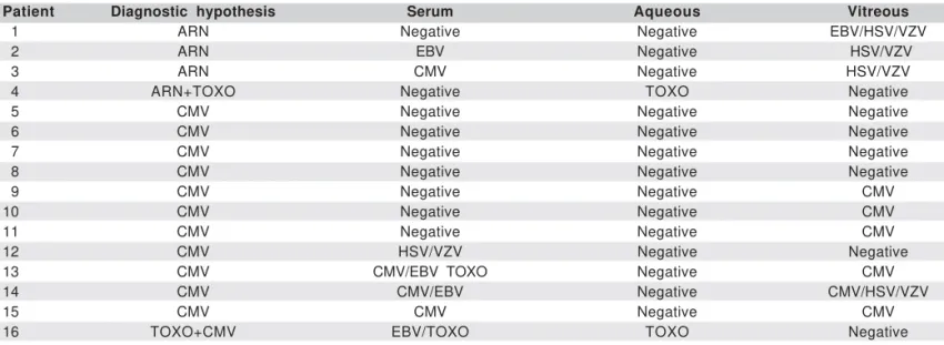
Toxoplasma gondii in the aqueous, all other individuals had Table 1. Correlation between diagnostic and PCR analysis of cytomegalovirus (CMV), toxoplasmosis (TOXO), Epstein-Barr virus (EBV), herpes simplex virus (HSV), varicella zoster virus (VZV) and mycobacteria (MB) in samples of serum, aqueous and vitreous in patients with intraocular
inflammation and AIDS
Patient Diagnostic hypothesis Serum Aqueous Vitreous
01 ARN Negative Negative EBV/HSV/VZV
02 ARN EBV Negative HSV/VZV
03 ARN CMV Negative HSV/VZV
04 ARN+TOXO Negative TOXO Negative
05 CMV Negative Negative Negative
06 CMV Negative Negative Negative
07 CMV Negative Negative Negative
08 CMV Negative Negative Negative
09 CMV Negative Negative CMV
10 CMV Negative Negative CMV
11 CMV Negative Negative CMV
12 CMV HSV/VZV Negative Negative
13 CMV CMV/EBV TOXO Negative CMV
14 CMV CMV/EBV Negative CMV/HSV/VZV
15 CMV CMV Negative CMV
16 TOXO+CMV EBV/TOXO TOXO Negative
ARN: acute retinal necrosis; EBV: Epstein-Barr virus; HSV: herpes simplex virus; VZV: varicella zoster virus; CMV: cytomegalovirus; TOXO: toxoplasmosis
Table 2. Correlation between diagnostic and PCR analysis of cyto-megalovirus (CMV), toxoplasmosis (TOXO), Epstein-Barr virus (EBV), herpes simplex virus (HSV), varicella zoster virus (VZV), and myco-bacteriosis (MB) in samples of serum, aqueous, and vitreous in
patien-ts with intraocular inflammation without AIDS
Patient Diagnostic Serum Aqueous Vitreous hypothesis
01 ARN Negative Negative Negative
02 TB MB Negative MB/ TOXO
03 TOXO Negative Negative TOXO
04 TOXO Negative TOXO TOXO
ARN: acute retinal necrosis; TB: tuberculosis; TOXO: toxoplasmosis; MB: mycobacteriosis
Table 3. Correlation between diagnostic and PCR analysis of cyto-megalovirus (CMV), toxoplasmosis (TOXO), Epstein-Barr virus (EBV), herpes simplex virus (HSV), varicella zoster virus (VZV) and myco-bacteriosis (MB) in samples of serum, aqueous and vitreous in
pa-tients without intraocular inflammation nor AIDS
Patient Diagnostic Serum Aqueous Vitreous hypothesis
01 Control Negative Negative Negative
02 Control Negative Negative Negative
03 Control Negative Negative Negative
04 Control Negative Negative Negative
Table 4. Primer sequences for detection of cytomegalovirus, Epstein-Barr virus, Herpes simplex virus, varicella zoster virus (VZV), Toxoplasma gondii, Mycobacterium sp
Primers Sense Antisense
Cytomegalovirus TGAGGATAAGCGGGAGATGT ACTGAGGCAAGTTCTGCAGT
Epstein-Barr Virus CTCTCTCTGTCCTTCAGAGG GTGGCTCCCCTCAGACATTC
Herpes simplex Virus ATCACGGTAGCCCGGCCGTGTGACA CATACCGGAACGCACCACACAA
Varicella Zoster Virus
Toxoplasma gondii TCTTTAAAGCGTTCGTGGTC GGAACTGCATCCGTTATGAG
negative PCR results for aqueous samples. Of the 12 indivi-duals with a putative diagnosis of CMV retinitis, 6 (50%) had a positive PCR for CMV in the vitreous. Three of these patients also had positive PCR reactions for CMV in the serum. Sensi-tivity in the serum and vitreous were 25% and 50% respecti-vely, and the specificity in the serum and vitreous were 87.5% and 100% respectively.
When we compared the two groups regarding accuracy of diagnosis in vitreous samples, in the group with AIDS the diagnosis was confirmed in 9 (56%) of 16 patients, while in the immunocompetent group the diagnosis was made in 3 (75%) of 4 patients. Of the five individuals with a clinical diagnosis of ARN, 3 (60%) had a positive HSV/VZV PCR in the vitreous. One case also had a positive EBV PCR in the vitreous, and this case had the worst outcome, becoming blind in both eyes within two months despite aggressive therapy. Another indi-vidual had a positive PCR for EBV in the serum but vitreous PCR was positive for HSV/VZV, and another was positive for CMV in the aqueous sample but vitreous PCR samples sho-wed positive HSV/VZV PCR. Concomitant infections are com-mon in AIDS, and often PCR testing of serum will reveal them; thus the serum results may not always represent correctly the infectious picture in the eye. Interestingly, one of the indivi-duals with a putative diagnosis of ARN had toxoplasmosis as a differential diagnosis, however aqueous PCR was positive only for toxoplasmosis. The sensitivities for toxoplasmosis in aqueous and vitreous were 75 and 50% respectively, and the specificities in aqueous and vitreous were 100 and 93.7% respectively. Once more the accuracy of using PCR as a diag-nostic method when we compared the two groups with and without AIDS was markedly distinct. In the two AIDS patients who were PCR positive in the aqueous, PCR confirmed the clinical diagnosis which in these cases was doubtful. In the group without AIDS there were 3 PCR positive vitreous sam-ples, and one aqueous samples. Ocular toxoplasmosis was a clinical diagnosis or was part of the differential diagnosis in four patients. All four had positive PCR reactions for toxo-plasmosis. One of them, cited above, was positive only in the aqueous samples. Another was positive in the aqueous and serum samples; this patient was also considered for CMV retinitis but CMV was not detected in any of the samples. However, EBV was detected in the serum of this patient.
Table 2 clearly shows that PCR was negative for all four control patients. Finally, two other patients had a positive PCR result for toxoplasmosis in the vitreous, once again con-firming the clinical diagnosis.
The only patient with a putative diagnosis of ocular tuber-culosis had a positive result for mycobacteriosis both in the serum and in the vitreous.
DISCUSSION
McCann et al.(28), studied patients with CMV retinitis
through PCR analysis and reported that sensitivity of PCR in treated patients was 48%, compared with 95% in non-treated
patients. All of our patients were treated; thus we cannot make any comparison between treated and non-treated groups.
In spite of several reports demonstrating a positive PCR analysis in aqueous humor specifically for herpes virus infec-tion(29-34), we found no positivity in aqueous samples.
Mean-while we found a positive PCR analysis in vitreous samples in three cases in a total of 5 cases of presumptive ARN. There-fore we conclude in our study that PCR analysis for these cases were more helpful when analyzed in vitreous samples.
Wiedbrauk et al(35), reported an inhibitory effect in PCR
ana-lysis of aqueous and vitreous, however in our study of the PCR analysis we found no evidence of this inhibitory effect; when PCR result was positive it confirmed the clinical diagnosis.
Chodosh(36), detected EBV DNA in cadaveric ocular tissue
at apparently normal ocular sites which raises the possibility of viral involvement, but emphasizes the need for specific criteria to implicate EBV in ocular pathology. In our study we found EBV in vitreous associated with HSV/VZV in a severe case of acute retinal necrosis, and in 4 patients whose sera was positive for EBV, [all were HIV positive, 2 with CMV retinitis, one with acute retinal necrosis, and an other with a clinical diagnosis of CMV and toxoplasmosis]. None of the control group samples showed any evidence of EBV in sera, aqueous or vitreous.
The confirmation of clinical diagnosis is highest in PCR of vitreous samples. In CMV retinitis and ARN, the positivity of the PCR analysis in vitreous was 50% and 60% respectively, in toxoplasmosis it was 50%, and in our only patient with myco-bacteriosis, the accuracy was 100%.
In patients with acute retinal necrosis, two of them had positive PCR results more than 3 months after onset the di-sease, and the other two had positive PCR result one month after the onset of the disease. In our study, duration of the disease did not interfere with the result of PCR analysis, in spite of the conclusions reached by de Boer et al.(37). There is
a good correlation between clinical diagnosis and PCR ampli-fication of infectious DNA from vitreous.
CONCLUSIONS
RESUMO
Objetivos: Avaliar a aplicabilidade (especificidade,
sensibili-dade) do teste da reação da cadeia de polimerase (PCR) na detecção de citomegalovírus (CMV), herpes vírus e varicela zoster (HSV, VZV), Epstein-Barr vírus (EBV), Mycobacterium sp e Toxoplasma gondii no diagnóstico de pacientes com ou
semAIDS, com uveíte infecciosa presumível, utilizando amos-tras de humor aquoso, humor vítreo e soro. Métodos: Vinte pacientes com uveíte infecciosa presumível foram estudados. Dezesseis destes apresentavam AIDS e quatro eram imuno-competentes. Foram utilizados quatro pacientes como grupo controle que se submeteram a vitrectomia. A avaliação clínica foi feita conjuntamente com três oftalmologistas. O exame do PCR do soro, aquoso e vítreo foi feito sem o conhecimento da hipótese diagnóstica pela equipe do laboratório. Resultados: Doze pacientes tinham o diagnóstico clínico de retinite por CMV. Deste subgrupo 6 (50%) eram PCR positivo para CMV no vítreo, 3 (25%) eram PCR positivos para CMV no soro e nenhum destes foi positivo no aquoso. Cinco pacientes ti-nham o diagnóstico clínico de necrose aguda de retina (ARN). Três (60%) destes eram PCR positivos para HSV/VZV no ví-treo. Um destes pacientes era PCR positivo tanto para EBV e HSV/VZV na amostra do vítreo. Um destes pacientes com herpes zoster cutâneo era PCR positivo para HSV/VZV no soro. Quatro pacientes tinham o diagnóstico de toxoplasmose ocular presumida, um paciente (25%) era PCR positivo para
Toxoplasma gondii no soro, 3 (75%) eram positivos no
aquo-so e 2 (50%) eram positivo no vítreo. Um paciente com tuber-culose ocular presumível era PCR positivo tanto no soro quan-to no vítreo. Nenhum dos pacientes do grupo controle era PCR positivo em qualquer amostra. Conclusão: O exame do PCR é procedimento diagnóstico auxiliar que deve ser utilizado con-juntamente com os aspectos clínicos.
Descritores: Reação em cadeia da polimerase; Uveíte;
Infla-mação; Oftalmopatias
REFERENCES
1. Orefice F, Belfort Jr R. Uveites. São Paulo: Roca; 1987.
2. Belfort Jr R, Couto CA, Castro FM. Uveites: Sinopsis diagnostica y terapeu-tica. Buenos Aires: Ciba Vision; 1997.
3. Abreu MT, Hirata PS, Belfort Jr R, Domingues Neto S. Uveítes em São Paulo: estudo epidemiológico, clínico e terapêutico. Arq Bras Oftalmol. 1980;43(1):10-6.
4. Muccioli C, Belfort Júnior R, Lottenberg C, Lima J, Santos P, Kim M, et al. Achados oftalmológicos em AIDS: avaliação de 445 casos atendidos em um ano. Rev Assoc Med Bras(1992). 1994;40(3):155-8.
5. Nussenblatt RB, Whitcup SM, Palestine AG. 2nd ed. Uveitis: fundamentals and clinical practice. St. Louis: Mosby; c1996.
6. De Smet MD. Differential diagnosis of retinitis and choroiditis in patients with acquired immunodeficiency syndrome. Am J Med. 1992;92(2A):17S-21S. Review.
7. Holland GN, O’Connor GR, Belfort R Jr, Remington JS. Toxoplasmosis. In: Pepose JS, Holland GN, Wilhelmus KR, editors. Ocular infection and immu-nity. St. Louis: Mosby; 1996. p.1183-223.
8. Holland GN, Tufail A, Jordan MC. Cytomegalovirus diseases. In: Pepose JS,
Holland GN, Wilhelmus KR, editors. Ocular infection and immunity. St. Louis: Mosby; 1996. p.1088-129.
9. Margolis TP, Atherton SS. Herpes simplex virus diseases: posterior segment of the eye. In: Pepose JS, Holland GN, Wilhelmus KR, editors. Ocular infection and immunity. St. Louis: Mosby; 1996. p.1155-68.
10. Culbertson WW, Blumenkranz MS, Pepose JS, Stewart JA, Curtin VT. Varicella zoster virus is a cause of the acute retinal necrosis syndrome. Ophthalmology. 1986;93(5):559-69.
11. Clarkson JG, Blumenkranz MS, Culbertson WW, Flynn HW Jr, Lewis ML. Retinal detachment following the acute retinal necrosis syndrome. Ophthal-mology. 1984;91(12):1665-8.
12. Sato EH, Machado VHT, Farah Neto ME, Abreu MT, Belfort Jr R. Necrose aguda de retina. Rev Bras Oftalmol. 1985;44(1):28-31.
13. Newman NM, Mandel MR, Gullett J, Fujikawa L. Clinical and histologic findings in opportunistic ocular infections. Part of a new syndrome of acquired immunodeficiency. Arch Ophthalmol. 1983;101(3):396-401.
14. Oréfice F, Carvalho AZ, Pinheiro SRA. Controvérsias nas uveítes tubercu-losas. Arq Bras Oftalmol. 1987;50(6):237-45.
15. Muccioli C, Belfort R Jr. Presumed ocular and central nervous system tuber-culosis in a patient with the acquired immunodeficiency syndrome. Am J Ophthalmol. 1996;121(2):217-9.
16. Mitchell SM, Fox JD. Aqueous and vitreous humor samples for the diagnosis of cytomegalovirus retinitis. Am J Ophthalmol. 1995;120(2):252-3. 17. Davis JL, Feuer W, Culbertson WW, Pflugfelder SC. Interpretation of
intra-ocular and serum antibody levels in necrotizing retinitis. Retina. 1995;15(3): 233-40.
18. Paya CV, Smith TF, Ludwig J, Hermans PE. Rapid shell vial culture and tissue histology compared with serology for the rapid diagnosis of cytomega-lovirus infection in liver transplantation.Mayo Clin Proc. 1989;64(6):670-5. 19. Schmitt-Graff A, Neuen-Jacob E, Rettig B, Sundmacher R. Evidence for cytomegalovirus and human immunodeficiency virus infection of the retina in AIDS. Virchows Arch A Pathol Anat Histopathol. 1990;416(3):249-53. 20. Bloch-Michel E, Nussenblatt RB. International Uveitis Study Group
recom-mendations for the evaluation of intraocular inflammatory disease. Am J Ophthalmol. 1987;103(2):234-5.
21. Holland GN. Standard diagnostic criteria for the acute retinal necrosis syndro-me. Executive Committee of the American Uveitis Society. Am J Ophthalmol. 1994;117(5):663-7.
22. Boom R, Sol CJ, Salimans MM, Jansen CL, Wertheim-van Dillen PM, van der Noordaa J. Rapid and simple method for purification of nucleic acids. J Clin Microbiol. 1990;28(3):495-503.
23. Mahalingam R, Wellish M, Wolf W, Dueland AN, Cohrs R, Vafai A, Gilden D. Latent varicella-zoster viral DNA in human trigeminal and thoracic gan-glia. N Engl J Med. 1990;323(10):627-31.
24. Nahass GT, Goldstein BA, Zhu WY, Serfling U, Penneys NS, Leonardi CL. Comparison of Tzanck smear, viral culture, and DNA diagnostic methods in detection of herpes simplex and varicella-zoster infection. JAMA. 1992;268 (18):2541-4.
25. Cathomas G, Morris P, Pekle K, Cunningham I, Emanuel D. Rapid diagnosis of cytomegalovirus pneumonia in marrow transplant recipients by bronchoal-veolar lavage using the polymerase chain reaction, virus culture, and the direct immunostaining of alveolar cells. Blood. 1993;81(7):1909-14.
26. Innis MA, Gelfand DH, Sninsky JJ. White TJ, editors. PCR protocols: a guide to methods and applications. San Diego: Academic Press; c1990. 27. Longo MC, Berninger MS, Hartley JL. Use of uracil DNA glycosylase to
control carry-over contamination in polymerase chain reactions. Gene. 1990; 93(1):125-8.
28. McCann JD, Margolis TP, Wong MG, Kuppermann BD, Luckie AP, Schwartz DM, et al. A sensitive and specific polymerase chain reaction-based assay for the diagnosis of cytomegalovirus retinitis. Am J Ophthalmol. 1995; 120(2):219-26.
29. Kumano Y, Manabe J, Hamamoto M, Kawano Y, Minagawa H, Fukumaki Y, Inomata H. Detection of varicella-zoster virus genome having a PstI site in the ocular sample from a patient with acute retinal necrosis. Ophthalmic Res. 1995;27(5):310-6.
30. van den Horn GJ, Meenken C, Troost D. Association of progressive outer retinal necrosis and varicella zoster encephalitis in a patient with AIDS. Br J Ophthalmol. 1996;80(11):982-5.
32. Chatzoulis DM, Theodosiadis PG, Apostolopoulos MN, Drakoulis N, Marko-michelakis NN. Retinal perivasculitis in an immunocompetent patient with systemic herpes simplex infection. Am J Ophthalmol. 1997;123(5):699-702. 33. Garweg J, Bohnke M. Varicella-zoster virus is strongly associated with
atypi-cal necrotizing herpetic retinopathies.Clin Infect Dis. 1997;24(4):603-8. 34. Yamamoto S, Pavan-Langston D, Kinoshita S, Nishida K, Shimomura Y,
Tano Y. Detecting herpesvirus DNA in uveitis using the polymerase chain reaction.Br J Ophthalmol. 1996;80(5):465-8.
35. Wiedbrauk DL, Werner JC, Drevon AM. Inhibition of PCR by aqueous and vitreous fluids. J Clin Microbiol. 1995;33(10):2643-6.
36. Chodosh J, Gan YJ, Sixbey JW. Detection of Epstein-Barr virus genome in ocular tissues.Ophthalmology. 1996;103(4):687-90.
37. de Boer JH, Verhagen C, Bruinenberg M, Rothova A, de Jong PT, Baarsma GS, Van et al. Serologic and polymerase chain reaction analysis of intraocular fluids in the diagnosis of infectious uveitis. Am J Ophthalmol. 1996;121(6): 650-8.