O
r i g i n a la
rt i c l e8 4 Arq Bras Oftalmol. 2017;80(2):84-7 http://dx.doi.org/10.5935/0004-2749.20170021
INTRODUCTION
Infectious keratitis occurs worldwide, and despite recent develop-ments, it remains a potentially blinding condition(1). Often, a specific
diagnosis is not clinically possible. Early diagnosis and immediate appropriate treatment can reduce disease morbidity(2,3).Laboratory
iden tification is important because different etiological agents can manifest themselves with similar clinical profiles, such as Acantha-moeba spp., herpes viruses, bacteria, and fungi(4-6).
The development of nucleic acid amplification tests, such as the polymerase chain reaction (PCR), has provided more specific and sensitive methods for diagnosis(7-9). Quantitative real-time PCR (qPCR)
has advantages over conventional methods because it is quicker and more sensitive(10). It can also detect genes that are expressed only in
the replication state and can relatively quantify the DNA sample(11,12).
The current study assesses the presence of herpes simplex virus-1 and -2 (HSV-1 and -2) and varicella zoster virus (VZV) by qPCR in ABSTRACT
Objective: Bacterial keratitis occurs worldwide, and despite recent developments, it remains a potentially blinding condition.This study assesses the presence of herpes simplex virus (HSV-1 and -2) and varicella zoster virus (VZV) by quantitative real-time polymerase chain reaction (qPCR) in corneal scrapings from patients with bacterial keratitis.
Methods: A total of65patients with clinical diagnoses of infectious corneal ulcers prospectively underwent clinical eye examinations. Corneal scrapings were investi-gated by Gram staining, Giemsa staining, culture, and qPCR (the study group). Risk factors and epidemiological data were recorded. The control group comprising 25 eyes with typical herpes dendritic keratitis was also analyzed by qPCR.
Results: From the study group (n=65),nine patients (13.8%) had negative smears, cultures, and qPCR findings. Fifty-six (86.2%) patients had positive cultures: 51 for bacteria, 4 for fungi, and 1 for amoebae. Of the patients who had positive bacterial cultures, qPCR identified 10 patients who were also positive for virus: one for VZV and nine for HSV-1. Of the 25 patients in the control group, 21 tested positive for HSV-1 by qPCR analysis.
Conclusions: Herpes may be present in patients with bacterial corneal ulcers, and qPCR may be useful in its detection.
Keywords: Herpes simplex; herpesviridae; Keratitis, herpetic; Polymerase chain reaction
RESUMO
Objetivo: Ceratites bacterianas ocorrem mundialmente e apesar dos novos desen-volvimentos permanece como uma condição que pode levar à cegueira. Avaliar a presença de herpes simples (-1 e -2) e vírus varicella zoster (VZV) por reação em cadeia quantitativa de polimerase em tempo real (qPCR) em raspados corneanos de pacientes com ceratite bacteriana.
Métodos: Sessenta e cinco pacientes com ceratite infecciosa foram submetidos a raspados corneanos estudados para gram, Giemsa, cultura e qPCR (grupo de estudo). Foram avaliados fatores de risco e epidemiológicos. O grupo controle foi composto por 25 casos de úlcera dendrítica típica por herpes analisados por qPCR.
Resultados: Do grupo de estudo (n=65), nove pacientes (13,8%) apresentaram cultura, qPCR e raspado negativos. Cinquenta e seis (86,2%) pacientes apresentaram cultura positiva, 51 para bacteria, 4 para fungo e 1 para ameba. A qPCR identificou 10 pacientes do grupo de cultura positiva para bactéria que também foram positivos para vírus, um VZV e 9 para HSV-1. Dos 25 pacientes que compunham o grupo controle, 21 apresentaram qPCR positivo para HSV-1.
Conclusão: Herpes pode estar presente em pacientes com úlceras de córnea bacte-rianas e a qPCR pode ser útil na sua detecção.
Descritores: Herpes simples; Infecções por herpesviridae; Ceratite herpética; Reação em cadeia da polimerase
corneal scrapings from patients with infectious keratitis. Risk factors are also studied.
METHODS
Patients with clinical diagnoses of infectious corneal ulcers refer-red for diagnostic corneal scrapings were included in this study (the study group). Risk factors and epidemiological information, as well as the duration of disease and its clinical evolution, were recorded for each patient. All patients signed an informed consent form after approval by the Ethics Committee (1422/06).
All patients underwent a clinical examination that included slit lamp biomicroscopy and photography. Diseased corneas were scraped and specimens were sent for direct examination (Gram and Giemsa staining techniques) and microbiology investigation for bacteria, fungi, and Acanthamoeba as well as qPCR for viruses. At the time of
exa-Detection of herpes simplex-1 and -2 and varicella zoster virus by quantitative
real-time polymerase chain reaction in corneas from patients with bacterial keratitis
Detecção de vírus herpes simplex tipo 1 e 2 e varicella zoster por reação em cadeia de
polimerase em tempo real em córneas de pacientes com ceratites bacterianas
Heloisa NascimeNto1, aripuaNã WataNabe2, aNa caroliNa cabreira Vieira1, aNdrea pelegriNi2, maria cecília Yu1, paulo José martiNs bispo2, celso FraNcisco HerNaNdes graNato2, aNa luisa HöFliNg-lima1
Submitted for publication: October 7, 2016 Accepted for publication: October 26, 2016
1 Department of Ophthalmology and Visual Sciences, Escola Paulista de Medicina (EPM), Universidade Federal de São Paulo (UNIFESP), São Paulo, SP, Brazil.
2 Infectology Discipline, Department of Medicine, Escola Paulista de Medicina (EPM), Universidade Federal de São Paulo (UNIFESP), São Paulo, SP, Brazil.
Funding: This study was supported by São Paulo State Research Foundation (FAPESP).
Disclosure of potential conflicts of interest: None of the authors have any potential conflict of interest to disclose.
Corresponding author: Heloisa Nascimento. Rua Dr. José Estéfno, 125 - São Paulo, SP - 04116-060 Brazil - E-mail: helomn@gmail.com
Na s c i m e N t o H, e t a l.
8 5 Arq Bras Oftalmol. 2017;80(2):84-7 mination, the following information was recorded for each patient:
gen der, age, occupation, history of ocular trauma, previous intraocular surgery, presence of local or systemic risk factors (such as contact lens use, diabetes, HIV, advanced age, and corneal exposition), current medications, and symptom duration.
Corneal scrapings were obtained from the edge of the ulcer after application of topical anesthesia and the ocular surface was washed with physiologic saline to avoid false-positive results during qPCR(9).
A portion of the sample was quickly frozen (-80°C) and sent for molecular diagnostic techniques. Cultures were considered positive when there was heavy growth on stria blood or chocolate agar and a positive correlation with the results of smears.
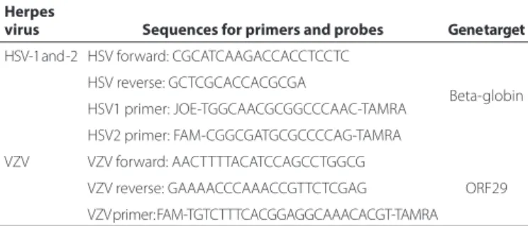
Total DNA extraction was performed using a QIAamp DNA Mini Kit (Qiagen, Valencia, CA), according to the manufacturer’s protocol. Molecular detection was carried out using TaqMan-based techno-logy, and reactions for HSV-1 and -2 and VZV (sequences of primers and probes are listed in Table 1) were tested for analytical specificity using positive (known viral DNA from HSV-1 and -2, and VZV) and negative (molecular grade water) controls(13). The reaction was
stan-dardized with primers and probes for HSV-1/2 in a duplex format and a VZV target in a separate reaction. Briefly, 5 mL of DNA was added to a final reaction volume of 25.0 mL (12.5 mL of the 2X TaqManTM
univer-sal PCR master mix, 0.3 mM of each primer, 0.3 mM of each probe, and 5.7 mL of molecular grade sterile water), and 45 PCR cycles were run: denaturation step (20 sec at 95°C), primer annealing and extension (1 min at 60°C). The beta-globin gene was used as the endogenous control(14) (10.0 mL of 2X TaqManTM universal PCR mix, 0.4 mM of each
primer, 0.2 mM of the probe, 7.0 mL molecular grade sterile water, and 2.0 mL of DNA sample to a final volume of 20.0 mL) and the same PCR conditions described above were used. All reactions were carried out in an ABI Prism 7500 device (Applied Biosystems, Carlsbad, CA, USA). The clinical samples were then analyzed for the presence of HSV-1 and -2 and VZV DNA. The molecular test was designed to detect only actively replicating virus, through collection of corneal scrapings only from symptomatic patients. The alpha subfamily of the herpes viruses replicates efficiently in skin and mucosa, in which the mucosa sore is a known site of active replication(14). The control
group comprised 25 eyes with typical herpes dendritic keratitis that were also analyzed by qPCR.
RESULTS
Sixty-five eyes of 65 patients (38 females; mean age 44.9 years, range 8-93 years) who had been clinically diagnosed with bacterial keratitis (the study group) and 25 eyes of 25 patients presenting with typical herpes dendritic keratitis (the control group) were tested from May 2008 to December 2010 (Table 2).
From the study group, 9 of 65 eyes (13.8%) had negative smears, cultures, and PCR findings. From these nine patients, eight had cor-neal ulcers <2 mm and one had a 3.5 × 3 mm ulcer. Six patients were contact lens wearers and two reported previous ocular trauma. All eyes with negative cultures were also negative for viruses by qPCR analysis.
The most frequent etiologic agent was coagulase-negative Sta-phylococcus spp., in 21 of 56 culture-positive eyes (37.5%). From the culture-positive eyes, gram-positive microorganisms were responsi-ble for the infections in 37 eyes (66.1%). Gram-negative microorganisms were present in 13 eyes (23.2%) and fungi were present in four eyes (7.1%). One eye (1.8%) presented Acanthamoeba growth on cultures. Most of the patients with positive cultures had ulcers >2 mm with va-riable clinical aspects. Risk factors for keratitis were present in 21 eyes (37.5%) and included trauma, previous intraocular surgery, bullous keratopathy, contact lens wear, and Steven-Johnson syndrome.
In 5 of 56 eyes with positive cultures, more than one etiological agent was detected: two eyes presented two Gram-positive organisms and three presented one Gram-positive and one Gram-negative agent. One of the patients with two Gram-positive organisms was also po-sitive for HSV-1 by qPCR analysis. From these five patients, four were
contact lens wearers and one had a history of several previous intrao-cular surgeries. All had ulcers >2 mm.
Four eyes harbored fungi and one eye presented Acanthamoeba
growth in cultures. Although these eyes were clinically diagnosed as being potential bacterial corneal ulcers, microbiology studies were able to reveal the causative microorganisms and guide appropriate management. None of those patients were positive for herpes viruses by qPCR analysis.
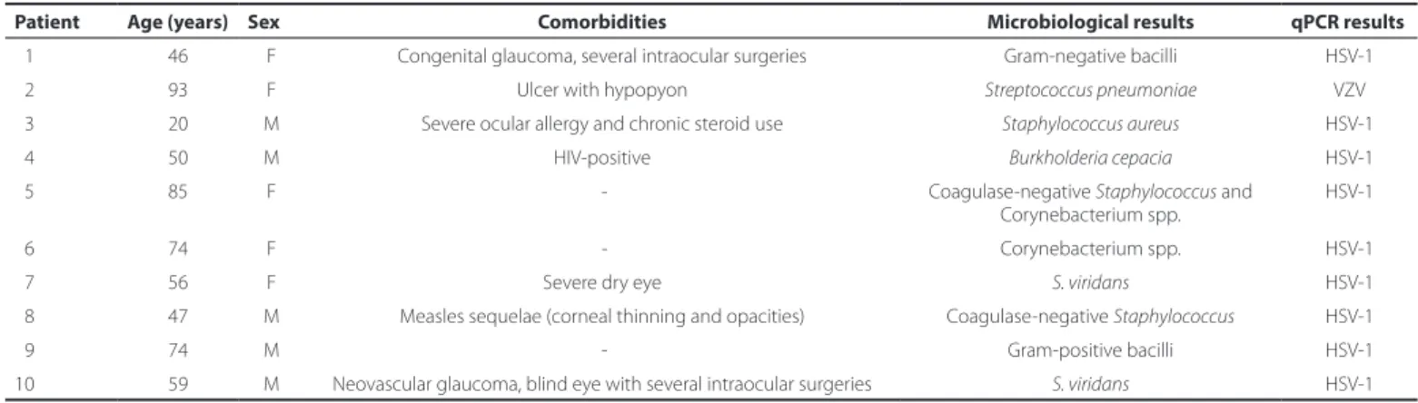
Ten of 65 eyes (15.3%) were positive for viruses in the qPCR analy-sis. Of those, VZV was identified in one eye of one patient (a 93-year-old female), and HSV-1 was identified in nine eyes. All had ulcers >2 mm. Ocular comorbidities or severe systemic diseases were present in 7 of the 65 eyes (70%) (Table 3).
The mean age of patients who had positive qPCR results for herpes was higher than that of those who were negative for herpes: 60.4 and 42.2 years, respectively (p=0.01) (Table 4).
The control group comprised 25 patients with clinical diagnoses of herpes epithelial keratitis. All had dendritic lesions at presentation. Twenty-one of these patients were positive for HSV-1 by qPCR analysis (84%).
DISCUSSION
HSV-1 and VZV were identified in this study using qPCR of samples of infectious keratitis with bacterial growth in cultures, indicating a potential co-infection. Systemic and ocular comorbidities were found more frequently (70%) in cases that were positive for both virus and bacteria than in cases that were positive only for bacteria (32%). Neo vascular glaucoma with several previous intraocular surgeries, systemic immunosuppression due to HIV infection, chronic steroid use, severe allergy, and dry eye disease were found in 7 of 10 patients with a confirmed co-infection, and 4 of the 10 patients (40%) from this group were older than 70 years. This is a large proportion compared with the general study population, in which 9 of the 65 patients (14%) were older than 70 years. Statistical analysis revealed that the mean age of patients with positive qPCR results was significantly higher than that of patients with negative results (60.4 and 42.18 years, res pectively; p=0.01, Table 3). In addition, there was a tendency to a higher prevalence of positive PCR results in patients older than 70 years (p=0.07). Therefore, advanced age could be a factor for herpes infection.
A possible study limitation was the detection of latent HSV-1 and -2, as well as VZV. However, the target of the real-time primers and probes were amplified genes (the beta-globin gene in HSV-1 and -2, and the gene for the ORF29 protein in VZV), which are expressed almost ex clusively during active viral replication. HSV-1 latency in the nuclei of neurons of regional ganglia is well described(2,3).As such,
when clinical samples are collected from lesions located in the pe-riphery of an axon, in the conjunctiva or mucosal surfaces, the virus is actively replicating and not latent. Besides, there are limited genes expressed during latency, and in designing the primers for this study,
Table 1. Primers and probes targeting herpes simplex virus-1 and -2, and varicella zoster virus
Herpes
virus Sequences for primers and probes Gene target
HSV-1 and -2 HSV forward: CGCATCAAGACCACCTCCTC
Beta-globin HSV reverse: GCTCGCACCACGCGA
HSV1 primer: JOE-TGGCAACGCGGCCCAAC-TAMRA
HSV2 primer: FAM-CGGCGATGCGCCCCAG-TAMRA
VZV VZV forward: AACTTTTACATCCAGCCTGGCG
ORF29 VZV reverse: GAAAACCCAAACCGTTCTCGAG
VZV primer: FAM-TGTCTTTCACGGAGGCAAACACGT-TAMRA
De t e c t i o no fh e r p e ss i m p l e x- 1 a n D - 2 a n D va r i c e l l az o s t e rv i r u sb yq ua n t i tat i v er e a l-t i m ep o ly m e r a s ec h a i nr e a c t i o ni nc o r n e a s
f r o mpat i e n t sw i t hb a c t e r i a lk e r at i t i s
8 6 Arq Bras Oftalmol. 2017;80(2):84-7
Table 2. Microbiology and quantitative real-time polymerase chain reaction results from patients with clinical diagnoses of infectious corneal ulcers
Patient Age (years) Sex Microbiological result qPCR
01 49 F Negative Negative
02 40 M Coagulase-negative Staphylococcus Negative
03 46 F Gram-negative Bacillus HSV-1
04 50 M Aspergillus flavus Negative
05 93 F Streptococcus pneumonia VZV
06 28 M Fusarium dimerum Negative
07 58 M Serratia nonliquefaciens Negative
08 32 F Negative Negative
09 23 F Negative Negative
10 70 F Corynebacterium spp. Negative
11 41 M Negative Negative
12 20 M Staphylococcus aureus HSV-1
13 62 M Streptococcus pneumonia Negative
14 48 M Moraxella nonliquefaciens and Corynebacterium spp. Negative
15 50 M Burkholderia cepacia HSV-1
16 66 F Enterobacter aerogenes Negative
17 23 M Negative Negative
18 62 F Serratia marcescens Negative
19 15 M Pseudomonas aeruginosa Negative
20 68 F Beta hemolytic Streptococcus Negative
21 59 M Coagulase-negative Staphylococcus Negative
22 73 F Staphylococcus aureus Negative
23 29 M Beta hemolytic Streptococcus Negative
24 26 M Staphylococcus aureus Negative
25 50 M Corynebacterium spp. Negative
26 13 M Coagulase-negative Staphylococcus Negative
27 14 F Gram-negative cocci Negative
28 31 F Acanthamoeba spp. Negative
29 37 F Coagulase-negative Staphylococcus Negative
30 85 F Coagulase-negative Staphylococcus, Corynebacterium spp. HSV-1
31 74 F Corynebacterium spp. HSV-1
32 16 F Coagulase-negative Staphylococcus, Streptococcus group viridians Negative
33 41 M Fusarium solani Negative
34 28 F Negative Negative
35 16 F Coagulase-negative Staphylococcus Negative
36 56 F Streptococcusviridians HSV-1
37 47 M Coagulase-negative Staphylococcus HSV-1
38 79 F Negative Negative
39 28 F Streptococcus pneumonia Negative
40 56 F Coagulase-negative Staphylococcus Negative
41 77 F Staphylococcus aureus Negative
42 21 M Serratia SSP. and coagulase-negative Staphylococcus Negative
43 27 F Staphylococcus aureus Negative
44 8 F Fusarium solani Negative
45 83 F Gram-positive cocci Negative
46 74 M Gram-positive bacillus HSV-1
47 59 M Streptococcusviridians HSV-1
48 51 M Coagulase-negative Staphylococcus Negative
49 28 F Pseudomonas oryzihabitans and coagulase negative Staphylococcus Negative
50 42 M Serratia spp. Negative
51 37 F Coagulase-negative Staphylococcus Negative
52 42 F Moraxella nonliquefaciens Negative
53 35 M Negative Negative
54 70 M Coagulase-negative Staphylococcus Negative
55 47 F Coagulase-negative Staphylococcus Negative
56 23 F Staphylococcus aureus Negative
57 76 F Coagulase-negative Staphylococcus Negative
58 70 F Enterobacter aerogenes Negative
59 24 F Staphylococcusaureus Negative
60 37 M Coagulase-negative Staphylococcus Negative
61 27 F Negative Negative
62 09 F Coagulase-negative Staphylococcus Negative
63 29 F Coagulase-negative Staphylococcus Negative
64 59 M Coagulase-negative Staphylococcus Negative
65 67 F Coagulase-negative Staphylococcus Negative
Na s c i m e N t o H, e t a l.
8 7 Arq Bras Oftalmol. 2017;80(2):84-7 Table 3. Characteristics of patients with positive bacterial cultures and positive quantitative real-time protein chain reaction results
Patient Age (years) Sex Comorbidities Microbiological results qPCR results
01 46 F Congenital glaucoma, several intraocular surgeries Gram-negative bacilli HSV-1
02 93 F Ulcer with hypopyon Streptococcus pneumoniae VZV
03 20 M Severe ocular allergy and chronic steroid use Staphylococcus aureus HSV-1
04 50 M HIV-positive Burkholderia cepacia HSV-1
05 85 F - Coagulase-negative Staphylococcus and
Corynebacterium spp.
HSV-1
06 74 F - Corynebacterium spp. HSV-1
07 56 F Severe dry eye S. viridans HSV-1
08 47 M Measles sequelae (corneal thinning and opacities) Coagulase-negative Staphylococcus HSV-1
09 74 M - Gram-positive bacilli HSV-1
10 59 M Neovascular glaucoma, blind eye with several intraocular surgeries S. viridans HSV-1
F= female; M= male; HSV= herpes simplex virus; qPCR= quantitative real-time polymerase chain reaction; VZ= varicella zoster virus.
Table 4. Mean age of patients with positive and negative quantitative real-time protein chain reaction results
Positive Negative
Number 10 55
Mean age 60.40 42.18
Standard deviation 21.57 20.52
p value 0.01276
sequences that are expressed only during the active phases of infection and not during latency were chosen. Therefore, it is fairly certain that replicating HSV that was causing the disease was detected rather than latent viruses(15-17).
The control group, which was composed of eyes with only active dendritic epithelial keratitis, showed a high positivity in qPCR analysis (84%). This strengthens the hypothesis of positive PCR results during active viral replication. Most of the eyes from the control group that had negative cultures and qPCR results had ulcers <2mm. The small amount of material collected could be responsible for the negative results from both culture and qPCR.
CONCLUSION
Microbiology studies are essential in infectious keratitis because different etiologic agents can manifest with similar clinical profiles. Molecular techniques show that herpes virus may be present in pa -tients with bacterial corneal ulcers and qPCR may be useful in its detection and treatment, so as to avoid steroids, or appropriately admi-nister antiviral drugs to patients who are positive for herpes by PCR. Cost-effective studies should be carried out to evaluate the impact of such molecular techniques.
ACKNOWLEDGEMENTS
Dr. Melissa Tomimassu for her help in sending patients in the be -ginning of the study and Dr. Rubens Belfort Jr. for the advices con cer-ning publication.
REFERENCES
1. McDonald EM, Ram FS, Patel DV, McGhee CN. Topical antibiotics for the management of bacterial keratitis: an evidence-based review of high quality randomised controlled trials. Br J Ophthalmol. 2014;98(11):1470-7.
2. Kaye S, Choudhary A. Herpes simplex keratitis. Prog Retin Eye Res. 2006;25:355-80. 3. Toma HS, Murina AT, Areaux RG Jr, Neumann DM, Bhattacharjee PS, Foster TP, et al.
Ocular HSV-1 latency, reactivation and recurrent disease. Semin Ophthalmol. 2008; 23(4):249-73.
4. Liesegang TJ. Herpes zoster ophthalmicus natural history, risk factors, clinical presen-tation, and morbidity. Ophthalmology. 2008;115(2):S3-12.
5. Claoué C. Preliminary data suggest a role for bacterial superinfection of a viral keratitis after zosteriform spread of herpes simplex virus to the eye of the mouse. J R Soc Med. 1988;81(8):452-5.
6. Rumelt S, Cohen I, Rehany U. Spontaneous corneal graft perforation due to mixed Acanthamoeba and herpes simplex keratitis: a clinicopathologic study. Cornea. 2000; 19(2):240-2.
7. Kamimura A, Takata MI, Fernandes AC, Neves JP, Viegas MT, Murata VY, et al. Molecular de tection of herpes simplex virus by polymerase chain reaction in patients with typical and atypical herpetic keratitis. Arq Bras Oftalmol. 2008;71(6):827-30. 8. Goldschmidt P, Rostane H, Saint-Jean C, Batellier L, Alouch C, Zito E, et al. Effects of topical
anesthetics and fluorescein on the real-time PCR used for the diagnosis of herpes viruses and Acanthamoeba keratitis. Br J Ophthalmol. 2006;90(11):1354-6. 9. Seitzman GD, Cevallos V, Margolis TP. Rose Bengal and lissamine green inhibit
detec-tion of herpes simplex virus by PCR. Am J Ophthalmol. 2006;141(4):756-8. 10. Kennedy DP, Clement C, Arceneaux RL, Bhattacharjee PS, Huq TS, Hill JM. Ocular HSV-1:
Is the Cornea a Reservoir for Viral Latency or a Fast Pit Stop? Cornea. 2011;30(3):251-9. 11. Kaufman HE, Azcuy AM, Varnell ED, Sloop GD, Thompson HW, Hill JM. HSV-1 DNA in
tears and saliva of normal adults. Invest Ophthalmol Vis Sci. 2005;46(1):241-7. 12. Kakimaru-Hasegawa A, Kuo CH, Komatsu N, Komatsu K, Miyazaki D, Inoue Y. Clinical
application of real-time polymerase chain reaction for diagnosis of herpetic diseases of the anterior segment of the eye. Jpn J Ophthalmol. 2008;52(1):24-31.
13. Sugita S, Shimizu N, Watanabe K, Mizukami M, Morio T, Sugamoto Y, et al. Use of mul-tiplex PCR and real-time PCR to detect human herpes virus genome in ocular fluids of patients with uveitis. Br J Ophthalmol. 2008;92(7):928-32.
14. Grinde B. Herpesviruses: latency and reactivation - viral strategies and host response. J Oral Microbiol. 2013 Oct 25;5. doi: 10.3402/jom.v5i0.22766.
15. Bispo PJ, de Melo GB, Hofling-Lima AL, Pignatari AC. Detection and gram discrimination of bacterial pathogens from aqueous and vitreous humor using real-time PCR assays. Invest Ophthalmol Vis Sci. 2011;52(2):873-81.
16. Nicoll MP, Proenca JT, Efstathiou S. The molecular basis of herpes simplex virus latency. FEMS Microbiol Rev. 2012;36(3):684-705.