Brain Correlates of Self-Evaluation Deficits in
Schizophrenia: A Combined Functional and
Structural MRI Study
Shuping Tan1,2☯, Yanli Zhao2☯, Fengmei Fan2, Yizhuang Zou2
*, Zhen Jin3, Yawei Zen3, Xiaolin Zhu2, Fude Yang2, Yunlong Tan2, Dongfeng Zhou1*
1Institute of Mental Health, Peking University, Beijing, 100191, China,2Center of Psychiatry Research, Beijing Huilongguan Hospital, Beijing, 100096, China,3Magnetic Research Imaging Unit, The 306th Hospital of PLA, Beijing, 100101, China
☯These authors contributed equally to this work. *[email protected](YZZ);[email protected](DFZ)
Abstract
Self-evaluation plays an important role in adaptive functioning and is a process that is typi-cally impaired in patients with schizophrenia. Underlying neural mechanisms for this dys-function may be associated with manifested psychosis. However, the brain substrates underlying this deficit are not well known. The present study used brain blood oxygen level dependent (BOLD) functional magnetic resonance imaging (fMRI) and gray matter voxel-based morphometry to explore the functional and structural brain correlates of self-evalua-tion deficits in schizophrenia. Eighteen patients with schizophrenia and 17 healthy controls were recruited and asked to judge whether a set of personality-trait adjectives were appro-priate for describing themselves, a familiar other, or whether the adjectives were of positive or negative valence. Patients had slower response times for negative trait attributions than controls did; responses to positive trait attributions were faster than those for negative traits among the patient group, while no differences were observed in the control group. Control subjects showed greater activation within the dorsal medial prefrontal cortex (dMPFC) and the anterior cingulate cortex (ACC) than the patient group during the self-evaluation>
semantic positivity-evaluation contrast. Patients showed greater activation mainly within the posterior cingulate gyrus (PCC) as compared to controls for the other-evaluation>semantic positivity-evaluation contrast. Furthermore, gray matter volume was reduced in the MPFC, temporal lobe, cuneus, and the dorsal lateral prefrontal cortex (DLPFC) among the patient group when compared to controls. The present study adds to previous findings regarding self- and other-referential processing in schizophrenia, providing support for neurobiological models of self-reflection impairment.
OPEN ACCESS
Citation:Tan S, Zhao Y, Fan F, Zou Y, Jin Z, Zen Y, et al. (2015) Brain Correlates of Self-Evaluation Deficits in Schizophrenia: A Combined Functional and Structural MRI Study. PLoS ONE 10(9): e0138737. doi:10.1371/journal.pone.0138737
Editor:Xi-Nian Zuo, Institute of Psychology, Chinese Academy of Sciences, CHINA
Received:May 18, 2015
Accepted:September 1, 2015
Published:September 25, 2015
Copyright:© 2015 Tan et al. This is an open access article distributed under the terms of theCreative Commons Attribution License, which permits unrestricted use, distribution, and reproduction in any medium, provided the original author and source are credited.
Data Availability Statement:All relevant data supporting our results are within the paper and due to ethical restrictions imposed by the ethics committee at at Beijing Huilongguan Hospital related to protecting patient confidentiality, our original fMRI data can be only available upon request for interested researchers by contacting: Dr. Shuping Tan ([email protected]), Yanli Zhao ([email protected]), or Fengmei Fan ([email protected]).
Introduction
Self-evaluation refers to a conscious process whereby a decision is made regarding oneself [1], or it can refer to the capacity for consciously reflecting on one’s sense of self. This process can be drawn from schemata reflecting one’s abilities, traits, and attitudes [2]. Self-evaluation is used interchangeably with terms such as“self-reflection,” “self-referential processing,” “ self-aware-ness,” “self-appraisal, and the like. Self-evaluation is also a core part of both meta- [3] and social cognition [1,4], which are crucial for adaptive functioning in the social environment [4].
Research suggests that capacities for self-referential processing are compromised in schizo-phrenia [4,5], and self-reflection deficits may be associated with manifested psychosis [6]. More importantly, inability to self-reflect may cause impaired insight [1,7], a common feature and important predictor of treatment adherence and prognosis in schizophrenia [8]. One recent study has shown significant relationship between self-reflection and insight in schizo-phrenia in brain areas related to self-reflection [8]. Additionally, compared to neurocognitive assessments or psychopathology, capacities for self-reflection are better predictors of social competence [3]. For these reasons, studies on impaired self-awareness in schizophrenia have been attracting more and more attention [9]. However, at present, the neural correlates of impaired self-reflection in schizophrenia are not well understood, and only a handful of studies have examined the neural networks underlying self-evaluation in schizophrenia.
A common paradigm for addressing self-evaluation processes in neuroscience research is to present subjects with personality-trait adjectives. Here, subjects are asked to judge whether the adjectives describe themselves or a familiar other. Functional magnetic resonance imaging (fMRI) research [1] examining normal, adult subjects has found that self-referential processing mainly relies upon the Cortical Midline Structures (CMS), which encompass the medial pre-frontal cortex (MPFC), the anterior cingulate cortex (ACC), and the posterior cingulate cortex (PCC). Shad and his colleague [9] reviewed the available literature on schizophrenia and pro-posed a hypothetical model for deficits in self-awareness. They suggested that there is an ante-rior-to-posterior shift in CMS among patients with schizophrenia during self-evaluation, which was probably mediated by deficits in functional connectivity between the dorsal medial prefrontal cortex (dMPFC) and the posterior CMS. This model has been further refined based on findings from a few other studies with relatively small sample sizes [9]; however, not all studies support this model. For example, some studies [10,11] do not observe any abnormal activation in the CMS during self-evaluation but do observe oddities in other brain areas (i.e., the temporal gyrus). Based on the aforementioned evidence, additional studies are needed to further test and refine this proposed model.
The present study utilized whole-brain fMRI, voxel-based morphometry (VBM), and struc-tural magnetic resonance imaging (sMRI) to evaluate the functional and strucstruc-tural correlates of self-referential processing in schizophrenia. It was hypothesized that there would be differ-ential neural responses during self-referdiffer-ential processing between patients with schizophrenia and healthy controls. These differences would be mainly observed in the CMS; these areas should also reveal differential gray matter volume between the two groups.
Methods
Subjects
Eighteen adult patients at Beijing HuiLongGuan Hospital in China with a DSM-IV diagnosis of schizophrenia [12] were recruited. All patients were screened to exclude for a history of major head trauma, significant medical or neurological illness, and substance abuse in the last 6 months. Patients’current clinical symptoms were assessed with the Positive and Negative
Technology Commission (Z141107002514016)(SPT), Beijing Municipal Science & Technology Commission (D121100005012005)(FDY), International Cooperation and Exchange of the National Natural Science Foundation of China (81461130016) (YLT), Beijing Municipal Administration of Hospitals Clinical medicine Development of special funding (zy201409) (FDY) and the National Natural Science Foundation of China (81401133) (YLZ). The funders had no role in study design, data collection and analysis, decision to publish, or preparation of the manuscript.
Syndrome Scale (PANSS) using 5 component scores: positive, negative, cognitive, hostility, and emotional discomfort [13]. All patients received antipsychotic medication (5 received typical neuroleptics, 12 atypical neuroleptics and medication use were missing for 1 patient.). The mean chlorpromazine equivalent dosages (CPZ) were 277.9±126.8 mg per day [14]. An addi-tional 17 healthy controls were recruited from the surrounding community through poster advertisements. Exclusion criteria for healthy controls were (1) any mental disorders by direct psychiatric interview, (2) history of major head trauma or significant medical or neurological illness, (3) substance abuse or dependence in the past 6 months, (4) and first-degree relatives diagnosed with schizophrenia. There were no significant differences between the 2 groups with respect to age, sex, handedness, or education (Table 1). All subjects gave written informed con-sent, and the ethics committee at Beijing Huilongguan Hospital approved the experimental protocol. The study was conducted in accordance with the Declaration of Helsinki.
Self-evaluation task
The present study included three different judgment tasks, and subjects were asked to judge whether or not each personality-trait adjective was appropriate for describing the self, a famil-iar other (Jia Bao Wen, the premier of the People’s Republic of China from 2003 to 2012), or adjective valence (positive or negative).
Twelve lists of 9 personality-trait adjectives were selected from a pool of normalized person-ality-trait adjectives [15]. All adjectives consisted of two-four Chinese characters. Lists were assigned to three conditions with four lists per condition. Each list consisted of four 2-character adjectives, three 3-character adjectives, and two 4-character adjectives. Adjective valence was counterbalanced in each condition (half of the words in each condition were positive traits, the remaining half were negative traits) and was nearly counterbalanced in each list (four or five words in each list were positive traits, the remaining five or four words were negative traits). The task consisted of one scanning run with 12 blocks. Each block was associated with one of three conditions (self-evaluation/other-evaluation/semantic positivity-evaluation), which contained 9 adjectives. Each condition was repeated 4 times, and condition order was counterbalanced as to avoid presenting the same condition during two consecutive blocks. The semantic positivity-evaluation task was used as the baseline to control for neural activa-tion due to emoactiva-tional stimulus quality, language/motoric abilities, and the task’s attentional/
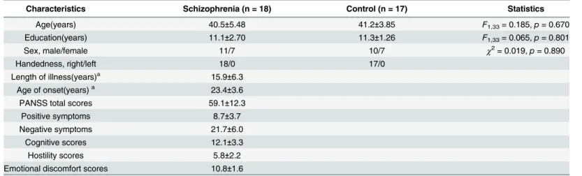
Table 1. Demographic and clinical characteristics of schizophrenia and controls.
Characteristics Schizophrenia (n = 18) Control (n = 17) Statistics
Age(years) 40.5±5.48 41.2±3.85 F1,33= 0.185,p= 0.670
Education(years) 11.1±2.70 11.3±1.26 F1,33= 0.065,p= 0.801
Sex, male/female 11/7 10/7 χ2= 0.019,p= 0.890
Handedness, right/left 18/0 17/0
Length of illness(years)a 15.9±6.3
Age of onset(years)a 23.4±3.6
PANSS total scores 59.1±12.3
Positive symptoms 8.7±3.7
Negative symptoms 21.7±6.0
Cognitive scores 12.1±3.3
Hostility scores 5.8±2.2
Emotional discomfort scores 10.8±1.6
aDate of illness duration andage of onset were missing for 1 patient.
decision-making demands [10]. Prior to the functional run, participants were given practice trials to become familiarized with the tasks. An instruction was given before each block (e.g.,
“Please judge whether the words below are appropriate for describing yourself”) for 3 s, fol-lowed by 9 trials that each lasted for 3 s (Fig 1). During each trial, a small cue word (“self,” “other,”or“valence”) remained in the upper part of the screen in order to promote differen-tiation between the referential conditions [10] or to reduce memory demands [4]. The trial sequence in each block was randomized. Participants indicated their responses via a left- or right-handed key press mounted on a joystick. The left-/rightness of responses were counter-balanced across subjects. All text was presented in Song font, in white letters, on a black background. Visual stimuli were presented using the DMDX software package and were viewed on a back-projected screen via a head coil-mounted mirror.
Behavioral data measures and analysis
Reaction times (RT) and responses for each of the three task conditions were recorded using DMDX. Data analyses were performed using SPSS statistics 16.0 (IBM, USA). Two 2 × 2 × 2 repeated measures ANOVAs were performed with group as a between-subjects factor and task
Fig 1. Schematic diagram of the self-reflection task.Each block started with the instruction presented in the middle of the screen (3s), followed by 9 successive pictures of personality-trait adjectives (3s each). Subjects were required to judge whether each adjective presented was appropriate for describing themselves (A), a familiar other(B), or whether the adjectives were of positive or negative valence(C).
conditions and valence as within-subjects factors to compare groups on RTs and mean propor-tions of self-and other-attribution.
Image acquisition and processing
Imaging was performed on a 2-T GE/Elscint Prestige MRI scanner at Beijing 306 Hospital. Functional images were acquired during one run of 6 minutes using a gradient echo-planar image (EPI) sequence with the following parameters: repetition time (TR)/echo delay time (TE)/flip angle = 3000 ms/45 ms/90°, in-plane resolution = 2.9 mm × 2.9 mm, 6 mm slice thickness. Twenty contiguous axial slices were acquired parallel to the AC-PC line covering the whole brain with no gap. Anatomical images were acquired using a T1 weighted 3D gradient-echo sequence with TR/TE/flip angle = 25 ms/6 ms/28°, slice thickness = 2 mm with no gap, in-plane resolution = 1 mm1 mm.
fMRI data were analyzed using Statistical Parametric Mapping software (SPM5, Wellcome Department of Cognitive Neurology, London, UK) [16]. For functional images, data were pre-processed to remove sources of noise and artifacts. Functional data were corrected for differ-ences in acquisition time between slices for each whole-brain volume, realigned within to correct for head movement and co-registered with each subject’s anatomical data using a 6-degrees-of-freedom linear affine transformation. Functional data were then transformed into a standard anatomical space (2-mm isotropic voxels) based on the parameter obtained by nor-malizing its own T1 image to the ICBM 152 brain template (Montreal Neurological Institute). Normalized data were then spatially smoothed (8 mm full width half maximum [FWHM]) using a Gaussian kernel. The inclusion criteria were a maximum absolute head motion displace-ment of<3 mm and rotation<3° in x/y/z; three patients and two controls were excluded.
For the first-level fMRI analyses after preprocessing, functional images were submitted to a General Linear Model regression analysis to estimate task activation for the self-and other-evaluation conditions compared to the semantic positivity-other-evaluation conditions using SPM. The maps were generated using regressors convolved with a hemodynamic response function for the self-evaluation, other-evaluation, and semantic positivity-evaluation task conditions, which also included the six motion parameters as covariates of no interest. For the second-level analyses, a group analysis was performed for whole brain activity to identify general task-related activations, including self-evaluation vs. semantic positivity-evaluation, other-evaluation vs. semantic positivity-other-evaluation, and self-other-evaluation vs. other-other-evaluation condi-tions. AlphaSim was used to correctp-values for multiple comparisons in the whole brain to p-corrected<0.01 two-tailed.
The T1 images were analyzed using the VBM5 (http://dbm.neuro.uni-jena.de/vbm.html) toolbox as part of the SPM5 software package in the following sequence: (1) A customized (i.e., a study population and tissue-type specific) template using individual T1 images from the con-trol group was created. (2) Images were bias-corrected, tissues were classified, and then regis-tered using linear (12-parameter affine) and non-linear transformations (warping) within a unified model20. (3) Gray matter (GM) segments were multiplied by the non-linear compo-nents derived from the normalization matrix in order to preserve actual local GM values (GM density). (4) The modulated densities were smoothed with a Gaussian kernel of 8 mm full width at half maximum (FWHM).
Results
Behavioral results
Reaction times. We excluded trials (1.96% and 9.98% of all trials, respectively, for controls and patients) where RTs were below 200 ms or above 2,500 ms. The three-way RT ANOVA revealed a significant interaction effect,F(1, 31) = 7.358,p= 0.011. Simple effects analyses observed that compared with controls (1289.21±57.48), patients had slower RTs
(1,529.96 ± 59.25,p= 0.007) toward negative trait attributions. Moreover, for the patient group, attributions toward positive traits (1,150.39 ± 96.77) were faster than for negative traits (1529.96 ± 59.25,p<0.0001), while no differences were found among the control group (p= 0.231).
Attribution style for personality traits. An analysis of attribution style revealed a signifi-cant“condition × valence”interaction effect,F(1, 31) = 8.01,p= 0.008. A simple effects analysis demonstrated that there were more positive trait attributions than negative attributions for both self- (87.0% vs. 25.9%,p<0.001) and other-referential processing (90.4%vs.15.8%, p<0.0001) conditions. Moreover, more negative trait attributions for the self (25.9%) were observed than negative attributions for others (15.8%),p<0.0001. Additionally, there was a significant“condition × group”interaction effect,F(1, 31) = 4.541,p= 0.041. A simple effects analysis indicated that control subjects gave more“yes”answers in the self-referential condi-tion (56.4%) than in the other-referential condicondi-tion (49.9%),p= 0.004, while no differences were found between the two conditions among the schizophrenic group.
Imaging results
Functional MRI. One-sample analyses indicated that the self-evaluation vs. semantic pos-itivity-evaluation contrast revealed activation of the MPFC and PCC, as well as deactivation within the postcentral gyrus (Fig 2A) among controls and revealed deactivation within the postcentral gyrus among patients with schizophrenia (Fig 2B).
Further, among controls, the other-referential processing condition induced hyper-activa-tion in the MPFC and PCC, as well as hypo-activahyper-activa-tion in the cerebellum relative to the seman-tic positivity-evaluation condition (Fig 3A); among patients, the other-evaluation vs. semantic positivity-evaluation contrast revealed hyper-activation in the precuneus and bilateral super frontal gyrus (Fig 3B).
Finally, the self-evaluation vs. other-evaluation contrast revealed stronger activation in the insula and hyper-activation in the dMPFC (Fig 4) among healthy subjects, but it did not reveal any significant activation among patients.
Fig 2. Brain regions showing self-semantic activation in patients with schizophrenia and the control group.One-sample tests produced the spatial patterns of brain regions showing self-semantic activation for both healthy controls (A) and patients with schizophrenia (B). Warm colors represent positive while cool colors indicate negative activation. A two-sample t-test assessed the differences between the two groups (controlsversuspatients, C). The above results were obtained with thresholdp<0.01, AlphaSim correction
p<0.01.
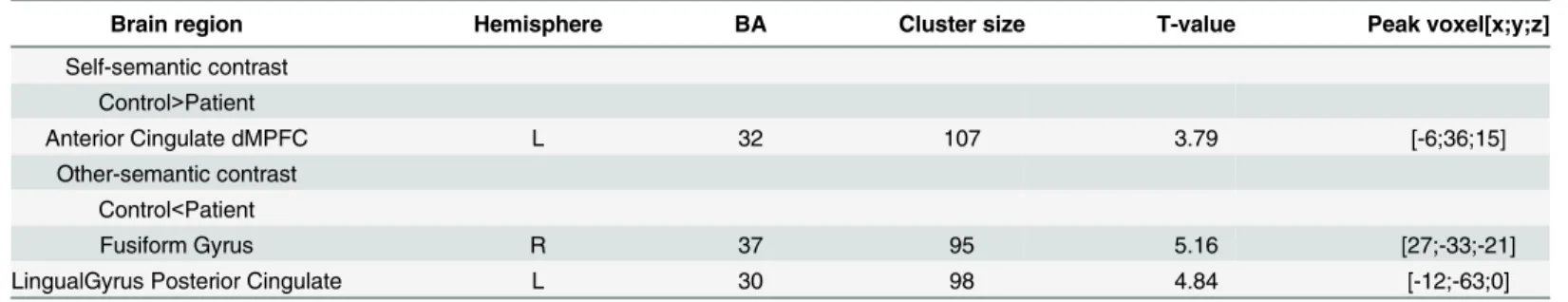
Between-groups analysis showed that there were decreased activations of the dMPFC (i.e., dorsal areas of the anterior CMS) and the ACC in the self-evaluation vs. semantic posi-tivity-evaluation contrast (Fig 2CandTable 2) in the patient group. Additionally, compared to controls, the patient group showed increased activation in the PCC, fusiform gyrus, and lingual gyrus in the other-semantic contrast (Fig 3CandTable 2). Finally, no significant dif-ferences were found between the two groups in the self- vs. other-referential processing contrast.
Fig 3. Brain regions showing other-semantic activation in patients with schizophrenia and the control group.One-sample tests produced the spatial patterns of brain regions showing other-semantic activation for both healthy controls (A) and patients with schizophrenia (B). Warm colors represent positive while cool colors indicate negative activation. A two-sample t-test assessed the differences between the two groups (controlsversuspatients, C). The above results were obtained with thresholdp<0.01, AlphaSim correction p<0.01.
doi:10.1371/journal.pone.0138737.g003
Fig 4. Brain regions showing self-other activation in the control group.One-sample tests produced the spatial patterns of brain regions showing self-other activation for both healthy controls. Warm colors represent positive while cool colors indicate negative activation. The above result was obtained with thresholdp<0.01, AlphaSim correctionp<0.01.
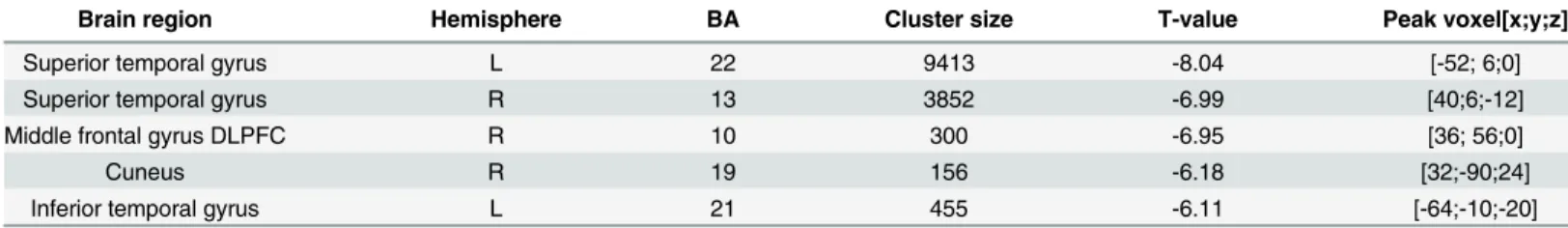
Structural MRI. Compared to controls, patients with schizophrenia showed a significant gray matter volume reduction in the MPFC, temporal lobe, cuneus and the dorsal lateral pre-frontal cortex (DLPFC) (Fig 5andTable 3).
Correlation analysis. No significant correlations were observed between the imaging find-ings and insight, positive symptoms or negative symptoms subscale from the PANSS in the patient group.
Discussion
The present study combined fMRI and sMRI to evaluate the neural and structural correlates of self-and other-referential processing deficits among patients with schizophrenia. Additionally, a model outlining the anterior-to-posterior shift in midline cortical activity related to schizo-phrenia during self-reflection [9] was tested.
Behaviorally, patients had slower RTs for negative trait attributions compared to control subjects; RTs were faster for positive traits than negative traits in the patient group, while no differences were observed in the control group. These findings revealed that patients with schizophrenia need more time to process negative trait information. Additionally, there were more attributions of positive traits than negative traits in both the self- and other-referential conditions across subjects. In contrast to our study, Liu and colleagues [17] found that patients with schizophrenia were less likely and slower to endorse positive self-attributes, and more likely and quicker to endorse negative self-attributes, than control subjects. Such differences may be due to patients in the present study being inpatients with more serious negative symp-toms while patients in Liu and colleagues’study were stable outpatients with more serious posi-tive symptoms.
At a neural level, this is one of the first studies that revealed significant hypoactivation in the dMPFC between patients and controls when self-evaluation was contrasted with non-self evaluation. To our knowledge, only one study has reported significant group differences in response to a similar contrast [18]. One of the most important limitations of that study from Bedford et al. (2012) was the small sample size, which including only 11 patients and 8 normal controls. The present study confirmed the existence of altered self-referential activity in the dMPFC with a larger sample. More importantly, the present study provided evidence regarding the role of the dMPFC in mediating self-referential activity in schizophrenia. This is in contrast to prior research [19,20,21] demonstrating decreased activation in the ventral medial prefron-tal cortex (vMPFC), and a hypothetical model [9] of potential changes in the dMPFC during self-reflection being primarily based on data from healthy subjects. Moreover, previous studies revealed decreased activation in the vMPFC, but not the dMPFC, perhaps due to methodologi-cal differences between fMRI analytimethodologi-cal techniques, such as an ROI-based approach [19] or a cortical surface-based analysis [20], rather than a whole brain voxel-wise analysis. It is also
Table 2. Significant brain activation differences in the task between controls and patients.
Brain region Hemisphere BA Cluster size T-value Peak voxel[x;y;z]
Self-semantic contrast Control>Patient
Anterior Cingulate dMPFC L 32 107 3.79 [-6;36;15]
Other-semantic contrast Control<Patient
Fusiform Gyrus R 37 95 5.16 [27;-33;-21]
LingualGyrus Posterior Cingulate L 30 98 4.84 [-12;-63;0]
worth noting that even if the methods used were the same, the results would still be inconsis-tent. For example, using a whole brain analysis, one study [22] found group differences in the posterior CMS while in another study [18] and the present study, significant group differences were observed in the anterior CMS. This is likely due to differences in sample characteristics. Nevertheless, as the dMPFC monitors the“cognitive self”[9,23,24], decreased dMPFC activity among patients with schizophrenia in the present study may indicate that cognitively oriented decision-making, specifically when referring an object to the self, would be deficient. This may, in turn, cause inappropriate self-referential judgments and decision-making.
Beyond the dMPFC, control subjects also showed stronger activation in the ACC than patients did during self-evaluations compared to semantic positivity-evaluations. The ACC plays an important role in directing attention toward the self [1] and in emotion processing and regulation [25,26]. Modinos et al [6] investigated the neural mechanisms underlying self-referential processing among high psychosis-proneness (PP) subjects, reporting increased acti-vation in the ACC, insula, and dMPFC for negative self-related traits in the high PP compared
Fig 5. Brain regions shown significant grey volume differences between normal subjects and patients with schizophrenia.The above result was obtained with thresholdp<0.001, AlphaSim correctionp<0.01.
doi:10.1371/journal.pone.0138737.g005
Table 3. Brain regions showing significant dGM differences between patients and controls.
Brain region Hemisphere BA Cluster size T-value Peak voxel[x;y;z]
Superior temporal gyrus L 22 9413 -8.04 [-52; 6;0]
Superior temporal gyrus R 13 3852 -6.99 [40;6;-12]
Middle frontal gyrus DLPFC R 10 300 -6.95 [36; 56;0]
Cuneus R 19 156 -6.18 [32;-90;24]
Inferior temporal gyrus L 21 455 -6.11 [-64;-10;-20]
to the low PP subjects. This was interpreted as attempts to diminish (e.g., activity in the dMPFC and ACC) increased emotional responses (e.g., activity in the insula) elicited by self-relevant stimuli, Perhaps the mechanisms underlying self-evaluation are different between patients with schizophrenia and high PP subjects. For instance, increased activation in the ACC and dMPFC might be a compensatory process among high PP individuals that is not present in schizophrenia.
The contrast between other-evaluations and semantic positivity- evaluations mainly dem-onstrated greater activation in the PCC among patients with schizophrenia as compared to healthy controls. The PCC is involved in autobiographical memory [6] and provides further information regarding past instances of self- and other-referential information [1]. This increased PCC response during other-reflections in schizophrenia may suggest that more cog-nitive effort is needed to draw on autobiographical memories of past situations shared with others. Several previous studies [19,20] observed similar results to the current study, namely hyper-activation among patients with schizophrenia for other vs. baseline processing in the PCC. However, other studies [8] indicate hypoactivation in the PCC in schizophrenia for other vs. baseline contrasts. Such results may be associated with differences in baseline control condi-tions and experimental paradigms [8].
Finally, patients with schizophrenia showed a significant gray matter volume reduction in the MPFC, temporal lobe, cuneus and the DLPFC compared to controls, These findings con-firm previous reports [27], indicating that impaired self-referential processing in schizophrenia may be related to specific structural abnormalities.
The present study has a few limitations of note. First, the patient sample included those with chronic schizophrenia who were taking atypical antipsychotic; thus, any medication effects on the present results cannot be excluded. Self-referential processing studies assessing antipsychotic-naïve patients or patients recently experiencing their first schizophrenic episode could minimize the confounding effects of long-term medication use. Moreover, similar to prior research [20], the present study did not observe significant differences in the self vs. other-evaluation contrasts. Perhaps this issue could be addressed through modifications to the chosen imaging paradigm [9]. For instance, using the patient’s name rather than a personal pronoun has been done elsewhere [20,22]. Using trait adjectives incorporated the delusional themes of patients [22] or mental and physical illness terms [18]. Thirdly, self-reflection may be a cognitive process that underlies insight in psychosis [8]; accordingly, we performed a relation analysis between the imaging findings and insight scores. However, no significant cor-relations were observed. We think that this might be because insight is a multidimensional structure [28], including illness awareness, the ability to relabel symptoms, and treatment com-pliance. We only used one measure of awareness in the PANSS as a dependent variable. Future studies could include scales such as the Unawareness of Mental Disorder, the Schedule for the Assessment of Insight-Extended version, and the Beck Cognitive Insight Scale to measure ill-ness awareill-ness and explore its relationship with self-reflection in schizophrenia. Interesting results may emerge, which will be helpful in improving our understanding of the neural corre-lates of impaired insight.
Acknowledgments
We thank the helpful comments of the anonymous reviewers.
Author Contributions
Conceived and designed the experiments: YZZ DFZ. Performed the experiments: ZJ YWZ. Analyzed the data: FMF. Contributed reagents/materials/analysis tools: XLZ. Wrote the paper: YLZ SPT. Gave important suggestions for the revision of the manuscript: FDY YLT.
References
1. van der Meer L, Costafreda S, Aleman A, David AS. Self-reflection and the brain: a theoretical review and meta-analysis of neuroimaging studies with implications for schizophrenia. Neurosci Biobehav Rev. 2010; 34: 935–946. doi:10.1016/j.neubiorev.2009.12.004PMID:20015455
2. Johnson SC, Baxter LC, Wilder LS, Pipe JG, Heiserman JE, Prigatano GP. Neural correlates of self-reflection. Brain. 2002; 125: 1808–1814. PMID:12135971
3. Lysaker PH, Dimaggio G, Carcione A, Procacci M, Buck KD, Davis LW, et al. Metacognition and schizo-phrenia: the capacity for self-reflectivity as a predictor for prospective assessments of work perfor-mance over six months. Schizophr Res. 2010; 122: 124–130. doi:10.1016/j.schres.2009.04.024
PMID:19457645
4. Harvey PO, Lee J, Horan WP, Ochsner K, Green MF. Do patients with schizophrenia benefit from a self-referential memory bias. Schizophr Res. 2011; 127: 171–177. doi:10.1016/j.schres.2010.11.011
PMID:21147520
5. Silva JR, Torres WM, Ortiz MS. Abnormal electrophysiological activation in schizophrenics during a personal traits attribution task. Biol Res. 2008; 41: 143–150. PMID:18949131
6. Modinos G, Renken R, Ormel J, Aleman A. Self-reflection and the psychosis-prone brain: an fMRI study. Neuropsychology. 2011; 25: 295–305. doi:10.1037/a0021747PMID:21443341
7. Lysaker PH, Dimaggio G, Buck KD, Callaway SS, Salvatore G, Carcione A, et al. Poor insight in schizo-phrenia: links between different forms of metacognition with awareness of symptoms, treatment need, and consequences of illness. Compr Psychiatry. 2011; 52: 253–260. doi:10.1016/j.comppsych.2010. 07.007PMID:21497218
8. van der Meer L, de Vos AE, Stiekema AP, Pijnenborg GH, van Tol MJ, Nolen WA, et al. Insight in schizophrenia: involvement of self-reflection networks. Schizophr Bull. 2013; 39: 1288–1295. doi:10. 1093/schbul/sbs122PMID:23104865
9. Shad MU, Brent BK, Keshavan MS. Neurobiology of self-awareness deficits in schizophrenia: A hypo-thetical model. Asian J Psychiatr. 2011; 4: 248–254. doi:10.1016/j.ajp.2011.09.002PMID:23051156
10. Murphy ER, Brent BK, Benton M, Pruitt P, Diwadkar V, Rajarethinam RP, et al. Differential processing of metacognitive evaluation and the neural circuitry of the self and others in schizophrenia: a pilot study. Schizophr Res. 2010; 116: 252–258. doi:10.1016/j.schres.2009.11.009PMID:20051318
11. Pauly KD, Kircher TT, Schneider F, Habel U. Me, myself and I: temporal dysfunctions during self-evalu-ation in patients with schizophrenia. Soc Cogn Affect Neurosci. 2014; 9: 1779–1788. doi:10.1093/ scan/nst174PMID:24369435
12. APA. Diagnostic and Statistical Manual of Mental Disorders, 4th edn. American Psychiatric Associa-tion: Washington, DC;1994.
13. Bell MD, Lysaker PH, Beam-Goulet JL, Milstein RM, Lindenmayer JP. Five-component model of schizophrenia: assessing the factorial invariance of the positive and negative syndrome scale. Psychia-try Res. 1994; 52: 295–303. PMID:7991723
14. Woods SW. Chlorpromazine equivalent doses for the newer atypical antipsychotics. J Clin Psychiatry. 2003; 64: 663–667. PMID:12823080
15. Zhiyong Zhang, Lei Wang. Basic dimensions of Chinese personality traits: a factor analysis of the self description in a sample of Chinese college students. Acta Psychological Sinica. 1998, 30(1): 85–92.
16. Ashburner J, Friston KJ. Unified segmentation. Neuroimage. 2005; 26: 839–851. doi:10.1016/j. neuroimage.2005.02.018PMID:15955494
18. Bedford NJ, Surguladze S, Giampietro V, Brammer MJ, David AS. Self-evaluation in schizophrenia: an fMRI study with implications for the understanding of insight. BMC Psychiatry. 2012; 12: 106. doi:10. 1186/1471-244X-12-106PMID:22876974
19. Blackwood NJ, Bentall RP, Ffytche DH, Simmons A, Murray RM, Howard RJ. Persecutory delusions and the determination of self-relevance: an fMRI investigation. Psychol Med. 2004; 34: 591–596.
PMID:15099414
20. Holt DJ, Cassidy BS, Andrews-Hanna JR, Lee SM, Coombs G, Goff DC, et al. An anterior-to-posterior shift in midline cortical activity in schizophrenia during self-reflection. Biol Psychiatry. 2011; 69: 415–
423. doi:10.1016/j.biopsych.2010.10.003PMID:21144498
21. Modinos G, Ormel J, Aleman A. Activation of anterior insula during self-reflection. PLoS One. 2009; 4: e4618. doi:10.1371/journal.pone.0004618PMID:19242539
22. Shad MU, Keshavan MS, Steinberg JL, Mihalakos P, Thomas BP, Motes MA, et al. Neurobiology of self-awareness in schizophrenia: an fMRI study. Schizophr Res. 2012; 138: 113–119. doi:10.1016/j. schres.2012.03.016PMID:22480958
23. Northoff G, Bermpohl F Cortical midline structures and the self. Trends Cogn Sci. 2004; 8: 102–107.
PMID:15301749
24. Northoff G, Heinzel A, de Greck M, Bermpohl F, Dobrowolny H, Panksepp J Self-referential processing in our brain—a meta-analysis of imaging studies on the self. Neuroimage. 2006; 31: 440–457. PMID: 16466680
25. Lane RD, Reiman EM, Axelrod B, Yun LS, Holmes A, Schwartz GE. Neural correlates of levels of emo-tional awareness. Evidence of an interaction between emotion and attention in the anterior cingulate cortex. J Cogn Neurosci. 1998; 10: 525–535. PMID:9712681
26. Mohanty A, Engels AS, Herrington JD, Heller W, Ho MH, Banich MT, et al. Differential engagement of anterior cingulate cortex subdivisions for cognitive and emotional function. Psychophysiology. 2007; 44: 343–351. doi:10.1111/j.1469-8986.2007.00515.xPMID:17433093
27. Ellison-Wright I, Glahn DC, Laird AR, Thelen SM, Bullmore E The anatomy of first-episode and chronic schizophrenia: an anatomical likelihood estimation meta-analysis. Am J Psychiatry. 2008; 165: 1015–
1023. PMID:18381902doi:10.1176/appi.ajp.2008.07101562