Study of Vascular Reactivity in HIV Patients whether or not Receiving
Protease Inhibitor
Hamilton Nenrod Pereira Teixeira, Evandro Tinoco Mesquita, Mário Luiz Ribeiro, Anna Ricordi Bazin, Cláudio Tinoco
Mesquita, Manuel Pereira Teixeira, Rafael da Cunha Pellegrini, Antonio Claudio Lucas da Nóbrega
Universidade Federal Fluminense (UFF), Niterói, RJ - Brazil
Summary
Background: A great number of HIV-infected patients using antiretroviral drugs develop endothelial dysfunction and atherothrombosis, which lead to a high medical and social burden. Thus, it is important to identify pathophysiological mechanisms involved with the endothelial function in these patients, so that early intervention can be made to avoid disease progression.
Objective: To evaluate endothelial function using endothelium-dependent and independent vasodilation in HIV-positive patients and in a control group.
Methods: A total of 27 HIV-positive patients and 16 controls were evaluated. Endothelium-dependent (reactive hyperemia) and independent (SL nitroglycerine) vasodilation of the brachial artery was used to evaluate the endothelial function.
Results: HIV-positive patients receiving protease inhibitors (PI) showed significantly lower endothelium-independent vasodilation than the HIV-negative (p=0.020) and HIV-positive without PI (p=0.034) subgroups.
The change in brachial artery diameter during active hyperemia was not statistically significant in any subgroup. Multiple linear regression analysis showed that only PI was associated with the relative delta of brachial reactivity to vasodilator in HIV-positive patients at 60s and 90s.
Conclusion: positive patients receiving PI presented endothelium-independent dysfunction when compared to HIV-positive patients not receiving PI and to the control group. (Arq Bras Cardiol 2009; 93(3) : 340-346)
Key Words: Endothelial dysfunction; vascular Doppler; HIV.
Mailing address: Hamilton Nenrod Pereira Teixeira •
Rua dos Rouxinóis, 7 / Q 8 / 404, Ed. Gramado Renascença II, 65.075-630,São Luiz do Maranhão, MA - Brazil
E-mail: [email protected]
Manuscript received August 10, 2008; revised manuscript received November 09, 2008; accepted December 09, 2008.
Introduction
By the end of the 1980’s, Human Immunodeficiency Virus (HIV)-infected patients had short survival1. In contrast, in the
early 21st century, there was an increase in the life expectancy of these patients thanks to the impact of the treatment with new antiretroviral drugs - the Highly Active Antiretroviral Therapy (HAART)2,3. HAART improves the prognosis of
HIV-infected patients by reducing the incidence of opportunistic infections, hospitalizations and mortality4.
Several factors contribute to the increase in cardiovascular disease risk in HIV patients, such as chronic inflammation due to the viral infection, PI action, and interaction with traditional risk factors5. Therefore, the predisposition to
atherosclerosis results from cumulative exposure to the virus and from metabolic changes secondary to the use of PI, thus
determining an unfavorable metabolic profile characterized by hypertriglyceridemia, low HLD-c and insulin resistance observed in 25 to 60% of the patients6.
PIs are currently considered independent cardiovascular risk factors and are associated with an increase by 26% in the rate of acute myocardial infarction (AMI) per year of exposure in the patients who had started treatment for at least 4 to 6 years7. In 1998, the first two cases of premature coronary
artery disease were demonstrated in two young HIV-positive patients receiving PI8.
Several studies have demonstrated that endothelial dysfunction occurs in the early stages of insulin resistance found in patients receiving HAART. Hypoadiponectinemia is one of the factors responsible for lipodystrophy and metabolic disorder in HIV patients receiving PI9,10. Endothelial dysfunction
is a premature event in the development of atherosclerotic lesions and may be easily measured using non-invasive ultrasound study of the brachial artery flow11. The mechanisms
that lead to endothelial dysfunction in HIV-positive patients receiving PI remain not fully understood.
non-invasive method of flow-mediated vasodilation of the brachial artery (DILA), using ultrasound study in HIV-positive patients with and without HAART. DILA was compared in three groups: HIV-infected patients not receiving PI, HIV-infected patients receiving PI, and HIV-negative controls.
Methodology
A total of 43 patients of both genders aged between 20 and 55 years who had undergone clinical and laboratory assessment and the 1-day protocol for non-invasive ultrasound study of the endothelial function were included in the study.
Group I included 15 HIV-positive patients in chronic use (> 2 years) of PI, and group II included 12 HIV-positive PI-naïve patients. A control group of 16 HIV-negative volunteers of both genders was also evaluated.
Exclusion criteria were the presence of coronary artery disease; cerebrovascular disease; peripheral vascular disease; use of contraceptives;ventricular dysfunction (left ventricular ejection fraction < 45% using Simpson’s rule); plasma creatinine > 1.5 mg%; neoplastic diseases; and thyroid diseases.
Data from the patient’s clinical history included age, gender, weight (kg), body mass index (BMI), smoking and stage of HIV infection according to CD4 (T helper lymphocytes).
Endothelial function study – protocol
The technique used was that described by Celermajer et al and recommended by the International Brachial Artery Reactivity Task Force12.
The subjects fasted for at least eight hours, and were required not to exercise in the day before the test or ingest substances that might affect arterial vasodilation, such as caffeine, vitamin C and cigarette. Women should be assessed out of their menstrual period.
The subjects were placed in the supine position with the arm comfortably leaning on a proper support. The
brachial artery was imaged above the antecubital fossa in the longitudinal plane. The room was kept quiet, at a temperature of 21 degrees Celsius.
The tests were performed in a GE ultrasound scanner (VIVID 3) equipped with a vascular software for two-dimensional imaging, color and spectral Doppler, high-frequency (7-12 MHZ) linear vascular transducer, and internal electrocardiogram monitor for the record of a one-lead ECG tracing.
For the assessment of endothelium-dependent vasodilation, a sphygmomanometer was placed around the patient’s arm and the cuff was inflated with air up to 50 mmHg above the systolic blood pressure found. The cuff was deflated 5 minutes later, thus permitting that ischemia be followed by vasodilation via an autoregulatory mechanism and reactive hyperemia.
The brachial artery flow was continuously recorded by the color-Doppler from 30s to 90s after cuff deflation. After the reactive hyperemia phase recording, the patient rested for 10 minutes. Then, assessment of endothelium-independent vasodilation was started.
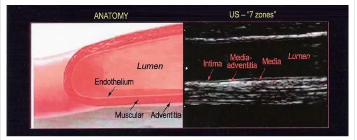
Figure 1 -Flow-mediated dilation of the brachial artery .
This was characterized by the administration of SL nitroglycerine spray (0.4 mg), with a 3-minute-long wait, which is the time necessary for vasodilation to occur. After this period of time, the brachial artery flow was recorded by color-Doppler. The use of nitroglycerin is contraindicated in individuals with hypotension and bradycardia.
The diameter of the brachial artery was measured from its longitudinal image, by calculating the distance between the near and far intima during diastole. Because of the wide variability in the measurement of the intima diameter, a single reference point was selected and the measurements were taken in five cardiac cycles. The measurements were taken at the same time in the cardiac cycle, using electrocardiographic gating during image acquisition. The R wave was used to identify end diastole, and the peak of the T wave, end systole.
DILA was calculated as the post-occlusion percentage increase in the diameter of the brachial artery (PODBA) in relation to baseline values.
DILA CALCULATION: (PODBA – BDBA) / BDBA X 100%
BDBA - baseline diameter of the brachial artery
Values higher than 10% of variation in brachial artery dilation in relation to baseline were considered normal.
Statistical analysis
Student’s t test for independent samples or the Mann-Whitney (non-parametric) test were used to analyze the statistical significance of the differences in continuous (numeric) variables between two subgroups. The chi square test (X²) or the Fisher’s exact test were used for the comparison of categorical (qualitative) variables.
The repeated measures analysis of variance (ANOVA) was used to study the behavior of brachial artery reactivity measurements throughout time (three measurements), separated by subgroups. Bonferroni’s multiple comparison test (adjusted for repeated measures) was used to identify which moments were different between themselves. One-factor repeated measures ANOVA was carried out to verify whether the behavior throughout time was different between the subgroups
The Spearman correlation coefficient was used to measure the degree of association between the relative delta and the metabolic profile; multiple linear regression analysis was used to simultaneously verify the influence of clinical and metabolic data on the relative delta of brachial reactivity to vasodilator.
Non-parametric methods were used since some variables (TG and relative deltas) were not normally distributed (Gaussian distribution) due to scattered data and/or skewness.
The significance level was set at 5%. The statistical analysis was carried out using the SAS 6.04 software (SAS Institute, Inc., Cary, North Carolina).
Ethics
This study was approved by the research ethics committee of the Universidade Federal Fluminense School of Medicine.
For data collection, all patients gave their informed consent.
Results
Twenty seven HIV-positive patients with mean age of 41.6 years (± 8.5) from the AIDS outpatient clinic of the discipline of Infectious and Parasitic Diseases of the Universidade Federal Fluminense School of Medicine were selected. Of these, 15 patients (55.6%) were receiving protease inhibitors.
A group of 16 patients (37.2%) with mean age of 43.8 (± 4.3) comprised the control group.
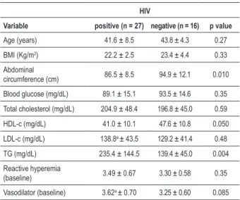
Demographic, clinical and laboratory variables are shown in Tables 1 and 2.
No significant difference was found in relation to age and gender between the HIV-positive and negative groups (p = 0.27).
Continuous variables such as waist circumference (WC) and HDL-c were significantly lower in the HIV-positive group, with p = 0.010 and p = 0.050, respectively.
HIV-negative patients with hypertension (93.8% versus 14.8%; p < 0.0001) had higher blood pressure levels than HIV-positive patients.
TG was the predominant metabolic variable in the subgroup of HIV-positive patients receiving PI with p = 0.004 and p = 0.009 in relation to HIV-negative patients not receiving PI.
Results of brachial artery ultrasound studies are shown in Table 3.
Using the repeated measures ANOVA, brachial reactivity to reactive hyperemia and vasodilators was evaluated in HIV-positive and HIV-negative patients at three timepoints (baseline,
Table 1 - Analysis of demographic, clinical and laboratory variables according to the HIV status
HIV
Variable positive (n = 27) negative (n = 16) p value
Age (years) 41.6 ± 8.5 43.8 ± 4.3 0.27
BMI (Kg/m2) 22.2 ± 2.5 23.4 ± 4.4 0.33
Abdominal
circumference (cm) 86.5 ± 8.5 94.9 ± 12.1 0.010
Blood glucose (mg/dL) 89.1 ± 15.1 93.5 ± 14.6 0.35
Total cholesterol (mg/dL) 204.9 ± 48.4 196.8 ± 45.0 0.59
HDL-c (mg/dL) 41.0 ± 10.1 47.6 ± 10.8 0.050
LDL-c (mg/dL) 138.8a ± 43.5 129.2 ± 41.4 0.48
TG (mg/dL) 235.4 ± 144.5 139.4 ± 45.0 0.004
Reactive hyperemia
(baseline) 3.49 ± 0.67 3.30 ± 0.58 0.35
Vasodilator (baseline) 3.62a ± 0.70 3.25 ± 0.60 0.085
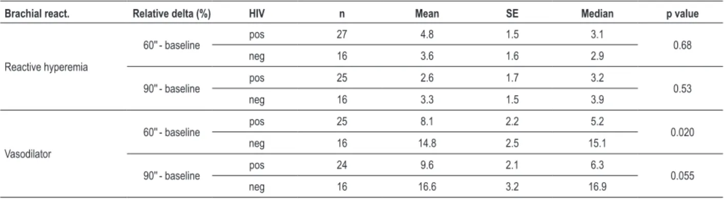
Table 4 - Analysis of the relative delta (%) of brachial reactivity according to the HIV status
Brachial react. Relative delta (%) HIV n Mean SE Median p value
Reactive hyperemia
60''- baseline pos 27 4.8 1.5 3.1 0.68
neg 16 3.6 1.6 2.9
90''- baseline pos 25 2.6 1.7 3.2 0.53
neg 16 3.3 1.5 3.9
Vasodilator
60''- baseline pos 25 8.1 2.2 5.2 0.020
neg 16 14.8 2.5 15.1
90''- baseline pos 24 9.6 2.1 6.3 0.055
neg 16 16.6 3.2 16.9
SE: Standard Error.
Table 2 – Analysis of demographic, clinical and laboratory variables according to the use of protease inhibitors
Protease Inhibitor
Variable with (n = 15) without (n = 12) p value
Age (years) 43.2 ± 8.2 39.6 ± 8.7 0.27
BMI (Kg/m2) 21.7 ± 2.4 22.9 ± 2.5 0.23
Abdominal circumference (cm) 85.9 ± 8.7 87.3 ± 8.6 0.68
Blood glucose (mg/dL) 90.1 ± 16.8 87.9 ± 13.5 0.72
Total cholesterol total (mg/dL) 206.8 ± 52.6 202.4 ± 44.9 0.82
HDL-c (mg/dL) 38.6 ± 12.0 44.1 ± 6.3 0.13
LDL-c (mg/dL) 139.4a ± 48.0 138.0a ± 39.3 0.94
TG (mg/dL) 265.6 ± 120.6 197.7 ± 167.5 0.009
Reactive hyperemia (baseline) 3.65 ± 0.58 3.29 ± 0.73 0.16
Vasodilator (baseline) 3.79 ± 0.64 3.38b ± 0.74 0.15
Data expressed as mean ± standard deviation; a one patient lost; b two patients lost.
60s, and 90s). A significant increase in brachial reactivity to reactive hyperemia was observed in the HIV-positive (p = 0.033) and HIV-negative (p =0.014) subgroups up to 60 s. No statistical significance was found for HIV-positive patients at 90s.
The endothelium-independent analysis (use of vasodilator) showed a significant increase in brachial reactivity in both the positive and negative subgroups, with p = 0.0001 and p = 0.0001, respectively.
Also using ANOVA, the subgroups of patients receiving and not receiving PI were compared at 60s and 90s of brachial reactivity, showing a significant increase to reactive hyperemia in the subgroup with PI (p = 0.038) only at 60s, with no statistical significance for this measurement at 90s.
Using SL nitrate, a significant increase in brachial reactivity was observed in the subgroups with and without PI, with p = 0.016 and p = 0.0001, respectively.
Although a significant increase in brachial reactivity measurements (reactive hyperemia and vasodilator) was observed, especially from baseline to 60s, both in the HIV-positive and in the PI subgroups, this increase was differentiated, that is, HIV-positive or with PI subgroups showed significantly lower increase to vasodilator than the HIV-negative or without PI subgroups.
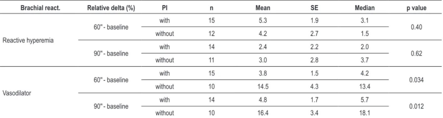
Based on the analysis of the relative delta (%) of brachial reactivity according to the HIV status and use of PI, we observed that, at 60s, both the HIV-positive and with PI groups had significantly lower relative delta to the use of vasodilator than the HIV-negative and without PI groups, with p = 0.020 and p = 0.034, respectively.
No statistical significance was found for the relative delta to reactive hyperemia in any of the subgroups.
The analysis of the relative delta of brachial reactivity according to the HIV status and use of protease inhibitor (PI) is shown in Tables 4 and 5, respectively.
Variables such as gender; body mass index (BMI); waist circumference (WC); blood glucose; total cholesterol (TC); HDL-c; LDL-c; and TG showed no statistical difference in
Table 3 - Longitudinal analysis of brachial reactivity to reactive hyperemia for the HIV and protease inhibitor subgroups
Reactivity to reactive hyperemia
Subgroup Baseline 60 sec 90 sec p valuea signiicant ≠ b
HIV positive (n = 25) 3.48 ± 0.68 3.60 ± 0.68 3.55 ± 0.68 0.033 baseline ≠ 60"
HIV negative (n = 16) 3.30 ± 0.58 3.42 ± 0.61 3.40 ± 0.59 0.014 baseline ≠ 60" and 90"
I. Protease with (n = 14) 3.62 ± 0.59 3.78 ± 0.54 3.69 ± 0.61 0.038 baseline ≠ 60"
I. Protease without (n = 11) 3.29 ± 0.77 3.37 ± 0.80 3.36 ± 0.74 0.54
relation to brachial reactivity.
Multiple linear regression analysis showed that only PI decreased the relative delta in HIV-positive patients in response to vasodilator at 60s and 90s, with p = 0.032 and p = 0.010, respectively.
Discussion
This study showed that HIV-positive patients receiving HAART, including PI, for more than two years, presented endothelial dysfunction in comparison to the group without PI and to the non-infected control group.
The main contribution of these findings in relation to previous studies is the demonstration of the impaired endothelium-independent vasodilation response in HIV-positive patients receiving PI.
Previous studies12 have assessed endothelial function in
HIV patients by means of the DILA technique, correlating it with the likelihood of development of coronary artery disease. DILA is a noninvasive, accurate and reproducible technique, albeit dependent on operational characteristics related to the ultrasound frequency and technical training.
Endothelial dysfunction to reactive hyperemia (endothelium-dependent) and to the use of nitroglycerine (endothelium-independent) was analyzed by means of the study of the brachial artery at different timepoints of dilation (60 and 90 seconds). According to a validated protocol12,
the nitric oxide (NO) peak release and artery dilation occur in this period.
Our study showed that both in the subgroup of HIV-positive patients and in that of patients using PI, there was increased brachial artery dilation to reactive hyperemia, with no difference in relation to the HIV-negative subgroup and the subgroup without PI.
Nolan et al13 did not find any difference in endothelial
function when they compared the brachial artery flow in 24 HIV-positive patients treated with PI and 24 HIV-negative patients of the control group13.
Stein et al14 showed decreased brachial artery dilation
in positive patients receiving PI in comparison to HIV-positive patients not receiving PI14.
Table 5 - Analysis of the relative delta (%) of brachial reactivity according to the use of protease inhibitor
Brachial react. Relative delta (%) PI n Mean SE Median p value
Reactive hyperemia
60''- baseline with 15 5.3 1.9 3.1 0.40
without 12 4.2 2.7 1.5
90''- baseline with 14 2.4 2.2 2.0 0.62
without 11 3.0 2.8 3.7
Vasodilator
60''- baseline with 15 3.8 1.5 4.2 0.034
without 10 14.5 4.3 13.4
90''- baseline with 14 4.8 1.7 5.7 0.012
without 10 16.4 3.4 18.1
SE: Standard Error.
Endothelium-independent vasodilation using exogenous NO is related to the smooth muscle function. In our study, only PI significantly influenced the relative delta to vasodilator at 60 and 90 seconds.
A study evaluated 800 asymptomatic HIV-negative patients with risk factors for cardiovascular disease by using exogenous nitroglycerin; it concluded that the reduced response to nitroglycerin was correlated with advanced age, increased total cholesterol, history of diabetes mellitus and smoking15.
Takase et al16 analyzed the interaction between
hypertension and DM in endothelium-independent dysfunction in four groups divided into type-2 DM, hypertension, hypertension / DM, and control. The results showed that nitroglycerine-induced vasodilation was decreased in the group of patients with hypertension and diabetes in comparison to the control group (p < 0.001), thus showing that the interaction between DM and hypertension caused deterioration of both the vascular endothelium and the smooth muscle responsible for endothelium-independent vasodilation16.
The mechanism of endothelial dysfunction is explained by both the viral action and the use of HAART. Shankar et al demonstrated a reduction in brachial artery dilation in HIV-positive children not receiving antiretroviral drugs17.
The virus acts on the endothelium by means of glycoprotein 120 (Gp120), leading to the production of adhesion molecules (ICAM-1), prothrombotic states, TNF alpha and interleukin 6 (IL-6).
Although PIs reduce endothelial nitric oxide synthase (eNOS), which leads to a decreased production of nitric oxide, reactive oxygen species (ROS) contribute to endothelial dysfunction, thus resulting in inflammation, cell damage, and endothelial cell apoptosis. PIs induce ROS formation18. Mondal et al19 demonstrated the presence
of ROS in aortic endothelial cells of patients receiving zidovudine and efavirenz19.
PIs also lead to mitochondrial DNA dysfunction, thus contributing to increased oxidative stress. Cote et al showed the first case of mitochondrial dysfunction in HIV-positive patients receiving PI and presenting symptoms of hyperlactatemia20.
References
1. Barbaro G. Pathogenesis of HIV-associated heart disease. AIDS. 2003; 17: S12-20.
2. Prendergast BD. HIV and cardiovascular medicine. Heart. 2003; 89: 793-800.
3. Barbaro G. Cardiovascular manifestations of HIV infection. J R Soc Med. 2001; 94: 384-90.
4. Murphy El, Collier AC, Kalish LA. Highly active antiretroviral therapy decrease mortality and morbidity in patients with advanced HIV disease. Ann Intern Med. 2001; 135: 17-26.
5. Samaras K, Wand H, Law M, Emery S, Cooper D, Carr A. Prevalence of Metabolic Syndrome in HIV-Infected Patients Receiving Highly Active Antiretroviral Therapy Using International Diabetes Foundation and Adult Treatment Panel III Criteria. Diabetes Care. 2007; 30: 113-9.
6. Constant J, Pellegrin Jl, Peuchant E. Plasma lipids in HIV-infected patients: a prospective study in 95 patients. Eur J Clin Invest. 1994; 24: 416-20.
7. The Data Collection on Adverse Events of Anti-HIV Drugs (DAD) Study Group. Combination antiretroviral therapy and the risk of myocardial infarction. N Engl J Med. 2003; 349: 1993-2003.
8. Henry K, Melroe H, Huebsch J, Hermundson J, Levine C, Swensen L. Severe premature coronary artery disease with protease inhibitors. Lancet. 1998; 351: 1328.
9. Chaparro J , Reeds D, Wen W, Xueping E, Klein S, Semenkovich C, et al. Alterations in thigh subcutaneous adipose tissue gene expression in protease inhibitor-based highly active antiretroviral therapy. Metabolism. 2005; 54: 561-7.
10. Xu A, Yin S, Wong L, Chan KW. Adiponectin ameliorates dyslipidemia induced by the human immunodeficiency virus protease inhibitor ritonavir in mice. Endocrinology. 2004; 145: 487-94.
11. Blanco JJ, Garcia IS, Cerezo JG, Rivera JM, Anaya PM, Raya PG, et al. Endothelial function in HIV-infected patients with low or mild cardiovascular risk. J Antimicrob Chemother. 2006; 58: 133-9.
12. Corretti MC, Anderson TJ, Benjamin EJ, Celermajer D, Charbonneau F,
Creager MA, et al. Guidelines for the ultrasound assessment of endothelial-dependent flow-mediated vasodilation of the brachial artery: a report of the International Brachial Artery Reactivity Task Force. J Am Coll Cardiol. 2002; 39: 257-65.
13. Nolan D, Watts GF, Herrmann SE, French MA, Jonh M, Mallal S. Endothelial function in HIV infected patients receiving protease inhibitor therapy: does immune competence affect cardiovascular risk? QJM. 2003; 96: 825-32.
14. Stein JH, Klein MA, Bellehumeur JL, McBride PE, Wiebe DA, Otvos JD, et al. Use of human immunodeficiency virus – 1 protease inhibitors in associated with atherogenic lipoprotein changes and endothelial dysfunction. Circulation. 2001; 104: 257-62.
15. Ma L, Zhao S, Li J, Zhou Q, Gao M. Interaction of hypertension and diabetes on impairment of endothelial function. Chin Med J (Engl). 2001; 114 (6): 563-7.
16. Takase B, Uehata A, Akima T, Nagai T, Nishioca T, Hamabe A, et al. Endothelium-dependent flow mediated vasodilation in coronary and brachial arteries in suspected coronary artery disease. Am J Cardiol. 1998; 82: 1535-9.
17. Shankar SS, Dube MP. Clinical aspects of endothelial dysfunction associated with human immunodeficiency virus infection and antiretroviral agents. Cardiovasc Toxicol. 2004; 4: 261-9.
18. Rush JW, Denniss SG, Graham DA. Vascular nitric oxide and oxidative stress: determinants of endothelial adaptations to cardiovascular disease and to physical activity. Can J Appl Physiol. 2005; 30 : 442-74.
19. Mondal D, Pradhan L, Ali M, Agrawal KC. HAART drugs induce oxidative stress in human endothelial cells and increase endothelial recruitment of mononuclear cells. Cardiovasc Toxicol. 2004; 4: 287-302.
20. Côté H, Brumme ZL, Craib K, Alexander CS, Wynhoven B, Wong H, et al. Changes in mitochondrial DNA as a marker of nucleoside toxicity in HIV-infected patients. N Engl J Med. 2002; 346: 811-20.
21. Wang X, Chai H, Yao Q, Chen C. Molecular mechanisms of HIV protease inhibitor-induced endothelial dysfunction. J Acquir Immune Defic Syndr. 2007; 44: 493-9.
endothelial dysfunction is related to protein kinase activation (MAPKs) by reactive oxygen species. PIs activate different types of MAPKs in different cells21.
ROS are also present in cells of the smooth muscle, which is responsible for endothelium-independent vasodilation, thus leading to endothelial dysfunction with the use of NTG in patients receiving PI. Chai et al22,23 demonstrated the presence
of the superoxide anion in endothelial smooth muscle cells of patients treated with PI22,23. Kim et al24 demonstrated that viral
proteins affect the proliferation of endothelial smooth muscle cells, which represents the greatest event in the formation of vascular lesion24.
Preliminary studies showed that antioxidants such as ginsenoside and curcumin may reverse the endothelial dysfunction caused by PIs, thus decreasing ROS25.
The major limitation of our study was the presence of hypertension in the group of volunteers, which, however, did not preclude the confirmation of our findings in relation to the endothelium-independent vascular dysfunction.
Conclusion
HIV-positive patients receiving PI present endothelium-independent dysfunction when compared to HIV-positive patients not receiving PI, as well as to the control group individuals.
Potential Conflict of Interest
No potential conflict of interest relevant to this article was reported.
Sources of Funding
This study was partially funded by CAPES.
Study Association
22. Chai H, Zhou W, Lin P, Lumsden A, Yao Q, Chen C. Ginsenosides block HIV protease inhibitor ritonavir-induced vascular dysfunction of porcine coronary arteries. Am J Physiol Heart Circ Physiol. 2005; 288: H2965 – H2971.
23. Chai H, Yan S, Peter L, Alan BL, Yao Q, Chen C. Curcumin blocks HIV protease inhibitor ritonavir-induced vascular dysfunction in porcine coronary arteries. J Am Coll Surg. 2005; 200: 820-30.
24. Kim J, Ruff M, Karwatowska-Prokopczuk E, Hunt L, Ji H, Pert CB, et al. HIV envelope protein gp120 induces neuropeptide y receptor-mediated proliferation of vascular smooth muscle cells: relevance to AIDS cardiovascular pathogenesis. Regul Pept. 1998; 75-76: 201-5.