Association between skin thickness and bone density in
adult women
*Associação entre espessura da pele e densidade óssea em mulheres adultas
Patrícia de Paula Yoneda1 Sckarlet Ernandes Biancolin2
Matheus Souza Martins Gomes3 Hélio Amante Miot4
Received on 24.06.2010.
Approved by the Advisory Board and accepted for publication on 20.11.10.
* Study conducted at the outpatient departments of the Teaching Hospital, Botucatu School of Medicine, Universidade Estadual Paulista Julio de Mesquita Filho, São Paulo, Brazil.
Conflict of interest: None / Confllito de interesse: Nenhum
Financial funding / Suporte financeiro: São Paulo State Foundation for the Support of Research (FAPESP), Grant # 08/54628-2
1 Resident Physician in Ophthalmology, Botucatu School of Medicine, Universidade Estadual Paulista Julio de Mesquita Filho, São Paulo, Brazil. 2
Undergraduate medical student, Botucatu School of Medicine, Universidade Estadual Paulista Julio de Mesquita Filho, São Paulo, Brazil. 3 Resident Physician in Anesthesiology, Botucatu School of Medicine, Universidade Estadual Paulista Julio de Mesquita Filho, São Paulo, Brazil.
4 PhD. Assistant Professor, Department of Dermatology and Radiotherapy, Botucatu School of Medicine, Universidade Estadual Paulista Julio de Mesquita Filho, São Paulo, Brazil.
©2011 by Anais Brasileiros de Dermatologia
Abstract:BACKGROUND: Osteoporosis mainly affects menopausal women and the elderly, predisposing these
individu-als to fractures that result in morbidity, mortality and costs to the healthcare system. Since dermal collagen reduces in parallel with a decrease in bone mass with aging, skin thickness may be indicative of a risk of osteoporosis.
OBJECTIVES: To evaluate the correlation between bone density and skin thickness on the backs of the hands of adult women.
METHODS: A cross sectional study involving adult women attending a university hospital outpatient clinic who were
interviewed individually and submitted to bone densitometry and measurement of skin thickness on the backs of their hands using skinfold calipers. Other risk factors for osteoporosis were also investigated.
RESULTS: A total of 140 patients were evaluated. Mean age (± standard deviation) was 57 ± 11 years. Mean skin thickness
on the backs of the hands was 1.4 ± 0.4 mm. There was a correlation between the right and left hands (R = 0.9; p<0.01). A direct correlation was found between skin thickness on the backs of the hands and bone density at the lumbar spine and femur (p<0.01). These results remained consistent even following adjustment for the covariables of age, skin pho-totype, body mass index, smoking, use of oral corticoids, anti-inflammatory use and time since menopause. Osteoporosis was inversely associated with the thickness of the skin on the back of the hands (odds ratio = 0.10; p<0.03).
CONCLUSIONS: An independent correlation was found between skin thickness and bone density, suggesting that these
events occur simultaneously. Skin signs may represent a non-invasive method of stratifying risk in these patients, help-ing identify cases requirhelp-ing early treatment.
Keywords: Collagen; Densitometry; Osteoporosis; Skin; Skin aging
Resumo:FUNDAMENTOS: Osteoporose acomete principalmente mulheres em menopausa e idosos, predispondo a
frat-uras que geram morbidade, mortalidade e custos ao sistema de saúde. Como o colágeno dérmico diminui paralela-mente à redução da massa óssea com o envelhecimento, a medida da espessura da pele pode ser indício do risco de osteoporose.
OBJETIVOS: Avaliar a correlação entre densidade óssea e espessura da pele do dorso das mãos de mulheres adultas.
MÉTODOS: Estudo transversal envolvendo mulheres adultas atendidas em ambulatório de hospital universitário
submeti-das à densitometria óssea, que foram avaliasubmeti-das individualmente e mensurada, por paquímetro, a espessura da pele no dorso das mãos, além de investigados demais fatores de risco para osteoporose.
RESULTADOS: Avaliaram-se 140 pacientes. A média (±dp) de idade foi de 57 (±11) anos; a média da espessura da pele
do dorso das mãos foi de 1,4 (±0,4) mm. Houve correlação entre as medidas das mãos direita e esquerda (R=0,9; p<0,01). Observou-se correlação direta entre as espessuras de pele do dorso das mãos e as densidades ósseas lom-bares e femorais (p<0,01). Tais resultados permaneceram consistentes mesmo quando ajustados pelas covariáveis: idade, fototipo, índice de massa corpórea, tabagismo, uso de corticoide oral, uso de anti-inflamatório oral e tempo de menopausa. Osteoporose se associou inversamente com a espessura da pele das mãos (Odds Ratio=0,10; p<0,03). CONCLUSÕES: Espessura da pele correlacionou-se, independentemente, com a densidade óssea, sugerindo
simultanei-dade dos eventos. Sinais cutâneos podem contribuir para a estratificação de risco não invasiva desses pacientes, e colaborar na identificação e tratamentos precoces.
INTRODUCTION
Osteoporosis is a systemic syndrome character-ized by a reduction in bone mass and microarchitec-tural damage to the skeleton, predisposing the indi-vidual to mechanical fragility and a risk of fracture. It results from the alterations in the process of bone remodeling that occur as a consequence of age, the use of certain drugs, hormonal stimuli and external factors. It affects both genders and all age-groups; however, the principal groups affected by the
condi-tion are the elderly and postmenopausal women. 1
In Brazil, more than 10 million individuals are estimated to be affected by the disease. The annual incidence of hip fractures, adjusted for age, is calculat-ed at 6-13/10,000 men and 12-28/10,000 women. Osteoporosis has a significant effect on quality of life and on the morbidity and mortality of the population. The high direct costs to the healthcare system and the indirect costs to the government and to society make the prevention and early detection of osteoporosis
priority action items in public health. 2-4
The diagnosis of osteoporosis is defined, according to the World Health Organization (WHO), as bone mineral density (BMD) < 2.5 standard devia-tions below the mean values found for healthy young adults of the same race and gender, using dual-energy
x-ray absorptiometry (DEXA). 5
In adults, osteoblastic activity decreases with age and in the menopause. Genetic factors are also involved, as is smoking, caffeine consumption, the chronic use of certain drugs (corticosteroids and antiin-flammatories), nutritional deficiencies (vitamin D and calcium), biotype (female, fair-skinned, slim), a seden-tary lifestyle and diseases such as malabsorption
syn-dromes, diabetes mellitus and hypoparathyroidism. 2,6
Most of the collagen in the human body is located in the skin and bone tissue, with over 70% being composed of type-I collagen. Irrespective of an individual’s exposure to the sun, a progressive decline in the extracellular matrix and in collagen occurs as a result of the aging process, with a similar process occurring in the bones. Some authors have demonstrated the simultaneity of these events, with a correlation between transparent skin and osteoporo-sis. This led to a hypothesis that there is a correlation between bone loss and skin thinning, suggesting col-lagenase activity in both tissues. Various studies have reported this relationship; however, skin measure-ments alone were unable to provide a precise
esti-mate of mineral density. 7-12
The objective of the present study was to eval-uate the correlation between skin thickness on the backs of the hands and bone density in adult women, as well as its relationship with the principal risk fac-tors for osteoporosis.
MATERIAL AND METHODS
A cross sectional study was conducted involving adult female patients attending outpatient clinics at the
Teaching Hospital of the Faculty of Medicine, University
Estadual Paulista (UNESP) between September 2008 and January 2010. Patients had to be able to give their informed consent to participate in the study and had to have been submitted to bone densitometry (Hologic Discovery bone densitometer) at the same institution within the year preceding the interview.
The skin examination and the individual views were conducted by the investigators. The inter-views were based on a standardized, structured form designed to collect data on demographical variables and the risk factors for osteoporosis. No specific systemati-zation was adopted for the collection and selection of the sample, with all the patients available who fulfilled the admission criteria being included consecutively.
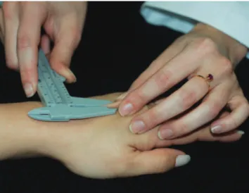
Skin thickness was estimated on the backs of both hands using a pachymeter (Black Bull/classic). The value used for analysis consisted of the mean thickness of the patient’s two hands except in cases in which the patient had lost one of her hands or had lesions on the skin at that site (Figure 1).
The dependent variables were bone density (T-score values) at the right femoral neck, at the Ward’s triangle and the total value of the femur, as well as at the lumbar spine (mean value of the L1-L4
verte-brae).5 The principal independent variable was the
skin thickness on the backs of the hands. However, demographic and constitutional aspects were also evaluated, as well as variables associated with an increased risk of osteoporosis such as: age, body mass index (BMI), skin phototype, diabetes mellitus, pur-pura of the upper limbs, smoking, daily caffeine intake, duration of antiinflammatory use, duration of corticosteroid use, time since menopause and
dura-tion of hormone replacement therapy. 2,6
Osteoporosis was defined in accordance with the World Health Organization’s classification as a BMD (T-score) below -2.5 standard deviations and
osteopenia as below -1.0 standard deviation. 5
Continuous variables were represented as means and standard deviations or medians and interquartile deviations if distribution was not para-metric. The normality of the samples was evaluated
using the Shapiro-Wilk test. 13 Categorical and ordinal
variables were represented by their percentage fre-quency.
each other using one-way analysis of variance (ANOVA) or the Kruskal-Wallis test if heterogeneity of variance (heteroscedasticity) was demonstrated by Levene’s test.13
The co-variables were then used in the statisti-cal adjustment of the correlations in a multivariate lin-ear regression model (rank ANCOVA) with the inclu-sion of all the covariates that achieved maximum
sig-nificance of 0.2 in the bivariate analysis. 13
The variables were tested with respect to their association with osteopenia (T<-1.0) and osteoporosis (T<-2.5) based on a multinomial logistic regression model, adjusted for the other covariables that achieved maximum significance of 0.2 in the bivariate analysis.
The measures of association were represented by the Spearman and Pearson correlation coefficients
and in the multivariate analysis as βregression
coeffi-cients or odds ratios, with significance defined as a two-tailed p-value of 0.05.
The ability of skin thickness measurement on the backs of the hands to classify osteoporosis was cal-culated using the receiver operating characteristics (ROC) curve. Data were tabulated in Microsoft’s Excel program and analyzed using the SPSS software
pro-gram, version 17.0.14
To calculate sample size, a preliminary analysis was performed on the first 100 patients,
contemplat-ing a type I (α) error of 0.05 and a type II (β) error of
0.2 in the direct correlations between the dependent and independent variables. It was found that a further 10 cases were required for each co-variable to
com-plete the final multivariate model. 13
RESULTS
A total of 140 female patients were interviewed, 118 (84%) of whom were in the menopause. The principal demographic and clinical characteristics of the patients are shown in Table 1. As shown, the sam-ple consisted of adult patients and there was a wide range in bone density and skin thickness measure-ments in these women.
There was a strong correlation between skin
thickness on the right and left hands (RPearson = 0.87,
p<0.01). A negative correlation was found between the mean skin thickness of the hands and the patient’s
age (RPearson = -0.21, p=0.01), and a positive
correla-tion between mean skin thickness and BMI (RPearson=
0.34, p<0.01).
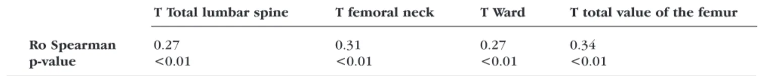
A direct significant correlation was found between the variation in skin thickness on the backs of the hands and the variation in all BMD measurements, with similar results when the menopausal patients were evaluated as a separate group (Table 2).
In the multivariate analysis, a correlation was found between bone density at the lumbar spine and
femur and skin thickness on the backs of the hands (p<0.05), irrespective of the other risk factors (Table 3). Other factors that proved significant in estimating BMD included age, BMI, time since menopause, skin photo-type and the use of nonsteroidal antiinflammatory drugs.
Qualitative analysis conducted using a multino-mial logistic regression model adjusted for age, time since menopause, BMI, skin phototype, smoking, duration of antiinflammatory use and duration of cor-ticosteroid use showed that mean skin thickness on the backs of the hands was associated with osteoporo-sis (odds ratio = 0.10; 95%CI: 0.01 – 0.75; p<0.03) but not with osteopenia (odds ratio = 0.67; 95%CI: 0.19 – 2.36; p>0.52).
When skin thickness on the backs of the hands alone was considered a marker of osteoporosis, the area under the ROC curve was 0.68 (p<0.01).
DISCUSSION
A statistically significant correlation was found between the variation in skin thickness on the backs of the hands of adult women and the variation in BMD at the lumbar spine and the femur, irrespective of the other risk factors. This finding is in agreement with results from other similar studies, suggesting that the bone alterations that occur in parallel with skin aging are probably due to collagenase activity in these organs (Table 4). 7,15
The small magnitude of the correlation between skin thickness and BMD highlights the multifactorial character of osteoporosis, including its genetic com-ponent and the strong effect of external elements in the development of the disease. The other risk factors identified in this study are similar to those found in epidemiological studies for this age group. No model has been found that is ideal for calculating the risk in this population. 6,16,17
Given the importance of an early diagnosis of osteoporosis and osteopenia, the development of a probabilistic model to calculate the risk in the popula-tion would facilitate early identificapopula-tion of the disease and contribute towards reducing morbidity and decreasing costs to the healthcare system. Noninvasive methods simplify population-based policies and should be proposed for use in research studies or even in campaigns to stratify risk, thus improving primary prevention. Measurement of skin thickness and the collection of factors concerning exposure and demo-graphic data can be performed by a paramedic, there-by increasing the capacity of healthcare services.
The pachymeter has been validated as an instru-ment for this type of evaluation and it allows skinfold
thickness to be measured at different sites. 18,19 Various
differ-ent anatomical sites and their findings of a correlation between skin thickness and bone density are in
agree-ment with the results of the present study. 15,20-22
Although skin thickness on the backs of the hands varies in accordance with occupational factors and sun exposure, the high correlation between the measurements on the right and left hands permitted mean skin thickness at this site to be used as a study
variable. Other studies have shown a correlation between skin thickness measured at different sites, reducing the likelihood of the hypothesis that sun damage is a factor that would activate skin collagenas-es and affect the thickncollagenas-ess of the skin of the backs of
the hands. 22,23 The authors adopted skin thickness of
the hands rather than any other anatomical site based on the results of previous studies that had adopted this methodology and concluded that there was an ade-quate correlation between the two measurements.
10,15,19-23
Furthermore, skin thickness does not vary with ethnicity; however, it varies significantly with age, body weight, the use of medication such as corticos-teroids, in the presence of systemic diseases and with
hormone replacement therapy. 8,23,24
On the other hand, BMD may vary significantly between different evaluation sites, with Ward’s trian-gle in the femur generally being the densest area and
the lumbar spine the least dense. 5 These
inconsisten-cies motivated an independent study of the correla-tion between skin thickness and bone density in these areas, although the final results were very similar.
There was a strong association in the present study between the time since menopause and bone density at the lumbar spine. This ratifies previous find-ings that bone alterations intensify from the time of
Variables Normal Osteopenia Osteoporosis Total p-value
n (%) 38 (27) 65 (47) 37 (26) 140 (100)
Age (years)a 53.7±6.9 54.4±10.4 65.5±10.8 57.1±10.9 <0.01*
Body mass index (kg/m2)a 29.4±4.9 27.3±4.2 26.2±4.5 27.6±4.6 <0.01**
Skin phototype - n (%) >0.16***
I 1 (3) 5 (8) 4 (11) 10 (7)
II 11 (29) 17 (26) 8 (22) 36 (26)
III 14 (37) 26 (40) 19 (51) 59 (42)
IV 7 (18) 6 (9) 6 (16) 19 (14)
V 5 (13) 7 (11) 0 (-) 12 (9)
VI 0 (-) 4 (6) 0 (-) 4 (3)
Diabetes mellitus - N (%) 8 (21) 9 (14) 4 (11) 21 (15) 0.22***
Purpura of the upper limbs - n (%) 19 (50) 33 (51) 20 (54) 72 (52) 0.73***
Coffee consumption (cups/day)a 3.2±3.6 2.6±2.2 2.2±1.9 2.7±6.6 0.23**
Smoking (packs/year)b 0.0±4.0 0.0±100.0 0.0±68.5 0.0±60.0 0.15*
Nonsteroidal anti-inflammatories (months)b 0.0±2.5 0.0±1.1 0.0±0.0 0.0±0.0 0.17*
Oral corticosteroids (months)a 1.7±7.1 5.8±28.9 3.6±19.7 4.1±22.4 0.66*
Time since menopause (years)b 3.0±6.8 5.0±10.5 18.0±16.5 6.0±15.0 <0.01*
Hormone replacement therapy - n (%) 10 (26) 15 (23) 12 (32) 37 (26) 0.55***
Skin thickness of the backs of the hands (mm)a 1.6±0.3 1.5±0.4 1.3±0.4 1.4±0.4 <0.01**
Total density of the lumbar spine (T)b 0.7±1.6 -1.0±1.0 -2.4±1.3 -0.9±1.8 <0.01*
Density at the femoral neck (T)b 0.3±1.1 -1.0±0.9 -2.0 ±0.9 -1.0±1.5 <0.01*
Density of the Ward’s triangle (T)b -0.2±1.1 -1.6±0.9 -2.8±0.8 -1.5±1.5 <0.01**
Total value of femur density (T)b 0.2±1.2 -0.9±0.8 -2.1±0.9 -0.9±1.4 <0.01*
FIGURE1:Standardized technique for measuring skin thickness on
the backs of the hands
Table 1:The principal clinical and demographic characteristics of the patients studied
T Total lumbar spine T femoral neck T Ward T total value of the femur
Ro Spearman 0.27 0.31 0.27 0.34
p-value <0.01 <0.01 <0.01 <0.01
menopause onwards due to decreased estrogen pro-duction. Known risk factors such as fair skin, low BMI and time since menopause were confirmed in this study and in this sample population these factors rep-resented an important parallel to skin thickness meas-urements in the hands.
Bateman’s purpura was not associated with bone density levels despite the fact that this associa-tion has been described in previous studies. This is justified by the greater sensitivity of the measurements of skin thickness in estimating the variation in bone density, since purpura is only present at more advanced stages of skin atrophy. Extreme forms of skin atrophy, transparent skin, purpura and skin fragility resulting principally from aging are grouped under the general term of dermatoporosis and are
strongly associated with osteoporosis. 8,25
The use of oral corticosteroids is one of the risk factors most associated with osteoporosis; however, this association was not found in this study group either in the bivariate or multivariate analyses. The small number of patients who reported the use of cor-ticoids (12.1%) may have reduced the power of
analy-sis; therefore, specific designs would be required to
evaluate the magnitude of this effect. 26
Likewise, in another study that investigated users of oral corticoids, no correlation was found between skin thickness and bone density, possibly because the osteoclastic activity induced by the corti-coid may have been more intense or accelerated than
the resulting skin atrophy. 21
The scientific value of this study resides in the finding that the correlation between skin thickness of the backs of the hands and bone density, analyzed in a Brazilian population, was substantiated by adjustment for various other risk factors for osteoporosis, showing its independence and consequently reiterating its potential for identifying individuals at risk. Use of a representative sample, clearly defined end-points, mean skin thickness measurements on the backs of both hands and multivariate analysis that took the prin-cipal risk factors for osteoporosis into consideration are examples of the care taken to minimize biases in meas-urement, selection or the addition of random errors.
Elements that may have limited the present study consisted of the fact that the sample was small,
T total lumbar T femoral necka
T Warda
T total value of spinea
the femur a
Variable βb
pc
βb
pc
βb
pc
βb
pc
Skin thickness on the backs of 0.17 0.03 0.16 0.03 0.15 0.05 0.15 0.03
the hands (mm)a
Age in yearsa -0.01 <0.01 -0.12 <0.01 -0.21 <0.01 -0.25 <0.01
Body mass index (kg/m2)a 0.17 <0.01 0.29 <0.01 0.14 <0.01 0.37 <0.01
Time since menopause (years)a -0.42 <0.01 -0.25 <0.01 -0.28 <0.01 -0.17 0.04
Nonsteroidal anti-inflammatory 0.27 0.03 0.12 0.21 0.10 0.32 0.05 0.62
drugs (months)a
Oral corticosteroids (months)a -0.18 0.21 0.10 0.45 0.11 0.42 0.03 0.80
Smoking (packs/year)a -0.04 0.20 -0.07 0.34 -0.12 0.12 -0.11 0.09
Skin phototype 0.02 <0.01 0.01 <0.01
I -4.57 -27.79 3.29 11.41
II 6.22 6.74 34.38 34.63
III -6.424 -1.25 33.57 20.93
IV 12.98 -1.76 32.14 23.57
V 6.80 8.16 35.69 30.16
VI - - -
-Table 3:Multivariate model of bone density (rank ANCOVA) and mean skin thickness on the backs of the
hands adjusted for selected covariables
aVariables transformed in ranks; bPartial regression coefficient β; cTest for global effects in model (type I)
TABLE2: Linear correlation between mean skin thickness measurements on the backs of the hands
restricted to only women and the women had to have been referred for densitometry in order to be includ-ed in the study. These factors resultinclud-ed in a very homogenous sample and reduced the possibility of analyzing sub-groups and forming a gradient of densi-ty measurements to be evaluated with respect to the covariables. Furthermore, the patients were predom-inantly housewives and it was difficult to evaluate sedentary lifestyle, an important risk factor for osteo-porosis, because of the imprecise dimension of physi-cal activity in the home.
Dermatologists should be alert to skin signs that may be indicative of systemic disorders, including
thin-ning of the skin as a risk marker for osteoporosis. 27-30
Studies on the correlation between collagen density, the activity of skin metalloproteinases, elastases
and collagenases, and bone density should supplement clinical findings to increase physiogenic and pathogen-ic knowledge on the process of bone and skin aging.
CONCLUSION
An independent correlation was found between the variability in skin thickness on the backs of the hands and the variation in bone density, suggesting that these events occur simultaneously and defining this measurement as a possible risk marker for osteo-porosis. The clinical perception of skin thinning asso-ciated with the evidence of other risk factors for osteo-porosis may contribute towards the noninvasive strat-ification of these patients, providing guidance on modifiable factors and collaborating in early
identifi-cation and treatment. ❑
Study Comments
McConkey, 196329 130 patients; correlation between transparent skin, skin thickness, purpura and osteoporosis;
skin on the back of the hands measured with a pachymeter; bivariate analysis; osteoporosis diagnosed by x-ray.
McConkey, 196525 102 women with rheumatoid arthritis and 200 with other diseases; correlation between
transparent skin, skin thickness, age, purpura, use of corticoids and osteoporosis; skin thickness on the back of the hands measured with a pachymeter; bivariate analysis; osteoporosis diagnosed by x-ray.
Chappard, 199110 133 women; correlation between skin thickness, age, body mass index and osteoporosis; skin
thickness on the back of the hand measured using a pachymeter; multivariate analysis; osteoporosis diagnosed by densitometry.
Orme, 199419 225 women; correlation between skin thickness, age, time since menopause, weight, height and
osteoporosis; skin thickness on the back of the fourth finger of the right hand measured using a pachymeter; bivariate analysis; osteoporosis diagnosed by densitometry.
Castello-Branco, 199411 76 women; correlation between dermal collagen, age, time since menopause and osteoporosis; abdominal skin collagen measured by spectrophotometry; bivariate analysis; osteoporosis diagnosed by bone densitometry.
Varilla, 199522 60 women; correlation between skin thickness, age, time since menopause, diabetes, body mass
index, skin phototype, smoking and osteoporosis; skin measured at the malleolus, abdomen and forearm by ultrasonography; bivariate analysis; osteoporosis diagnosed by densitometry.
Pedersen, 199524 40 patients; correlation between skin thickness, weight and osteoporosis; skin thickness
measured at the forearm by ultrasonography; bivariate analysis; osteoporosis diagnosed by densitometry.
Smeets, 199730 98 postmenopausal patients; no correlation between skin thickness and osteoporosis; skin
thickness measured at the forearm using ultrasonography; bivariate analysis; osteoporosis diagnosed by quantitative tomography.
Werth, 199821 14 patients in use of corticoids; no correlation between skin thickness and osteoporosis; skin
thickness measured on the arm by ultrasonography; bivariate analysis; osteoporosis diagnosed by densitometry.
Patel, 200715 603 patients; correlation between skin thickness, age, weight and osteoporosis; fracture risk
evaluated; skin thickness measured at the forearm using ultrasonography; multivariate analysis; osteoporosis diagnosed by densitometry.
Cagle, 200720 98 patients; correlation between skin thickness, age, weight and osteoporosis; skin thickness
measured on the arm by ultrasonography; multivariate analysis; osteoporosis diagnosed by densitometry.
Present study 140 women; correlation between skin thickness, age, time since menopause, diabetes, body
mass index, skin phototype, smoking and osteoporosis; skin thickness measured on the backs of the hands using a pachymeter, multivariate analysis; osteoporosis diagnosed by densitometry.
An Bras Dermatol. 2011;86(5):878-84.
How to cite this arti cle/Como citar este arti go: Poziomczyk CS, Köche B, Dornelles MA, Dornelles SIT. Yoneda PP,
Biancolin SE, Gomes MSM, Miot HA. Association between skin thickness and bone density in adult women . An Bras Dermatol. 2011;86(5):878-84.
REFERENCES
1. Pinheiro MM, Ciconelli RM, Martini LA, Ferraz MB. Clinical risk factors for osteoporotic fractures in Brazilian women and men: the Brazilian Osteoporosis Study (BRAZOS). Osteoporos Int. 2009;20:399-408.
2. Pinheiro Mde M, Ciconelli RM, Martini LA, Ferraz MB. Risk factors for recurrent falls among Brazilian women and men: the Brazilian Osteoporosis Study (BRAZOS). Cad Saude Publica. 2010;26:89-96.
3. Fleurence RL, Iglesias CP, Torgerson DJ. Economic evaluations of interventions for the prevention and treatment of osteoporosis: a structured review of the literature. Osteoporos Int. 2006;17:29-40.
4. Zethraeus N, Borgström F, Ström O, Kanis JA, Jönsson B. Cost-effectiveness of the treatment and prevention of osteoporosis--a review of the literature and a reference model. Osteoporos Int. 2007;18:9-23.
5. Zanette E, Stringari FF, Machado F, Marroni JB, Ng DPK, Canani LH. Avaliação do diagnóstico densitométrico de osteoporose/osteopenia conforme o sítio ósseo. Arq Bras Endocrinol Metab. 2003;47:30-47.
6. Guthrie JR, Dennerstein L, Wark JD. Risk factors for osteoporosis: A review. Medscape Womens Health. 2000;5:E1.
7. Shuster S. Osteoporosis, a unitary hypothesis of collagen loss in skin and bone. Med Hypotheses. 2005;65:426-32.
8. Kaya G, Saurat JH. Dermatoporosis: a chronic cutaneous insufficiency/fragility syndrome. Clinicopathological features, mechanisms, prevention and potential treatments. Dermatology. 2007;215:284-94.
9. Whitmore SE, Levine MA. Risk factors for reduced skin thickness and bone density: possible clues regarding pathophysiology, prevention, and treatment. J Am Acad Dermatol. 1998;38:248-55.
10. Chappard D, Alexandre C, Robert JM, Riffat G. Relationships between bone and skin atrophies during aging. Acta Anat (Basel). 1991;141:239-44.
11. Castelo-Branco C, Pons F, Gratacós E, Fortuny A, Vanrell JA, Gonzalez-Merlo J. Relationship between skin collagen and bone changes during aging. Maturitas. 1994;18:199-206.
12. Baronti G, Giusti U, Sullo B, Zucchelli G. [Transparent cutis as a symptom for the early diagnosis of senile osteoporosis]. G Gerontol. 1970;18:77-88.
13. Norman GR, Streiner DL. Biostatistics: the bare essentials. 3rd ed. Hamilton, ON: BC Decker Inc; 2008.
14. SPSS 17.0 for Windows. Statistical package for social sciences. 17th ed. Chicago (IL): SPSS; 2008.
15. Patel R, Blake GM, Fogelman I. Evaluation of osteoporosis using skin thickness measurements. Calcif Tissue Int. 2007;81:442-9.
16. Von Mühlen D, Visby Lunde A, Barrett-Connor E, Bettencourt R. Evaluation of the simple calculated osteoporosis risk estimation (SCORE) in older Caucasian women: the Rancho Bernardo study. Osteoporos Int. 1999;10:79-84.
17. McClung MR. Clinical risk factors and evaluation of the risk of osteoporosis in clinical practice. Ann Med Interne (Paris). 2000;151:392-8.
18. Dykes PJ, Francis AJ, Marks R. Measurement of dermal thickness with the Harpenden skinfold caliper. Arch Dermatol Res. 1976;256:261-3.
19. Orme SM, Belchetz PE. Is a low skinfold thickness an indicator of osteoporosis? Clin Endocrinol (Oxf). 1994;41:283-7.
20. Cagle PE, Dyson M, Gajewski B, Lukert B. Can dermal thickness measured by ultrasound biomicroscopy assist in determining osteoporosis risk? Skin Res Technol. 2007;13:95-100.
21. Werth VP, Kligman AM, Shi X, Pagnoni A. Lack of correlation of skin thickness with bone density in patients receiving chronic glucocorticoid. Arch Dermatol Res. 1998;290:388-93.
22. Varila E, Sievanen H, Vuori I, Oksanen H, Punnonen R. Limited value of ultrasound measured skin thickness in predicting bone mineral density in peri- and postmenopausal women. Maturitas. 1995;21:45-9.
23. Whitmore SE, Sago NJ. Caliper-measured skin thickness is similar in white and black women. J Am Acad Dermatol. 2000;42:76-9.
24. Pedersen H, Agner T, Storm T. Skin thickness in patients with osteoporosis and controls quantified by ultrasound A scan. Skin Pharmacol. 1995;8:207-10. 25. McConkey B, Fraser GM, Bligh AS. Transparent Skin and Osteoporosis: A Study in
Patients with Rheumatoid Disease. Ann Rheum Dis. 1965;24:219-23.
26. Khan YK, Kalaaji AN, Clarke BL. Glucocorticoid-induced osteoporosis in dermatologic practice: a review. J Drugs Dermatol. 2008;7:1053-9.
27. Tamega AA, Aranha AMP, Guiotoku MM, Miot LDB, Miot HA. Associação entre acrocórdons e resistência a insulina. An Bras Dermatol. 2010;85:25-31. 28. Miot HA, Medeiros LM, Siqueira CRS, Cardoso LC, Gumieiro JH, Pandini Filho MA,
et al. Associação entre doença arterial coronariana e as pregas lobular diagonal e anterotragal em homens. An Bras Dermatol. 2006;81:29-33.
29. McConkey B, Fraser GM, Bligh AS, Whiteley H. Transparent skin and osteoporosis. Lancet. 1963;1:693-95.
30. Smeets AJ, Kuiper JW, van Kuijk C, Berning B, Zwamborn AW. Skin thickness does not reflect bone mineral density in postmenopausal women. Osteoporos Int. 1994;4:32-5.
MAILINGADDRESS/ ENDEREÇO PARA COR RES PON DÊN CIA:
Hélio Amante Miot
Departamento de Dermatologia da Faculdade de Medicina da Unesp, S/N.
Campus Universitário de Rubião Jr. CEP: 18618-000 - Botucatu - SP, Brazil Phone/Fax: +55 14 3882 4922