AVALIAÇÃO DA EFETIVIDADE DE CIMENTOS RESINOSOS
AUTO-ADESIVOS APLICADOS SOB DIFERENTES PRÉ-TRATAMENTOS
DA DENTINA RADICULAR NA RESISTÊNCIA ADESIVA DE PINOS
DE FIBRA DE VIDRO
Lima, Luiz Rafael Calixto
Avaliação da efetividade de cimentos resinosos auto-adesivos aplicados sob diferentes pré-tratamentos da dentina radicular na resistência adesiva de ´pinos de fibra de vidro / Luiz Rafael Calixto Lima - Araraquara: [s.n.], 2013.
78 f. ; 30 cm.
Tese (Doutorado) – Universidade Estadual Paulista, Faculdade de Odontologia
Orientador: Prof. Dr. Marcelo Ferrarezi de Andrade
1. Cimentos de resina 2. Cimentos dentários 3. Pinos dentários I.
Título
AVALIAÇÃO DA EFETIVIDADE DE CIMENTOS RESINOSOS
AUTO-ADESIVOS APLICADOS SOB DIFERENTES PRÉ-TRATAMENTOS
DA DENTINA RADICULAR NA RESISTÊNCIA ADESIVA DE PINOS
DE FIBRA DE VIDRO
Tese apresentada ao Programa de Pós-Graduação em Ciências Odontológicas – Área de concentração: Dentística Restauradora, da Faculdade de Odontologia de Araraquara - Universidade Estadual Paulista, como parte dos requisitos para obtenção do título de Doutor em Ciências Odontológicas.
Orientador: Prof. Dr. Marcelo Ferrarezi de Andrade
AVALIAÇÃO DA EFETIVIDADE DE CIMENTOS RESINOSOS
AUTO-ADESIVOS APLICADOS SOB DIFERENTES PRÉ-TRATAMENTOS
DA DENTINA RADICULAR NA RESISTÊNCIA ADESIVA DE PINOS
DE FIBRA DE VIDRO
Tese para obtenção do grau de Doutor
Comissão Julgadora
Presidente e Orientador: Prof. Dr. Marcelo Ferrarezi de Andrade
2o examinador: Prof. Dr. Edson Alves de Campos
3o examinador: Prof. Dr. José Roberto Cury Saad
4o examinador: Prof. Dr. Paulo Sérgio Quagliatto
5o examinador: Prof. Dr. Mânio de Carvalho Tibúrcio
Luiz Rafael Calixto Lima
Filiação:
José Borges Lima Gislaine Calixto Lima
22/10/1980
Nascimento – Penedo/AL
1999 – 2003
Graduação em Odontologia pela Universidade Federal de Alagoas – UFAL.
2005 – 2006
Curso de Especialização em Dentística Restauradora pela Faculdade de Odontologia de Araraquara - Universidade Estadual Paulista – UNESP/SP.
2007 – 2009
Curso de Pós-Graduação em Dentística Restauradora – Nível Mestrado - Faculdade de Odontologia de Araraquara - Universidade Estadual Paulista – UNESP/SP.
2009 – 2013
À DEUS, por me proporcionar uma formação educacional digna. Pela
oportunidade de concretizar mais uma etapa da minha vida, de tornar esse sonho
uma realidade. Obrigado Senhor por estar sempre ao meu lado!
Aos meus PAIS e IRMÃOS, pelo companheirismo, confiança e incentivo na
busca do crescimento pessoal e profissional, me dando sempre apoio necessário
Ao meu orientador, Prof. Dr. Marcelo Ferrarezi de Andrade, pelo
aprendizado, apoio, confiança, pelas grandes oportunidades e, principalmente, pela
grande amizade. Obrigado por tudo Marcelão! Você me ensinou a ter prazer pela
profissão. É através da sua simplicidade que se constrói um grande futuro.
A Professora Maria Salete Machado Cândido (in memoriam). Obrigado
pelos ensinamentos, Professora. Você sempre será um exemplo de competência e
dedicação à Docência.
Aos meus grandes amigos, Tarcísio Anjos e Marcílio Moreira, pela amizade
sincera, pela humildade e competência na nossa profissão. Obrigado pelos
conselhos e apoio!!!
A minha namorada, Renata Leão, por ser uma pessoa que torna qualquer
À Faculdade de Odontologia de Araraquara - Universidade Estadual Paulista
Júlio de Mesquita Filho - pela oportunidade e crescimento pessoal e profissional.
Aos meus grandes amigos de pós-graduação Victor, William, Darlon,
Adriano e Renato pelos ensinamentos, amizade e companheirismo. Podem contar
sempre comigo! Torço sempre pelo sucesso de todos vocês!
Aos meus amigos de turma Matheus Bandeca, Esther Gomes e Fernando
Florez pela amizade proporcionada esses anos.
Ao meu amigo e parceiro Jorge Eustáquio, a pessoa que não tem tempo
ruim para nada.
Aos demais professores da Dentística Restauradora, José Roberto Cury
Saad, Osmir Batista Oliveira Júnior, Sizenando de Toledo Porto Neto, Edson
Alves de Campos, Andréa Abi Rached Dantas e Alessandra Nara de Souza
Rastelli, pela qualidade do ensino e formação profissional.
Ao Professor Luis Geraldo Vaz, por todo apoio e orientação nessa
pesquisa, pelo exemplo de competência e simplicidade.
Aos Funcinários da Dentística, Adriana, Conceição, Cida, Marinho e
Vanderlei, e em especial a Creusinha e Cida “Branca”, pela simpatia e amizade
com que nos recebem.
Aos funcionários da Pós-graduação, em especial a Mara, e aos funcionários
da Biblioteca, em especial a Maria Helena.
O degrau de uma escada não serve simplesmente para
que alguém permaneça em cima dele, destina-se a
sustentar o pé de um homem pelo tempo suficiente
para que ele coloque o outro um pouco mais alto.
de fibra de vidro [Tese de Doutorado]. Araraquara: Faculdade de Odontologia da
Unesp; 2013.
RESUMO
A retenção de pinos pré-fabricados de fibra de vidro utilizados na restauração
de dentes com tratamento endodôntico é baseada na sua união às superfícies das
paredes do canal radicular por meio dos agentes de fixação resinosos. Os cimentos
resinosos auto-adesivos surgiram recentemente com o objetivo de simplificar o
procedimento de cimentação, porém seu desempenho clínico ainda é controverso na
literatura. Assim, o propósito desse trabalho é avaliar a efetividade de cimentos
auto-adesivos sob diferentes condições de tratamento da dentina na resistência à
extrusão de pinos de fibra de vidro cimentados intrarradicularmente. Foi avaliada a
resistência adesiva (push-out) de pinos de fibra de vidro utilizando cimentos
resinosos auto-adesivos e, diferentes substâncias para o tratamento dentinário
(ácido poliacrílico 11,5%; EDTA 17% e hipoclorito de sódio) previamente ao
procedimento de cimentação. Foi avaliado também o tipo de falha entre o
pino/cimento/dentina através de microscopia óptica. Foram utilizados 216 unidades
experimentais, em forma de discos com aproximadamente 1 mm de espessura,
retirados de 72 raízes bovinas restauradas com retenções intra-radiculares. Foi
utilizada uma máquina de ensaios universais MTS 810 Material Test System, à
teste kruskall wallis com post-hoc com teste dunn, com nível de significância de 5%.
Os resultados mostraram que o pré-tratamento dentinário não influenciou na
resistência adesiva, e que o cimento auto-adesivo RelyX U100 parece ser uma
opção viável na cimentação de pinos de fibra.
de Doutorado]. Araraquara: Faculdade de Odontologia da Unesp; 2013.
ABSTRACT
The retention of prefabricated fiberglass posts used in the restoration of
endodontically treated teeth is dependent on their bond between the surface of the
post and the root canal walls by means of resin cements. The self-adhesive resin
cements have recently introduced to simplify the procedure for cementing, but their
clinical performance is still controversial in the literature. Thus, the objective of this
study is to evaluate the effectiveness of self-adhesive cements under different
pre-treatment of dentin in the resistance to extrusion of fiberglass posts. Prior to
cementing procedure, the pre-treatment of dentin was performed with 11.5%
polyacrylic acid, 17% EDTA or sodium hypochlorite. The type of failure between the
post/cement/dentin was evaluated by steriomicroscopy. Two hundred and sixteen
bovine dentin discs were used. The disks had approximately 1 mm of thickness and
they were taken from 72 bovine roots restored with intraradicular retentions. It was
employed a universal testing machine MTS 810 Material Test System at a speed of
0.5 mm/min-1 with a load cell of 50 kg to evaluate the push-out resistance in different
thirds of each sample . The values of resistance in kgf were converted to Mpa. Data
were submitted to Kruskal-Wallis test with Dunn post-hoc test with significance level
1 Introdução... 16
2 Objetivos...21
3 Capítulos... 23
3.1 Capítulo 1: Influence of different root dentin pretreatment in
self-adhesive cements to fiber posts………... 24
3.2 Capítulo 2: Self-adhesive cements and fiber posts: a review……..…... 43
4 Considerações Finais...59
5 Referências...63
1 INTRODUÇÃO
O tratamento restaurador de dentes tratados endodonticamente e com grande
destruição deve ser realizado por meio de procedimentos que os protejam de cargas
mastigatórias, devolvendo a função e a estética.
Destruições extensas devido a lesões cariosas, fraturas, acesso endodôntico
incorreto, substituições de restaurações ou reabsorções internas levam à
necessidade, normalmente, da utilização de pinos intrarradiculares e núcleos de
preenchimento para reter a restauração final2,8.
Os pinos não metálicos surgiram com a principal vantagem de permitir uma
adesão à dentina através de cimentos resinosos. Inicialmente surgiram os pinos
cerâmicos, posteriormente, os pinos de fibra, os quais apresentam propriedades
mecânicas próximas às da estrutura dentária, especialmente o módulo de
elasticidade semelhante ao da dentina, possibilitando uma melhor distribuição das
forças ao remanescente dentário25. Os pinos de fibra de vidro se destacam por sua
translucidez natural e proporcionar um excelente resultado estético9,25.
Devido as características de rigidez semelhante à dentina, os pinos de fibra
de vidro absorvem as tensões geradas pelas forças mastigatórias e protegem o
remanescente radicular. Também possibilitam a construção de uma unidade
mecanicamente homogênea, devido aos materiais que compõem esse conjunto
(estrutura dental, agente de fixação e pino)1,11.
Existem diferentes parâmetros a serem considerados para correta seleção
dos pinos que podem influenciar na sua retenção8,11, uma vez que sua resistência,
superficial, material e tratamento superficial são fatores que afetam diretamente
nessa retenção11.
Para a obtenção de sucesso na fixação dos pinos intrarradiculares estéticos,
a seleção do agente cimentante é fundamental. Segundo os fabricantes, a
cimentação dos pinos de fibra de vidro deve ser adesiva, já que sua natureza
química é semelhante ao Bis-GMA, comumente presente nos materiais resinosos,
não necessitando, dessa forma, tratamento prévio à cimentação1,5,22. Entretanto,
apesar das vantagens da cimentação adesiva, muitos fatores podem interferir na
formação da camada híbrida ao longo das paredes do canal radicular e na real
polimerização do agente cimentante, o que pode determinar o fracasso do
tratamento restaurador pela soltura do pino por falta de retenção. Dentre os fatores
que interferem na cimentação adesiva, pode-se citar: morfologia da dentina
intra-radicular, sistema adesivo, cimento resinoso e forma de polimerização do cimento
resinoso8,25. Roberts et al.19 também ressaltam a dificuldade de acesso da luz
fotoativadora nas áreas mais apicais do conduto radicular.
Nos procedimentos de cimentação de pinos intrarradiculares, cimentos
resinosos de ativação dupla vêm sendo utilizados em substituição aos de ativação
exclusivamente química por possibilitar um maior tempo de trabalho6. Idealmente, o
cimento resinoso de dupla ativação deve ser capaz de obter um grau de
polimerização, por meio somente da sua ativação química, similar à alcançada pela
sua dupla ativação. Porém, nas regiões mais apicais do canal radicular, a luz do
aparelho fotoativador pode não ser efetiva para desencadear a porção de ativação
Estudos recentes têm demonstrado que o principal mecanismo de retenção
dos pinos ao canal radicular não é adesivo, mas de natureza friccional7,10. Uma boa
retenção dos pinos de fibra de vidro parece ser dependente do grau de conversão
do cimento resinoso, o que irá influenciar nas suas propriedades mecânicas e na
sua interação com o sistema adesivo utilizado. Porém, nos últimos anos, têm surgido
novas formulações de cimentos resinosos com capacidade auto-adesiva. Esses
cimentos apresentam a vantagem de não necessitar da etapa de condicionamento
ácido e sistema adesivo, convencionalmente utilizados, facilitando o procedimento e
eliminando a possibilidade de falhas durante a aplicação do sistema adesivo. Essas
simplificação da técnica torna esse tipo de cimento uma alternativa vantajosa para
utilização clínica. Porém, a literatura ainda é controvérsia com relação a resistência
adesiva dos cimentos auto-adesivos comparados aos convencionais duais no
processo de cimentação de pinos intrarradiculares.
Radovic et al.18 mostraram que RelyX Unicem é o cimento mais estudado de
cimento auto-adesivo na literatura publicada e as suas características são
amplamente descritas pelo fabricante.
A deposição de restos de tecido dentinário provenientes do preparo do
conduto radicular para receber o retentor contribui para a formação de uma estrutura
amorfa, aderida às paredes do canal radicular, denominada lama dentinária (smear
layer). Quando esse material é depositado no interior dos canalículos, recebe a
denominação de smear plug, que pode dificultar a adesividade de alguns materiais.
Os cimentos resinosos auto-adesivos apresentam ácidos fracos em sua
composição, não sendo efetivos em dissolver completamente a camada de lama
convencionalmente em cimentos resinosos duais, tendo como conseqüência menor
resistência adesiva10,11.
A literatura mostra que a camada de smear layer pode ser removida de várias
maneiras, como a utilização de agentes quelantes como o EDTA14 ou a utilização de
ácidos, como o poliacrílico e o fosfórico3. Dependendo do tempo e concentração
dessas substâncias, ocorre a remoção total ou parcial da smear layer. Apesar dos
fabricantes da maioria dos cimentos auto-adesivos não recomendarem a remoção
prévia da smear layer, este procedimento pode ser vantajoso para uma melhor força
de adesão destes cimentos.
Além disso, com o crescente uso dos pinos estéticos e o surgimento destes
novos sistemas resinosos de cimentação com capacidade auto-adesiva, torna-se
necessário avaliar técnicas de limpeza prévia do canal radicular para proporcionar
2 OBJETIVO
1 Objetivo Geral
O propósito desta pesquisa foi avaliar efetividade de cimentos resinosos
auto-adesivos sob diferentes condições de tratamento prévio da dentina na resistência à
extrusão de pinos de fibra de vidro cimentados intrarradicularmente.
2 Objetivos específicos:
a. Avaliar a influência de diferentes pré-tratamentos da dentina radicular
previamente a aplicação de cimentos resinosos auto-adesivos na resistência
à extrusão de pinos de fibra de vidro, nos diferentes terços da raiz (cervical,
médio e apical)
b. Avaliar o tipo de fratura na interface pino/cimento/dentina com microscopia
óptica.
c. Fazer uma revisão atualizada da utilização de cimentos resinosos
3 CAPÍTULOS
3.1 Capítulo 1
Influence of different root dentin pretreatment in self-adhesive cements to fiber
posts
Luiz Rafael Calixto, DDS, MSc, Matheus C. Bandeca, DDS, MSc, PhD, Matheus Tonetto, DDS, MSc, Osmir Batista O. Junior, DDS, MSc, PhD Edson Alves de Campos, DDS, MSc, PhD and Marcelo F. Andrade, DDS, MSc, PhD
From the Department of Restorative Dentistry, Araraquara Dental School, São
Paulo State University, SP, Brazil;
Address requests for reprint to Luiz Rafael Calixto, Araraquara Dental School,
Department of Restorative Dentistry, Rua Humaitá 1680, Centro, Zip Code
14801-903 Araraquara, SP, Brazil. Phone Number: 16 3301-6388. e-mail address:
Submitted in: Journal of Adhesive Dentistry
ABSTRACT
The self-adhesive resin cements have recently introduced to simplify the
procedure for cementing. The objective of this study is to evaluate the effectiveness
of self-adhesive cements under different pre-treatment of dentin in the resistance to
extrusion of fiberglass posts. Prior to cementing procedure, the pre-treatment of
dentin was performed with 11.5% polyacrylic acid, 17% EDTA or sodium
hypochlorite. The type of failure between the post/cement/dentin was evaluated by
steriomicroscopy. Two hundred and sixteen bovine dentin discs were used. The
disks had approximately 1 mm of thickness and they were taken from 72 bovine roots
restored with intraradicular retentions. The values of push-out resistance in kgf were
converted to Mpa. Data were submitted to Kruskal-Wallis test with Dunn post-hoc
test with significance level of 5%. The results showed that pre-treatment had no
effect on dentin bond strength, and that the self-adhesive cement RelyX U100
appears to be a viable option in the cementation of fiber post.
Key-words: Resin Cement; fiber post; adhesive strength.
INTRODUCTION
Posts and core are frequently used in endodontically treated teeth that
suffered excessive loss of coronal tooth structure 1-3. According to manufacturer, the
fiber posts luting must be adhesive, as it have already BisGMA in its composition 4-6.
cure may interfere in the hybrid layer formation along the root canal walls, affecting
the post retention.
Various luting agents have been proposed for bonding FRC (fiber reinforced
composite) posts to root canal dentin. According to the adhesive strategy, the
currently available resin-based cements and accompanying bonding systems can be
classified as etch-and-rinse, self-etch, and self-adhesive luting agents7.
Self-adhesive cement does not require rinsing, decreasing the problem of
substrate moisture control, thus simplifying the clinical procedure. No dentin
pretreatment is indicated in this one-step technique. This simplification allowed by
self-adhesive resin cements is attractive to clinicians.
Radovic et al.8 showed that RelyX Unicem is the most investigated
self-adhesive cement in the published literature and its features are by far the most
extensively explained by the manufacturer
Although several studies have indicated that the bond-strength values of
self-adhesive cements are comparable to, or even higher than, those of conventional
luting strategies, their limited etching capability in the presence of the compact smear
layer created within the endodontic space is a matter of concern9.
The literature shows that smear layer can be removed in different ways, such
as using chelating agents - EDTA, or using acids - polyacrylic and phosphoric
acids10. The total or partial smear layer removal occurs according to time and
concentration of these substances. Although the most self-adhesive cements
manufacturers do not recommend the prior smear layer removal, this procedure may
Thus, after preparation of root canal for placement of a intraradicular retainer,
there is necessity of cleaning root canal walls. Therefore, it is convenient to evaluate
the optimal solution for cleaning root canals in cases of using self-adhesive cements.
MATERIALS AND METHODS
Freshly extracted bovine incisors (216) with mature apices and without root
curvature were obtained for this study. A digital pachymeter was employed to
measure the teeth in three root regions: cervical (RC), middle (RM) and apical (RA),
in mesion–distal (RMD) and buccolingual (RBL) direction, in all root length (RT). After
this analysis, an average of the root dimensions was determinated and 56 teeth were
selected.
For the endodontic treatment, a step-back preparation technique was used
with stainless steel K-files and Gates-Glidden (Moyco Union Broach, York, PA) (drills
#2 to #4). All enlargement procedures were followed by irrigation with 1% sodium
hypochlorite. Afterwards, the prepared root canals were obturated with gutta-percha
cones by using the lateral condensation technique and AH Plus resin sealer
(Dentsply). Subsequently, the filled roots were stored in distilled water at 37o for 48
hours.
After the storage period, the root canals were prepared to ensure a
standardized space for post insertion. The canal space of each root was firstly
enlarged with Gates-Glidden #3, permiting access for #3 post drill with a low-speed
Double conicity glass fiber translucent posts (#3 White post DC, FGM) and
different resins cements systems were utilized in this study, originating 9
experimental groups (n=8) (table 1).
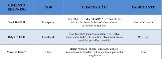
Tabela 1: Groups and composition of the luting agents by the manufactures.
In all groups, the posts were cleaned with 35% phosphoric acid for 60 seconds
followed by water rinsing and air drying. Then, a silane coupling agent (Ceramic
Primer – 3M/Espe) was applied in a single layer on the posts surface for 60 seconds
and then, dried with air. One coat of bond (Scotch bond Multi- Purpose - 3M/Espe)
was also applied, when necessary (group 1). The root canal pre-treatment was
performed de according with the different groups.
For the etch-and-rinse resin cements (group 1) the root canal dentine was
etched with 35% phosphoric acid for 15 seconds and rinsed for 30 seconds with
water. After removing the water excess from the root canal with paper points, one
layer of the primer (SBMP – 3M Espe) was applied with a microbrush and gently
air-dried for 5 seconds. Subsequently, the bond (SBMP – 3M Espe) was applied and
dried with paper points to remove the excess, and light-cured for 40 seconds by a
LED light-curing unit Elipar Freelight II (3M/Espe), with 900mW/cm2 intensity. RESIN
CEMENT COLOR COMPOSITION MANUFACTURE
Variolink® II Transparent BisGMA, UEDMA, TEGDMA, ytterbium trifluoride, Benzoyl Peroxide, initiators,
inorganic particles
Ivoclar/Vivadent
RelyXTM
U100 Translucent Phosphoric acid methacrylate ester, TEGDMA, silica, glass, calcium hydroxide, sodium
Toluenosulfinato, sodium persulfate
3M / Espe
Maxcem EliteTM
Clear Resin matrix (glycerol dimethacrylate), co-methacrylate monomers, photoinitiators,
particularly inorganic
For the cementation of glass fiber posts, equal amounts of resin cements
agents, base and catalyst, were mixed and applied onto the posts surface and into
the roots canal with a periodontal probe. Then, the post was inserted and cemented
into the root canal with light finger pressure, and the luting material excess was
immediately removed.
The cement was light cured for 40 seconds with the tip positioned parallel to
the pin (at its base) and over 40 seconds at 45° with the long axis of the pin.
For the self-adhesive resin cements groups (2, 3, 4, 5, 6, 7, 8 and 9), the pin
was cleaned with phosphoric acid followed by silane application, according to the
protocol described for group G1. It was not necessary adhesive application. The root
canal preparation was performed according to the different groups, with their
respective dentine pretreatments, as mentioned in table 2.
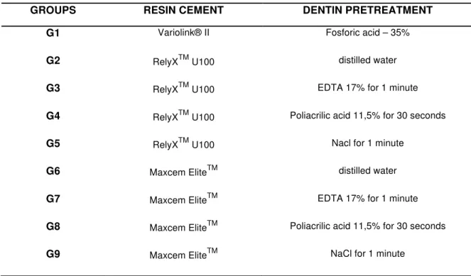
Tabela 2: Resin cement and the pre-treatment of root canal
GROUPS RESIN CEMENT DENTIN PRETREATMENT
G1 Variolink® II Fosforic acid – 35%
G2 RelyXTM U100 distilled water
G3 RelyXTM U100 EDTA 17% for 1 minute
G4 RelyXTM U100 Poliacrilic acid 11,5% for 30 seconds
G5 RelyXTM U100 Nacl for 1 minute
G6 Maxcem EliteTM distilled water
G7 Maxcem EliteTM EDTA 17% for 1 minute
G8 Maxcem EliteTM Poliacrilic acid 11,5% for 30 seconds
Different types of dentine pre-treatment were employed in order to remove or
modify the smear layer. For Groups 2 and 6 the root dentine surface was irrigated
with distilled water (no treatment groups). Prior to the resin cement application, for
groups G3 and G7, 17% EDTA (ethylenediaminetetraacetic acid) solution was
applied for a time of 1 minute and followed by rinsing with water for 1 minute. In the
groups G4 and G8, 11.5% polyacrylic acid was applied for 1 minute, followed by
rinsing with water at the same time. Sodium hypochlorite (NaCl) was used for groups
G5 and G9 during 1 minute, followed by rinsing with water.
The self-adhesive cements manipulation and pin insertion was similar to that
described for group 1.
After all cementation procedures, the specimens were stored in distilled water
for 24 hours at 37°C.
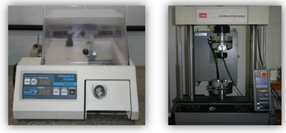
After the storage period, the specimens were sectioned by Isomet 1000 cutting
digital machine (Buehler UK LTD). The roots were divided in three parts, 1mm from
cervical surface. Three 1mm thick precise slabs, separated by 3mm space each,
were obtained per root and they were identified as cervical, middle and apical
specimens. The thickness of each slab was measured by the digital machine cutting
disc position along the root.
Immediately after the slabs were obtained, they were positioned on the
push-out jig (1mm diameter), which was placed on the Universal Testing Machine (MTS
810 Material Test System) with a cell load of 50Kg, at a crosshead speed of 0.5
mm/min until the post was dislodged.
The retentive strength of the post segment was expressed in MPa, by dividing
π (R + r) [(h2 + (R-r)2]0,5. Data were analyzed by using analysis of variance (ANOVA)
with GraphPad Prism 5 for Windows (GhaphPad Software Inc) statistical software,
followed by tukey test at 5% of significance (p<0.05).
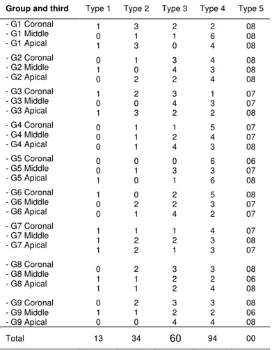
After the push-out testing, the specimens were analyzed by stereoscopic
microscope to determine the failure mode [Perdigão et al. 11]: type 1, adhesive
between post and resin cement (no resin cement visible around the post); type 2,
mixed with resin cement covering 0–50% of the post diameter; type 3, mixed with
resin cement covering between 50 and 100% of post surface; type 4, adhesive
between resin cement and root canal (post enveloped by resin cement); type 5,
cohesive in dentin.
RESULTS
Push-out test: The analysis of variance showed statistically significant
difference to resin cements evaluated (p<0 .05) and different thirds of root (p<0.05).
The results of the tukey test are displayed in table 2.
The highest values were found for the groups G3, G4 and G5, and there was
no bond strength significant difference to group G2. The G6 and G9 groups showed
the lowest values and had no statistically differences to G7 and G8. G1 had no
significant difference to G2. G1 and G5 demonstrated statistically lower resistence
values of the apical than the cervical third. G5 had lower values on the apical third
when compared to the middle third. The groups G2, G3, G4, G6, G7, G8 and G9 had
The failure modes of groups and level of the root are displayed in table 3. No
cohesive failure in dentin (type V) was observed. Higher incidence of failure type IV
(46,7%) and type III (29,9%) was observed in comparison to the failure type I (6,4%).
The failure type II occured in 17% of the cases.
Table 3: Means (standard deviation) of Push-out Bond Strength in MegaPascal (MPa)
Groups
Cervical
Third
Middle Apical
G1 11.51 (1.6) 10,49 (2.5) 8.46 (2.7)
G2 10.38 (2.2) 11.06 (1.7) 8.30 (2.4)
G3 10.80 (2.0) 10.40 (1.2) 9.28 (2.1)
G4 11.46 (2.5) 10.93 (1.4) 9.61 (1.9)
G5 12.61 (1.9) 12.02 (1.5) 8.25 (1.7)
G6 8.16 (1.4) 8.29 (2.0) 6.82 (2.1)
G7 9.94 (2.9) 9.35 (1.7) 8.11 (1.6)
G8 8.69 (1.5) 8.06 (1.3) 7.74 (2.8)
Table 4: Failure Modes of groups and level of the root canal after the push-out tests
DISCUSSION
The bovine dentine was used in this investigation due the limited availability
and the inhomogeneity of extracted human teeth. Moreover, the bioethical concerns
make it difficult to collect and use human teeth for researches12.
In the present study, the push-out test was performed 24 hours after
adhesive cementation procedures because bond strength can increase during this
period13.
The self-adhesive resin cement present a deficient hybridization of dentin
Group and third Type 1 Type 2 Type 3 Type 4 Type 5 - G1 Coronal
- G1 Middle - G1 Apical
1 0 1 3 1 3 2 1 0 2 6 4 08 08 08 - G2 Coronal
- G2 Middle - G2 Apical
0 1 0 1 0 2 3 4 2 4 3 4 08 08 08 - G3 Coronal
- G3 Middle - G3 Apical
1 0 1 2 0 3 3 4 2 1 3 2 07 07 08 - G4 Coronal
- G4 Middle - G4 Apical
0 0 0 1 1 1 1 2 4 5 4 3 07 07 08 - G5 Coronal
- G5 Middle - G5 Apical
0 0 1 0 1 0 0 3 1 6 3 6 06 07 08 - G6 Coronal
- G6 Middle - G6 Apical
1 0 0 0 2 1 2 2 4 5 3 2 08 07 07 - G7 Coronal
- G7 Middle - G7 Apical
1 1 1 1 2 2 1 2 1 4 3 3 07 08 07 - G8 Coronal
- G8 Middle - G8 Apical
0 1 1 2 1 1 3 2 2 3 2 4 08 06 08 - G9 Coronal
- G9 Middle - G9 Apical
0 1 0 2 1 0 3 2 4 3 2 4 08 06 08
along the root canal walls14-16. The application of RelyX Unicem and Maxcem to root
dentin does not result in the formation of hybrid layer or resin tags and inability to
etch through the smear layer formed in the root canal17. But, the self-adhesive resin
cements to root canal dentin seems to be related more to the friction along the canal
walls than to the adhesive bonding to root dentin18,19. The manufacturer of RelyX
U100 claims that the bonding mechanism of this self-adhesive cement is based on
micromechanical retention and chemical adhesion to hydroxyapatite. A recent study
showed an intense chemical interaction of RelyX U100 with hydroxyapatite20.
Rely-X U100 has limited etching potential when compared with
etch-and-rinse and self-etching adhesive systems. This could possibly be explained by the
methacrylated phosphoric esters present in this cement, which are not as effective as
phosphoric acid in dissolving the thick smear layer in the root canal walls during post
space preparation21-23. The information of the MaxCem cement is low in the literature.
The use of irrigants such as EDTA has been recommended as extremely
effectively in cleaning the root canal after post preparation and, as a result, improved
the bond strength in each regions of the root dentin24.
Another irrigant used to clean the root dentin is NaOCl because it has the
ability to remove the smear layer, which is created on the dentin surface during the
post space preparation25. The irrigation of root dentin with 5% NaOCl reduce the
bond strength of resin cements to dentin. This could be explained by an
oxygen-enriched dentin surface after application of NaOCl, which could act as a
polymerization inhibitor of resin materials26. The polyacrylic acid used as a
pretreatment in glass ionomer cements also has the ability to remove the smear layer
It is likely that phosphoric acid etching is more effective in highly tubular
areas of the coronal root dentin because it removes the thick surfaces smear layer
and the smear plug in dentinal tubules formed during post space preparation to allow
more effective micromechanical retention of resin cements29
In general, the root dentin should be irrigated with CHX or sterile saline
solution before post cementation in order to eliminate the negative effect of NaOCl on
the adhesive bond to dentine27. But, the study protocol followed the manufacturers’
instructions of RelyX Unicem (3M ESPE, Seefeld, Germany), which recommend the
irrigation of the root dentin with NaOCl followed by water.
The use of ultrasonic instrumentation in association with EDTA has been
suggested for a careful debridement of the post space walls improving performed
prior to cementation28.
Clinical investigations have reported that the most common cause of failure is
debonding of the fiber posts7,27-31.Adhesive failure between the dentine and cement
was the main failure mode12,32.In our results, the most samples had failures located
at the cement-dentin interface
The bond strengths were significantly affected by the root canal region, but
not for the self- adhesive resin cement12, 33, 34. According with our results. The
moisture tolerance is probably the factor responsible for the homogeneous bond
strength values of RelyX U100 in all root dentin regions33. Another aspect of the
self-adhesive cements bond strengths to root dentin seem to be related more to the area
of solid dentin than to the density of dentinal tubules30,34,35.
The results of this study showed the RelyX U100 showed the best
U100 without pretreatment of the dentin. Other studies show the superiority of
self-adhesive cements regarding etch-and-rinse cements7, 8, 15, 23, 30, 35, 36. The most of
these tests was performed with the self-adhesive cement (RelyX Unicem). Some
authors speculated that the moisture tolerance of the self-adhesive cement may
explain its favourable adhesion in root dentin15,37. Other studies revealed lower
bond-strength values for the self-adhesive resin cement RelyX Unicem compare to
etch-and-rinse cement7, 16, 32, 38, 39. The bond strength obtained for RelyX Unicem was in
the same range with that of Variolink II40.
The Maxcem Elite self-adhesive resin cement showed the lowest results of
bond strength, regardless of the type of pre-treatment performed on root dentin
before cementation of the fiberglass post. Other studies show the low performance of
Maxcem compared to RelyX Unicem12, 23. Soares et al., (2012) show in their studies
bubbles in the cement Maxcem, irrespective of the location, and the cement primarily
in the apical area did not appear to have polymerized12.
Regarding the irrigation solutions, the use of different products for partial
removal of smear layer does not influence on the bond strength of the self-adhesive
resin cements compared with the control distilled water. The RelyX U100 associated
with pretreatment with sodium hypochlorite (recommendation of the manufacturer)
showed the best results.
REREFENCES
1. Roberts HW, Leonard DL, Vandewalle KS, Cohen ME, Charlton DG. The
2. Teixeira EC, Teixeira FB, Piasick JR, Thompson JY. An in vitro assessment of prefabricated fiber post systems J Am Dent Assoc 2006;137:1006-12.
3. Goracci C, Grandini S, Bossù M, Bertelli E, Ferrari M. Laboratory assessment
of the retentive potential of adhesive posts: a review. J Dent. 2007 Nov;35(11):827-35. Epub 2007 Sep 4.
4. Boschian Pest L, Cavalli G, Bertani P, Gagliani M. Adhesive post-endodontic
restorations with fiber posts: push-out tests and SEM observations. Dent Mater 2002 Dec;18:596-602.
5. Ferrari M, Vichi A, García-Godoy F. Clinical evaluation of fiber-reinforced
epoxy resin posts and cast post and core. Am J Dent 2000 May; 13(Spec No):15B-18B.
6. Asmussen E, Peutzfeldt A, Heitmann T. Stiffness, elastic limit, and strength of
newer types of endodontic posts. J Dent 1999;27:275-8.
7. Zicari F, Couthino E, De Munck J, Poitevin A, Scotti R, Naert I, Van Meerbeek
B. Bonding effectiveness and sealing ability of fiber-post bonding. Dent Mater 2008; 24: 967–977.
9. Ferrari M. Immediate and 24-hour evaluation of the interfacial strengths of fiber posts Journal of Endodontics. 2006; 32(12) 1174-1177.
10. Bishara SE, Soliman M, Laffoon JF, Warren J. Bishara SE, Soliman M,
Laffoon JF, Warren J. Shear bond strength of a new high fluoride release glass ionomer adhesive.Angle Orthod. 2008; 78: 125-8.
11. Perdigão J, Gomes G, Lee IK. The effect of silane on the bond strengths of fiber posts. Dental Materials 2006:22;752–8.
12. Soares CJ, Pereira JC, Valdivia AD, Novais VR, Meneses MS. Influence of
resin cement and post configuration on bond strength to root dentine. Int Endod J. 2012 Feb;45(2):136-45.
13. Goracci C, Sadek FT, Fabianelli A, Tay FR, Ferrari M. Evaluation of the
adhesion of fiber posts to intraradicular dentin. Operative Dentistry 2005;30:627–35.
14. Monticelli F, Ferrari M, Toledano M. Cement system and surface treatment
selection for fiber post luting. Med Oral Patol Oral Cir Bucal. 2008 Mar 1;13(3):E214-21.
15. Bitter K, Paris S, Pfuertner C, Neumann K, Kielbassa AM. Morphological and
16. Calixto LR, Bandéca MC, Clavijo V, Andrade MF, Vaz LG, Campos EA. Effect of resin cement system and root region on the push-out bond strength of a translucent fiber post. Oper Dent. 2012 Jan-Feb;37(1):80-6. Epub 2011 Sep 26.
17. Sirimai S, Riis DN, Morgano SM. An in vitro study of the fracture resistance and the incidence ofvertical root fracture of pulpless teeth restored with six post-and-coresystems. J Prosthet Dent. 1999 Mar;81(3):
18. Wrbas KT, Altenburger MJ, Schirrmeister JF, Bitter K, Kielbassa AM. Effect of
adhesive resin cements and post surface silanization on the bond strengths of adhesively inserted fiber posts. J Endod. 2007;33:840-3.
19. Giachetti L, Grandini S, Calamai P, Fantini G, Scaminaci Russo D.
Translucent fiber post cementation using light- and dual-curing adhesive techniques and a self-adhesive material: push-out test. J Dent. 2009 Aug;37(8):638-42. Epub 2009 May 3.
20. Gerth HU, Dammaschke T, Zuchner H, Schafer E. Chemical analysis and
bonding reaction of RelyX Unicem and Bifix composites - a comparative study. Dent Mater 2006;22:934-941.
22. Kececi AD, Ureyen Kaya B, Adanir N. Micro-push-out bond strengths of four fiber-reinforced composite post systems and 2 luting materials. Oral Surg Oral Med Oral Pathol OralRadiol Endont. 2008: 105:121–128
23. Kahnamouei MA, Mohammadi N, Navimipour EJ, Shakerifar M. Push-out
bond strength of quartz fibre posts to root canal dentin using total-etch and self-adhesive resin cements. Med Oral Patol Oral Cir Bucal. 2012 Mar 1;17(2):e337-44.
24. Gu XH, Mao CY, Liang C, Wang HM, Kern M (2009) Does endodontic post
space irrigation affect smear layer removal and bonding effectiveness? European Journal of Oral Science 117, 597–603.
25. Demiryürek EÖ, Külünk S, Sarac D, Yüksel G, Bulucu B. Effect of different surface treatment on the push-out bond strength of fiber post to root canal dentin. Oral Surg Oral Med Oral Pathol Oral Radiol Endod. 2009. 108:e74–e8.
26. Ari H, Yasar E, Belli S. Effect of NaOCl on bond strengths of resin cements to
root canal dentin. J Endod 2003:248–251.
27. Bitter K, Kielbassa AM.Dentinadhäsive im Wurzelkanal. Quintessenz. 2005;
56:1045–1052.
28. Coniglio I, Magni E, Goracci C, et al. Post space cleaning using a new nickel
29. Cagidiaco MC, Goracci C, Garcia-Godoy F, Ferrari M. Clinical studies of fiber
posts: a literature review. Int J Pros- thodont 2008; 21: 328–336.
30. Bitter K, Meyer-Lueckel H, Priehn K, Kanjuparambil J, Neumann K, Kielbassa
AM. Effects of luting agent and thermocycling on bond strengths to root canal dentine. Int Endod J 2006; 39: 809–818.
31. Ferrari M, Cagidiaco MC, Grandini S, De Sanctis M, Goracci C. Post
placement affects survival of endodontically treated premolars. J Dent Res 2007; 86: 729–734.
32. Goracci C, Sadek FT, Fabianelli A, Tay FR, Ferrari M. Evaluation of the
adhesion of fiber posts to intraradicular dentin. Operative Dentistry 2005;30:627–35.
33. Gomes G, Gomes O, Reis A, Gomes J, Loguercio A, Calixto A.Effect of
Operator Experience on the Outcome of Fiber Post Cementation With Different Resin Cements. Oper Dent. 2012 Dec 5. [Epub ahead of print]
34. Bateman GJ, Lloyd CH, Chadwick RG, Saunders WP. Retention of
quartz-fibre endodontic posts with a self-adhesive dual cure resin cement. Eur J Prosthodont Restor Dent. 2005 Mar;13(1):33-7.
35. RadovicI,MonticelliF,GoracciC,VulicevicZR,Ferrari M. Self-adhesive resin
36. Mazzoni A, Marchesi G, Cadenaro M, Mazzotti G, Di Lenarda R, Ferrari M, Breschi L. Push-out stress for fibre posts luted using different adhesive strategies. Eur J Oral Sci. 2009 Aug;117(4):447-53.
37. Goracci C, Tavares AU, Fabianelli A, Monticelli F, Raffaelli O, Cardoso PC, Tay F, Ferrari M. The adhesion between fiber posts and root canal walls: comparison between microtensile and push-out bond strength measurements. Eur J Oral Sci 2004;112:353-61.
38. Dimitrouli M, Günay H, Geurtsen W, Lührs AK. Push-out strength of fiber posts
depending on the type of root canal filling and resin cement. Clin Oral Investig. 2011 Apr;15(2):273-81. Epub 2010 Jan 22.
39. Wang VJ, Chen YM, Yip KH, Smales RJ, Meng QF, Chen L. Effect of two fiber
3.2 Capítulo 2
Self-adhesive cements and fiber posts: a review
Luiz Rafael Calixto, DDS, MSc, Matheus C. Bandeca, DDS, MSc, PhD, Matheus Tonetto, DDS, MSc, José Roberto Cury Saad, DDS, MSc, PhD Edson Alves de Campos, DDS, MSc, PhD and Marcelo F. Andrade, DDS, MSc, PhD
From the Department of Restorative Dentistry, Araraquara Dental School, São
Paulo State University, SP, Brazil;
Address requests for reprint to Luiz Rafael Calixto, Araraquara Dental School,
Department of Restorative Dentistry, Rua Humaitá 1680, Centro, Zip Code
14801-903 Araraquara, SP, Brazil. Phone Number: 16 3301-6388. e-mail address:
lrcalixto@hotmail.com.
ABSTRACT
The self-adhesive resin cements have been introduced to simplify the procedure for
cementing, however their clinical performance to cementation of fiberglass posts is
still controversial in the literature. Over the years, several studies were made to
evaluate bond strength tests with push-out, pull-out and microtensile. The objective
of this paper is to review the self-adhesive cements and fiber posts, the main tests for
bond strength and the microscopic characteristics of the adhesive interface.
Key-words: Resin Cement; fiber post; self-adhesive cements.
INTRODUCION
Fibe-reinforced composite posts (FRC posts) have been commonly used in
endodontically treated teeth and in cases with great loss of dental structure, in order
to provide adequate support and retention, and re-establish aesthetic and function for
the final restoration. The fiber posts have similar rigidness characteristics to dentin
that absorb mastigatory strength and protect the radicular remnant. According to
manufactures, the cementation of fiber posts shall be adhesive, since its chemical
nature is similar to the Bis-GMA, usually found in the resinous material1-3.
The adhesive strategy recommended for the bonding of fiber posts, the
currently available resin-based cements and accompanying bonding systems can be
classified as etch- and-rinse, self-etch, and self-adhesive luting agents4.
Each-and-rinse cements require the separate use of phosphoric acid followed
by multi or two step each-and-rinse adhesives before the application of the resin
cement. Self-etch resin cements use an acidic primer, which without rinsing can alter
with a etch-and-rinse systems5. Self-adhesive cements do not require any
pretreatment of the tooth substrate: once the cement is mixed, application is
accomplished through a single clinical step6. The simplification allowed by
self-adhesive resin cements is attractive to clinicians7,8. This lack of pretreatment reduces
technique and operator sensitivity9.
The first self-adhesive cement was RelyX Unicem, which was launched into
the market in 2002. Since then, new products have been constantly introduced9.
These materials were designed with the purpose of overcoming some limits of both
conventional and resin cements. RelyX Unicem is the most investigated
self-adhesive cement in the current literature published in Medline cited journals9,10.
The retentive ability of adhesive posts has been assessed in numerous
laboratory studies over the last years. Microtensile, post-pull-out and push-out tests
have been performed in order to estimate the retention of luted posts or, selectively,
the strength at the post-cement or cement- dentin interfaces. Small-sized specimens
designs, such as the microtensile and thin-slice push-out tests, favour stress
uniformity, allow to discriminate regional differences and to limit the number of teeth
needed for data collection11.
The comparisons among the results from different in vitro studies may be
inappropriate because of the dependence of the collected data upon laboratory
set-ups and experimental conditions11. Therefore, the aim of this article is to review the
current use of self-adhesive resin cements in the retention of fiber posts using the
PUSH-OUT TESTS
Bond strength has been measured through the push-out test, conventional
tensile test on external root dentin12, or on the endodontic surface with the pull-out
13-16. However, the in vitro performance of self-adhesive cements in fiber post
cementation is frequently investigated using the push-out strength test17.
A variant of this method known to materials scientists as the ‘‘thin-slice’’
push-out test. The modification involve sectioning the posted root into a series of 1 mm
thick slices, and compressively loading the post-section within each slice with an
adequately sized plunger, until bond failure18. It has been shown to be more reliable
than the microtensile technique for measuring the adhesion of fiber posts. Another
advantage of using the push-out test method is the small standard deviation of the
mean obtained for each root canal site19.
It was suggested that a highly non-uniform stress may be developed at the
adhesive interface when the push-out test is performed on thick root sections.This
may explain the relatively low levels of bond strength that have been reported when
applying this method of adhesion testing20.
Several studies evaluated the immediate adhesive strength and/or 24h after
cementation of fiber post, through push-out tests, a self-adhesive cement, with
results quite controversial. Studies show the superiority of this cement in compare to
etch-and-rinse resin cements21-27, (etch and rinse) and the self–adhesive resin
cements22-26. The most of these studies were performed with the self-adhesive
cement called RelyX Unicem.
Other studies revealed lower bond-strength values for the self-adhesive resin
Moreover Zicari et al.28 in contrast, the push-out strength of Variolink II was
equal to the self-adhesive composite cement, according to another studies32-35.
Radovic et al.34 also tested an etch-and-rinse approach, a self-etch approach,
and a self-adhesive luting approach, and concluded that the self-etch approach may
offer less favourable adhesion to root dentin in comparison with etch-and-rinse and
the self-adhesive approaches
Bitter et al.38 investigated the effects of luting agent and thermocycling on
bond strengths (push-out test) to root dentin using six luting agents: Panavia F,
Multilink, Variolink II, PermaFlo DC, RelyX Unicem and Clearfil Core. Their results
showed that the self-adhesive resin cement RelyX Unicem obtained a better
performance after thermocycling compare to others cements. An increase in bond
strengths after thermocycling for RelyX Unicem was also observed in other
studies36,37.
The self-adhesive resin cements contain multifunctional hydrophilic monomers
with phosphoric acid groups, which can react with the hydroxyapatite and also
penetrate and modify the smear layer39,40. The chemical interaction between the
acidic monomers and hydroxyapatite ensures the adhesion of the self-adhesive
cements into dentin41.
RelyX Unicem has a limited etching potential compared with the
etch-and-rinse and self-etching adhesives42-44. RelyX Unicem exhibit an etching potential
insufficient to dissolve the thick smear layers created in post-space preparation with
burs45-46. The consequent opening of interfacial gaps may account for the relatively
low push-out strengths recorded for and for RelyX Unicem as compared with the
The self- adhesive simplified resin cement appears to have poor adhesion to
dentin in dry and aging conditions48,49.However, RelyX Unicem exhibits a moisture
tolerance because of water forming during the neutralization reaction of
phosphoric-acid methacrylate, basic fillers and hydroxy apatite (data provided by the
manufacturer). This could be an explanation for the good performance of the RelyX
Unicem, because moisture content after rinsing the root canal is difficult to control
because of the poor visibility50.
However, the methacrylated phosphoric esters in the RelyX Unicem probably
can not penetrate adequately through the retained partly dissolved smear layer on
the root canal walls, resulting in interfacial gaps and lower bond strengths in vitro51.
MICROTENSILE
The microtensile method was originally developed for ultimate tensile strength
testing of dental tissues, and later applied to bond strength measurements on crown
dentin and enamel47.
This method has already been applied to evaluate bond strength to root canals
that were treated with different irrigants and cement systems. However, laboratory
studies comparing bond tests of fiber posts using Variolink II cements and RelyX
Unicem self-adhesive found the almost specimens prepared through this method
failed prematurely during the cutting phase17, because of the lower bond strength
values with this substrate17,52. The results showed greater adhesion to Variolink II,
agreeing with the other studies4,47,53.
The displacement resistance value for self-adhesive resin luting cement
(Multilink)32.
Moreover, difficulties in conveying sufficient amount of primer-adhesive
solution to the apical region of canals and manipulation problems arising from
inadequate root canal access are the reasons for lower apical bond strength values
in each-and-rinse cements compared to self-adhesive ones52.
RelyX U100 and RelyX Unicem are highly adapted to the substrate and can
optimize physical interactions such as van der Waals forces, hydrogen bridges and
charge transfer, which enhance micromechanical retention and chemical bonding47.
Continuity of dentin/cement interface is necessary to promote sliding friction,
which according to many authors is the main factor for adhesive resistance in the
fiber post/resin cements system and for micromechanical interlocking54.
Some studies used the self-adhesive resin cement called MaxCem. This
cement contains several hydrophilic adhesive monomers of low molecular weight,
such as the glycerol phosphate dimethacrylate (GPDM), which provide the necessary
wettability for adhesion to dentin substrate. The cement is purportedly anhydrous
prior to mixing. The manufacturer does not specify which monomer is initially
hydrolyzed, the initial pH or the proprietary redox activator system47. This
self-adhesive cement generally have a performed poorly47,55 Provavelmente porque
despite the excellence of Maxcem monomers as bonding agents of good
compatibility with the humid dentin substrate, they do not provide an effective
cementation in deep areas of high humidity.
The fiber posts cemented with MaxCem Eite presented the lowest bond
strength values (push-out tests) compare to RelyX Unicem, with significant
mainly in the middle and apical third from the root canal55.
SEM ANALYSIS
As the ultrastructure of the interface between root dentin and simplified resin
cements was generally not suggestive of a solid micromechanical interlocking,
knowing that chemical reactions are marginally involved in the adhesion mechanism
of resin-based cements, the hypothesis may be raised that a contribution to fiber
post-retention is provided also by sliding friction between cement and dentin along
the canal walls11,56. The use of a flexible root-canal-shaped application aid reduces
the number of imperfections within the self-adhesive cement interface compared to
the conventional application technique10.
adhesive resin luting cement a recent laboratory study reported that
self-adhesive resin luting cement was unable to dissolve the smear layer completely10,43.
SEM analysis revealed many air bubbles at the cement–dentin interface, which must
be ascribed to a poor adaptation to the root-canal walls rather than the mixing
process4.
When using RelyX Unicem no distinct hybrid layer was seen. The cement
interacted only superficially with the smear layer, that was substantially retained
along with smear plugs. Gaps were visible between the smear layer and the
underlying intact dentin11. RelyX Unicem was found to result in a significantly lower
number of penetrated dentinal tubules compared with all other materials
(etch-and-rinse or self-etching cements)58.
Hybridization of dentin was only detected sporadically for the material RelyX
that also described a superficial morphological interaction29,41.
The self-adhesive cement Maxcem formed a discontinuous interface with
many gaps. However, the interfacial continuity produced by U100 self-adhesive
cement was as satisfactory as that formed by the dual-cure cements7.
A visualization of stress distribution among the root canals can be performed
using several finite element analysis (FEA) studies. Among them, the analyses using
three-dimensional (3-D) models are considered more reliable7.
REFERENCES
1. Asmussen E, Peutzfeldt A, Heitmann T. Stiffness, elastic limit, and strength of newer types of endodontic posts. J Dent. 1999 May;27(4):275-8
2. Boschian Pest L, Cavalli G, Bertani P, Gagliani M. Adhesive post-endodontic restorations with fiber posts: push-out tests and SEM observations. Dent Mater. 2002 Dec; 18(8):596-602.
3. Scotti R, Ferrari M. Sistemas de adesão. In:______ Pinos de fibra: considerações teóricas e aplicações clínicas. 1ed. São Paulo. Artes Médicas, 2003. Cap. 7, p.67-74.
5. Duarte S Jr, Botta AC, Meire M, Sadan A. Microtensile bond strengths and scanning electron microscopic evaluation of self-adhesive and self-etch resin cements to intact and etched enamel. J Prosthet Dent. 2008 Sep;100(3):203-10.
6. Monticelli F, Ferrari M, Toledano M. Cement system and surface treatment selection for fiber post luting.Med Oral Patol Oral Cir Bucal. 2008 Mar 1;13(3):E214-21
7. Silva RA, Coutinho M, Cardozo PI, Silva LA, Zorzatto JR. Conventional dual-cure versus self-adhesive resin cements in dentin bond integrity.J Appl Oral Sci. 2011 Aug;19(4):355-62. Epub 2011 Jun 24.
8. Goracci C, Ferrari M. Current perspectives on post systems: a literature review.Aust Dent J. 2011 Jun;56 Suppl 1:77-83
9. Baena E, Fuentes M, Garrido M, Rodríguez J, Ceballos L. Influence of Post-cure Time on the Microhardness of Self-Adhesive Resin Cements Inside the Root Canal. Oper Dent. 2012 Feb 15. [Epub ahead of print]
10. Watzke R, Blunck U, Frankenberger R, Naumann M. Interface homogeneity of adhesively luted glass fiber posts. Monticell. Dent Mater. 2008 Nov;24(11):1512-7. Epub 2008 May 7.
12. De Munck J, Van Landuyt K, Peumans M, Poitevin A, Lambrechts P, Braem M, et al. A critical review of the durability of adhesion to tooth tissue: methods and results. Journal of Dental Research 2005;84:118–32.
13. NikaidoT,TakanoY,SasafuchiY,BurrowMF,TagamiJ. Bond strengths to endodontically treated teeth. Am J Dent 1999; 12: 177–180.
14. QualthroughAJ,ChandlerNP,PurtonDG.Acomparisonof the retention of tooth-colored posts. Quint Int 2003; 34: 199–201.
15. Garcia Varela S, Bravos Rabade L, Rivas Lombardero P, Linares Sixto J, Gonzalez Bahillo J, Ahn Park S. In vitro study of endodontic post cementation protocols that
use resin cements. J Prosthet Dent 2003; 89: 146–153.
16. Prisco D, De Santis R, Mollica F, Ambrosio L, Rengo S, Nicolais L. Fiber post adhesion to resin luting cements in the restoration of endodontically treated teeth. Oper Dent 2003; 28: 515–521.
17. Goracci C, Tavares AU, Fabianelli A, Monticelli F, Raffaelli O, Cardoso PC, Tay F, Ferrari M. The adhesion between fiber posts and root canal walls: comparison between microtensile and push-out bond strength measurements. Eur J Oral Sci. 2004;112:353-61.
19. Wang VJ, Chen YM, Yip KH, Smales RJ, Meng QF, Chen L. Effect of two fiber post types and two luting cement systems on regional post retention using the push-out test. Dent Mater. 2008 Mar;24(3):372-7. Epub 2007 Jul 25.
20. Ngoh EC, Pashley DH, Loushine RJ, Weller N, Kimbrough F. Effects of eugenol on resin bond strengths to root canal dentin. J Endod 2001; 27: 411–414.
21. Radovic I, Mazzitelli C, Chieffi N, Ferrari M. Evaluation of the adhesion of fiber posts cemented using different adhesive approaches. Eur J Oral Sci. 2008 Dec;116(6):557-63.
22. Bateman GJ, Lloyd CH, Chadwick RG, Saunders WP. Retention of quartz-fibre endodontic posts with a self-adhesive dual cure resin cement. Eur J Prosthodont Restor Dent 2005; 13: 33–37.
23. RadovicI,MonticelliF,GoracciC,VulicevicZR,Ferrari M. Self-adhesive resin cements: a literature review. J Adhes Dent 2008; 10: 251–258.
24. Fokkinga WA, Kreulen CM, Vallittu PK, Creugers NH. A structured analysis of in vitro failure loads and failure modes of fiber, metal, and ceramic post-and-core systems. Int J Prosthodont 2004;17:476–482.
26. Zicari F, De Munck J, Scotti R, Naert I, Van Meerbeek B. Fact ors affecting the cement-post interface. Dent Mater. 2012 Mar;28(3):287-97. Epub 2011 Dec 12.
27. Kahnamouei MA, Mohammadi N, Navimipour EJ, Shakerifar M. Push-out bond strength of quartz fibre posts to root canal dentin using total-etch and self-adhesive resin cements. Med Oral Patol Oral Cir Bucal. 2012 Mar 1;17(2):e337-44
28. Zicari F, Couthino E, De Munck J, Poitevin A, Scotti R, Naert I, Van Meerbeek B. Bonding effectiveness and sealing ability of fiber-post bonding. Dent Mater. 2008 Jul;24(7):967-77. Epub 2008 Jan 3.
29. Goracci C, Sadek FT, Fabianelli A, Tay FR, Ferrari M. Evaluation of the adhesion of fiber posts to intraradicular dentin. Oper Dent 2005; 30: 627–635
30. Calixto LR, Bandéca MC, Clavijo V, Andrade MF, Vaz LG, Campos EA. Effect of resin cement system and root region on the push-out bond strength of a translucent fiber post. Oper Dent. 2012 Jan-Feb;37(1):80-6. Epub 2011 Sep 26.
31. Wang VJ, Chen YM, Yip KH, Smales RJ, Meng QF, Chen L. Effect of two fiber post types and two luting cement systems on regional post retention using the push-out test.Dent Mater. 2008 Mar;24(3):372-7. Epub 2007 Jul 25.
32. Toman M, Toksavul S, Sarikanat M, Firidinoğlu K, Akin A. The evaluation of
33. Rathke A, Haj-Omer D, Muche R, Haller B. Effectiveness of bonding fiber posts to root canals and composite core build-ups. Eur J Oral Sci. 2009 Oct;117(5):604-10.
34. Radovic I, Mazzitelli C, Chieffi N, Ferrari M. Evaluation of the adhesion of fiber posts cemented using different adhesive approaches. Eur J Oral Sci 2008;116:557–63.
35. Huber L, Cattani-Lorente M, Shaw L, Krejci I, Bouillaguet S. Push-out bond strengths of endodontic posts bonded with different resin-based luting cements. Am J Dent 2007;20:167–72.
36. Abo-Hamar SE, Hiller K-A, Jung H et al. Bond strength of a new universal self-adhesive resin luting cement to dentin and enamel. Clinical Oral Investigations. 2005; 9:61–7.
37. Reich SM, Wichmann M, Frankenberger R, Zajc D. Effect of surface treatment on the shear bond strength of three resin cements to a machinable feldspatic ceramic. Journal of Biomedical Materials Research. Part B: Applied Biomaterials. 2005; 74, 740–6.
38. Bitter K, Meyer-Lueckel H, Priehn K, Kanjuparambil JP, Neumann K, Kielbassa AM. Effects of luting agent and thermocycling on bond strengths to root canal dentine. Int Endod J. 2006 Oct;39(10):809-18.
40. Hikita K, Van Meerbeek B, De Munck J, Ikeda T, Van Landuyt K, Maida T et al. Bonding effectiveness of adhesive luting agents to enamel and dentin. Dent Mater. 2007; 23:71–80
41. Gerth HU, Dammaschke T, Zuchner H, Schafer E. Chemical analysis and bonding reaction of RelyX Unicem and Bifix composites – a comparative study. Dent Mater 2006; 22: 934–941.
42. Sirmai S, Riis DN, Morgano SM. An in vitro study of the fracture resistance and the incidence of vertical root fracture of pulpless teeth restored with six post-and-core systems. J Pros- thet Dent 1999; 81: 262–269.
43. Sadek FT, Goracci C, Monticelli F, Grandini S, Cury AH, Tay FR. Immediate and 24-hour evaluation of the interfacial strengths of fiber posts. J Endod 2006; 32: 1174– 1177.
44. Behr M, Rosentritt M, Regnet T, Lang R, Handel G. Marginal adaptation in dentin of a self-adhesive universal resin cement compared with well-tried systems. Dent Mater 2004;20:191–7.
45. De Munck J, Vargas M, Van Landuyt K, Hikita K, Lambrechts P, Van Meerbeek B. Bonding of an auto-adhesive luting material to enamel and dentin. Dent Mater 2004;20:963–71.
47. Goracci C, Cury AH, Cantoro A, Papacchini F, Tay FR, Ferrari M. Microtensile bond strength and interfacial properties of self-etching and self-adhesive resin cements used to lute composite onlays under different seating forces. J Adhes Dent. 2006;8(5):327-35.
48. Hikita K, Van Meerbeek B, De Munck J, Ikeda T, Van Landuyt K, Maida T, Lambrechts P & Peumans M .Bonding effectiveness of adhesive luting agents to enamel and dentin Dental Materials. 2007: 23(1) 71-80.
49. Baldissara P, Valandro LF, Monaco C, Ferrari M, Bottino MA & Scotti R. Fatigue resistance of the bond of a glass- infiltrated alumina ceramic to the human dentin Journal of Adhesive Dentistry. 2006; 8(2) 97-104.
50. De Munck J, Vargas M, Van Landuyt K, Hikita K, Lambrechts P, Van Meerbeek B. Bonding of an auto-adhesive luting material to enamel and dentin. Dent Mater 2004;20: 963–71.
51. Yang B, Ludwig K, Rainer A, Kern Matthias. Micro-tensile bond strength of three luting resins to human regional dentin. Dent Mater 2006;22:45–56.
53. Günay H, Geurtsen W, Lührs AK. Push-out strength of fiber posts depending on the type of root canal filling and resin cement. Clin Oral Investig. 2011 Apr;15(2):273-81. Epub 2010 Jan 22.
54. Teixeira CS, Felippe MCS, Silva-Sousa YTCM, Sousa-Neto MDInterfacial evaluation of experimentally weakened roots restored with adhesive materials and fibre posts: an SEM analysis. 2008;36(9):672-82.
55. Soares CJ, Pereira JC, Valdivia AD, Novais VR, Meneses MS. Influence of resin cement and post configuration on bond strength to root dentine. Int Endod J. 2012 Feb;45(2):136-45.
56. Giachetti L , Calamai P, Fantini G, Scaminaci Russo D. Translucent fiber post cementation using light- and dual-curing adhesive techniques and a self-adhesive material: push-out test. J Dent. 2009 Aug;37(8):638-42. Epub 2009 May 3.
57. Watzke R, Blunck U, Frankenberger R, Naumann M. Interface homogeneity of adhesively luted glass fiber posts. Monticell. Dent Mater. 2008 Nov;24(11):1512-7. Epub 2008 May 7.