321
CAKE KIDNEY DRAINED BY SINGLE URETER Case Report
International Braz J Urol
Official Journal of the Brazilian Society of Urology
Vol. 30 (4): 321-322, July - August, 2004
CAKE KIDNEY DRAINED BY SINGLE URETER
ADRIANO A. CALADO, ANTONIO MACEDO JR., MIGUEL SROUGI
Section of Urology, Paulista School of Medicine, Federal University of São Paulo, UNIFESP, São Paulo, Brazil
ABSTRACT
Cake kidney is a rare congenital anomaly of the urogenital tract, with a few more than 20 cases described in the literature. It can be diagnosed at any age range. Normally, drainage is achieved by 2 ureters, and there are only 5 reports in the literature of cake kidney drained by a single ureter. The authors describe one more case of this rare malformation of the urinary tract.
Key words: kidney; abnormalities; ureter; hydronephrosis Int Braz J Urol. 2004; 30: 321-2
INTRODUCTION
Cake kidney is a rare congenital anomaly of the urogenital tract, and it is normally drained by 2 ureters. There are only 5 reports in the literature of cake kidney drained by single ureter (1). The early diagnosis of potential complications that can accom-pany this anomaly must be always made in order to prevent permanent renal damage (2).The authors re-port one case of cake kidney manifested as hydro-nephrosis associated with urinary infection, and dis-cuss the therapeutic approach.
CASE REPORT
Male, 5-months old child, presented a his-tory of recurrent urinary tract infections. There was already a previous history of 2 hospitalizations due to acute pyelonephritis. At the moment, he was under antimicrobial prophylaxis with nitrofurantoin.
The patient underwent renal ultrasonography that revealed bilateral hydronephrosis, more pro-nounced on the right side. Voiding urethrocystography was normal. Renal scintigraphy (Figure-1) revealed the presence of a cystic renal mass with pelvic
local-ization, with an obstructive pattern for clearance of radiopharmaceutical (T1/2 = 39 min.).
The child was operated due to suspicion of ureteropelvic junction (UPJ) stenosis in a single
322
CAKE KIDNEY DRAINED BY SINGLE URETER
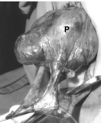
ney. The surgical finding was cake kidney with pel-vic location, presenting extra-renal calices. The ure-ter was single and presented moderate dilation (Fig-ure-2). Due to the child’s young age, a decompres-sive pyelotomy was performed. The child had a good outcome postoperatively, being discharged from the hospital on the fourth postoperative day. Currently he is under prophylaxis without new episodes of uri-nary infection.
COMMENTS
Cake kidney is a rare congenital malforma-tion of the urogenital tract, which can be diagnosed at any age group, from childhood to the eightieth de-cade of life (3).
This anomaly occurs at an early phase in the embryological development (2). Under normal con-ditions, the 2 masses of metanephrogenic tissue arise in the pelvis and ascend to their definitive position in the lumbar region, bilaterally. During such migration, they undergo a lateral deviation, with axial deflec-tion and internal rotadeflec-tion.
During the formation of a cake kidney, the nephrogenic blastemas would be compressed between the umbilical arteries at the beginning of the cranial migration of the ureteral buds, and this would leads to their fusion (2). Fused kidneys, such as the cake kidney, are prevented from ascending and remain in an ectopic pelvic position. The rare occurrence of a single ureter draining the fused renal mass can be caused by the regression of the second ureteral bud following the fusion of the metanephric blastemas (3). The majority of diagnosed cases have been reported to present malformations in other organs or in their blood supply, such as abnormal testicular mi-gration, Fallot’s tetralogy, vaginal or sacral agenesia, anal abnormalities, among others (3).
The diagnosis of cake kidney is not neces-sarily associated with a poor prognosis. However, complications that can be associated with anatomic malformations such as urinary stasis, infection, for-mation of stones, and vascular involvement, can cause serious clinical problems (2). Therefore, cases of cake kidney must be investigated in order to exclude con-comitant anomalies and to prevent complication.
Figure 2 – Intra-operative finding of cake kidney. Note the di-lated extra-renal pelvis (P) and single ureter.
REFERENCES
1. Martinez-Lazaro R, Cortes-Blanco A.: A. Cake kid-ney drained by single ureter: MAG3 renogram for di-agnosis and function follow-up. Nephrol Dial Trans-plant. 2000; 15: 1700-1.
2. Brock JW 3rd, Braren V, Phillips K, Winfield AC: Caudal regression with cake kidney and a single ure-ter: a case report. J Urol. 1983; 130: 535-6.
3. Goren E, Eidelman A: Pelvic cake kidney drained by single ureter. Urology. 1987; 30: 492-3.
Received: March 16, 2004 Accepted after revision: June 6, 2004