Pancake kidney with bladder herniation
_______________________________________________
Ihsan Yuce
1, Mecit Kantarci
1, Suat Eren
1, Akin Levent
11 Ataturk University, School of Medicine, Department of Radiology, Erzurum, Turkey
_______________________________________________________________________________________
1232
RADIOLOGY PAGE
CASE PRESENTATION
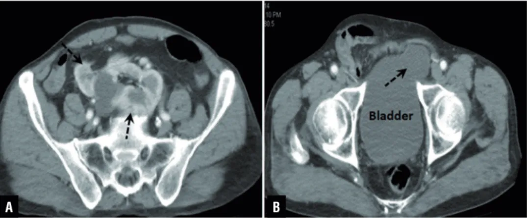
A 61-year-old man presented to the Emer-gency Department with vomiting and progres-sively worsening abdominal pain. A computed tomography (CT) was performed. The diagnosis of patient was acute cholecystitis and the patient was referred to general surgery clinic. In addi-tion CT scan showed bilateral ectopic kidneys with urinary bladder herniation (Figures 1 and 2). Both kidneys were fused at the medial borders of each pole. To our knowledge, the case of pancake kidney with bladder herniation was not published yet in the literature.
Pancake kidney is very rare type of conge-nital fusion anomaly of the kidney. It is described as a renal mass located in the pelvis which is for-med by complete for-medial fusion of renal parenchy-ma without intervening septum (1). Each kidney
has its own collecting system and anteriorly placed short ureters entering the bladder orthotopically (1). The presence of a pancake kidney may predispose the formation of stones due to the probable rotation anomaly of the collecting system and short ureters which are prone to stasis and obstruction. Patients with pancake kidney are usually asymptomatic, but may present with features of urinary tract infec-tion, fever and vague lower abdominal pain (1). If a pancake kidney has to undergo surgery, division of the parenchyma presents potential problems such as renal vascular damage, postoperative re-nal failure and eventual rere-nal failure (2). Asymp-tomatic cases can be managed conservatively with long-term follow-up of renal function (1). If there are symptoms of renal failure, surgery is warran-ted. Ultrasonography, excretory urography and CT were efficient in detection and evaluation of pan-cake kidney anomaly (1).
Figure 1 - Axial CT images show pancake kidney and bladder herniation (dashed arrows).
A B
Vol. 41 (6): 1232-1233, November . December, 2015
IBJU| RADIOLOGY PAGE
1233
ARTICLE INFO
Int Braz J Urol. 2015; 41: 1232-33
_____________________
Submitted for publication: December 16, 2014
_____________________
Accepted after revision: September 04, 2015
REFERENCES
1. Tiwari AK, Choudhary AK, Khowal H, Chaudhary P, Arora MP. Pancake kidney: A rare developmental anomaly. Can Urol Assoc J. 2014;8:E451-2.
2. Eze AR, White JV, Pathak AS, Grabowski MW. “Pancake kidney”: a renal anomaly complicating aortic reconstruction. Ann Vasc Surg. 1998;12:278-81.
_______________________ Correspondence address:
Ihsan Yuce, MD
H. Avni Ulaş Mh. Karataş
Ap. A blok. Kat:4, No :20 Erzurum, Turkey Fax:+ 90 442 236-1301 E-mail: [email protected]