Design of a Papain Immobilized
Antimicrobial Food Package with Curcumin
as a Crosslinker
Cynthya Maria Manohar☯, Veluchamy Prabhawathi☯, Ponnurengam
Malliappan Sivakumar, Mukesh Doble*
Department of Biotechnology, Bhupat & Jyoti Mehta School of BioSciences, IIT Madras, Chennai, 600036, India
☯These authors contributed equally to this work.
*mukeshd@iitm.ac.in
Abstract
Contamination of food products by spoilage and pathogenic microorganisms during post process handling is one of the major causes for food spoilage and food borne illnesses. The present green sustainable approach describes the covalent immobilization of papain to LDPE (low density polyethylene), HDPE (high density polyethylene), LLDPE (linear low density polyethylene) and PCL (polycaprolactam) with curcumin as the photocrosslinker. About 50% of curcumin and 82-92% of papain were successfully immobilized on these poly-mers. After 30 days, the free enzyme retained 87% of its original activity, while the immobi-lized enzyme retained more than 90% of its activity on these polymers. Papain crosslinked to LLDPE exhibited the best antibiofilm properties againstAcinetobactersp. KC119137.1 andStaphylococcus aureusNCIM 5021 when compared to the other three polymers, be-cause of the highest amount of enzyme immobilized on this surface. Papain acts by damag-ing the cell membrane. The enzyme is able to reduce the amount of carbohydrate and protein contents in the biofilms formed by these organisms. Meat wrapped with the modified LDPE and stored at 4°C showed 9 log reduction of these organisms at the end of the sev-enth day when compared to samples wrapped with the bare polymer. This method of cross-linking can be used on polymers with or without functional groups and can be adopted to bind any type of antimicrobial agent.
Introduction
Foods are spoiled by the spoilage microflora, whereas the occurrence of outbreaks of foodborne diseases due to pathogen contaminated food is a global phenomenon. Therefore antimicrobial food packaging technologies have become more intense. Microbial contamination of food oc-curs mainly at their surface due to post process handling [1]. Antimicrobial active packages are those which are in contact with the food aiding in extending its shelf life by preventing the mi-crobial growth [2]. Bacteriocins [3], organic acids [4], potassium sorbate [5] or pimaricin [6]
a11111
OPEN ACCESS
Citation:Manohar CM, Prabhawathi V, Sivakumar PM, Doble M (2015) Design of a Papain Immobilized Antimicrobial Food Package with Curcumin as a Crosslinker. PLoS ONE 10(4): e0121665. doi:10.1371/journal.pone.0121665
Academic Editor:George-John Nychas, Agricultural University of Athens, GREECE
Received:October 6, 2014
Accepted:February 3, 2015
Published:April 23, 2015
Copyright:© 2015 Manohar et al. This is an open access article distributed under the terms of the
Creative Commons Attribution License, which permits unrestricted use, distribution, and reproduction in any medium, provided the original author and source are credited.
Data Availability Statement:All relevant data are within the paper and its Supporting Information files.
Funding:The authors have no support or funding to report.
exhibit antimicrobial activity within the food packaging materials. Immobilization of antimi-crobials on food package rather than coating it on the surface of the wrapper reduces their amount required to achieve the antimicrobial effect as well as prolongs their activity.
Titanium dioxide, iron oxide, silver, gold and silver dioxide are examples of nanoparticle-based antimicrobials used in food wrap applications [7]. These are toxic [8] and affect the tis-sues in the human body. So, the use of natural antimicrobial agents including enzymes is in great demand. Peptides are emerging as new group of antibiotics and the antimicrobial nature of the one isolated from ovalbumin hydrolysate is reported [9]. Natural compounds including essential oils [10] and other herbal extracts have been tested as antimicrobial agents [11]. Per-oxides, eugenol, nisin, lactoferrin, sodium diacetate, sorbic acid [12,13], potassium sorbate, ly-sozyme [14], glucose oxidase [15] thymol, carvacrol [16], linalool and methylchavicol [17] are representative antimicrobial agents that have been found to inhibit the growth of food borne pathogens. Lysozyme loses its activity after immobilization on polyamide and ionomer films [15] which limits its use in food packs. Peroxides are toxic to humans and potassium sorbate shows toxicity towards animals. Essential oils and nisin exhibit poor antimicrobial activity against Gram negative bacteria. So there is a need for identifying novel compounds from natu-ral sources which exhibit high activity and stability over a long duration of time. In addition there is a need to identify a non toxic cross linker to immobilize these compounds on the food wrap.
Acinetobacterspp. are aerobic and encapsulated Gram-negative bacilli which are contami-nants found in a wide variety of products including pasteurized milk, frozen foods and chilled poultry [18].Acinetobacterspp. biofilms play an important role in infectious diseases including periodontitis, bloodstream infections, and urinary tract infections. They are resistant to most of the commonly used antimicrobials and are recognized as one of the most difficult health as-sociated infections to control and treat [19]. There are very few studies on preventing their con-tamination in frozen foods [20].Staphylococcus aureusis a very common food borne pathogen which causes illness by producing heat stable enterotoxins [21].
Papain is an endolytic plant cysteine protease enzyme with high stability and activity under varying environmental conditions [22]. It exhibits proteolytic activity towards proteins, short-chain peptides, amino acid esters and amide links. Papain has an active site consisting of three residues namely, Cysteine-25, Histidine-159 and Asparagine-175 [23]. Initially, the substrate containing a peptide bond binds to the active site. The cys-25 gets deprotonated by His-159 and attacks the carbonyl carbon of the peptide chain. His-159 acts as a general acid, protonat-ing the nitrogen in the peptide bond, which serves as the leavprotonat-ing group. After two more steps the carbonyl reforms to regenerate the enzyme. Asparagine-175 helps to orient the imidazole ring of His-159 to allow the deprotonation of Cys-25. Papain exhibits antifungal, antibacterial, anti-inflammatory and antibiofilm activities due to its proteolytic and elastolytic properties [24]. Hence it is used in several applications including debris removal in wound, chemo me-chanical dental caries removal, to overcome allergies associated with leaky gut syndrome, hypochlorhydria (insufficient stomach acid) and gluten accumulation in the intestine as a re-sult of insufficient pancreatic enzyme and stomach acid secretion [23]. Papain acts only in in-fected tissues in the tooth and breaks the partially degraded collagen present there, thereby removing dental caries [25]. Papain is mainly used in milk industry especially during cheese ripening for flavor development and milk coagulation [26], as a digestive and as an animal feed supplement, Literature reports on papain tested against food contaminant as food packages are minimal.
such substances to the surface of a polymer helps anchoring them to the material thereby prevent-ing their movement into the food and hence, sustainprevent-ing their activity and stability over a long pe-riod of time [28]. Covalent immobilization of an enzyme prevents its aggregation, proteolysis and interaction with the hydrophobic surface [28]. Currently, there is a strong interest in the use of re-newable and nontoxic supports for immobilization to make the process more ecofriendly [29].
In this study, the immobilization of papain to LDPE (low density polyethylene), HDPE (high density polyethylene), LLDPE (linear low density polyethylene) and PCL (polycaprolac-tam) using curcumin as the cross linker is reported [30]. The current study is a green sustain-able solution. LDPE is the most commonly used polymer in commercial films, carrier bags, protective foams and some flexible lids and bottles. It is a widespread material used for packing food on a daily basis [31]. Practically very little research is carried out on modifying surface of polyethylene food wrappers to impart antibacterial properties. One study describes the use of a bacteriocin produced byEnterococcus casseliflavusIM 416K1 entrapped in an organic—inor-ganic hybrid coating and applied to a LDPE film and tested as a food wrapper [32]. LDPE and LLDPE are flexible, while HDPE is rigid. All the three are used in food boxes. LDPE is used for making cling films and milk carton lining while LLDPE is used for stretch film. LDPE is more transparent than LLDPE and it is ideal for wrapping products which require visual observation. According to the 2011 data the global annual production of LDPE and LLDPE are 23.3 and 7.4 million tonnes indicating the easy availability of the former [33]. So experiments with food were performed with only one polymer, namely LDPE.
Photocrosslinking is a type of immobilization which involves the use of UV or visible light and has been extensively applied in metallic surface coatings, biomedical applications, drug de-livery and tissue engineering [34]. High cost, as well as the toxic nature of currently used cross-linkers restricts their use in food packages since they come in contact with the food.
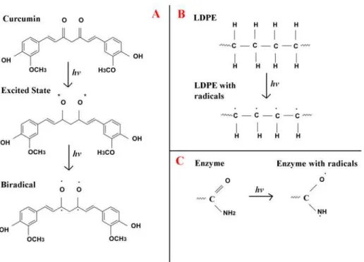
Commonly used photochemical reactive groups include aryl azides and diazirines which are explosophores and toxic [35]. Curcumin is tested as a crosslinking agent in the current study, since it is safe and widely used as a food flavoring agent. This is the first report in which it is studied for its potential as a photocrosslinker. It has several functional groups and is a well known antioxidant and anti-inflammatory agent and has medicinal benefits against several dis-eases including cancer and diabetis [36]. Even though crosslinking of polymers with high ener-gy radiation has been tested for many other applications, use of photocrosslinking technoloener-gy for food pack applications is minimal. In the present study, when curcumin and polymer are UV treated, they form biradicals. Papain crosslinks to these biradicals in the presence of UV light to form papain immobilized polymer with curcumin as the linker.
Materials and Methods
Bacterial strains and chemical materials
Laboratories (SRL), and HiMedia (Mumbai, India). Polycaprolactam was purchased from marine industrial polymers, Chennai, India., HDPE, LDPE and LLDPE sheets (0.175 micron thickness) were purchased from Industrial Insulations Ltd, Chennai, India.
Papain and curcumin estimation
The activity of papain was determined by using a reported procedure [37] using casein (SRL, item no: 034023) as the substrate. Curcumin was dissolved in ethanol (China Changshu Yang yuan Chemical, batch no: 20140720) and its concentration was estimated by using a reported method [38] with the help of an UV spectrophotometer (Perkin-Elmer, Lambda 35, Shelton, CT).
Determination of Minimum inhibitory concentration (MIC)
The MIC values of curcumin, papain and a mixture of curcumin and papain (1:2.5 by wt) against both the bacterial strains were determined by the microdilution broth assay method [39] with slight modifications as reported by Sarker et al [40] using resazurin (Sigma-Aldrich, item no: R7017) as an indicator. The colour change was assessed visually and the highest dilu-tion that remained blue (inhibidilu-tion of growth) indicated the minimum inhibitory concentra-tion of the compound. A colour change from blue to pink showed the growth of the organism.
UV crosslinking and characterization
The photochemical cross-linking of papain to LDPE, HDPE, LLDPE and PCL surfaces (1x1cm) was performed in two stages at 30°C in a rectangular cabinet (Superfit, India) in the presence of air, by exposing them to UV light at 365 nm and 500 W. The distance between the UV source and the film was 20 cm. Curcumin was dissolved in ethanol and 200μl of this
solu-tion containing 5.43μM of it was spread on these polymer films using a spin coater (Apex
in-struments co pvt ltd, India), followed by UV treatment for 24 hours to form CC (curcumin cross linked)-LDPE, CC-HDPE, CC-LLDPE and CC-PCL. In order to calculate the amount of curcumin crosslinked to these polymers, the curcumin crosslinked polymers were taken sepa-rately in four different tubes and washed with 25mM of phosphate buffer solution at a pH of 7 and the curcumin left in the washing solution was quantified. This was then subtracted from the curcumin initially taken for crosslinking. 0.1 mM of papain solution at a pH of 7 was then spin coated onto these surfaces and UV treated for 10 minutes to form PCC (Papain immobi-lized curcumin crosslinked)-LDPE, PCC-HDPE, PCC-LLDPE and PCC-PCL. The efficiency of the cross linking process and the activity of the enzyme retained after immobilization were esti-mated from the following formulae.
Immobilzation ef f iciency ¼ 1 Ps Po
x 100
Po is the initial concentration of papain prepared to coat the polymers; Ps is the papain con-centration in the washing solution left after washing the PCC-polymers.
Activity recovery or retainedð Þaf ter immobilization
¼ ðActivity of immobilized enzyme=Initial activity of f ree enzymeÞ x100
The polymers were stored at 4°C for 30 days to study their stability i.e. the activity retained by the enzyme after storage for 30days. The papain stability was calculated as,
papain stability
The polymers were washed in PBS buffer (pH of 7.0), and then the enzyme activity was esti-mated. This was repeated for seven cycles and the enzyme activity was calculated at the end of this washing process as follows.
Recycling ef f iciency ¼ ðEnzyme activity in the 7th cycle=activity in the f irst cycleÞ100
Physicochemical characterization of the films
The changes in the structure of the polymers, and the effect of photo crosslinking and the im-mobilization of the enzyme were identified from the Fourier Transform Infrared (FTIR) spectra recorded in the frequency range of 500–4000 cm-1using a Perkin-Elmer PE 1600 FTIR spec-trometer. The elemental composition of the polymers’surfaces after the immobilization were determined using a scanning electron microscope (SEM) equipped with a energy dispersive x-ray spectroscope (EDAX) (JEOL JSM 5600 LSV model, supplied by JEOL, Tokyo, Japan).
Contact angle of these polymers were measured using a Goniometer (Kruss germany) with Milli-Q water (Millipore grade). The images obtained were analyzed with a Digital Scrapbook Artist 2 Software (DSA2) to determine the static and dynamic contact angles (SW4001), with an accuracy of ±0.1°.
Biofilm formation and characterization
Each bacterial strain was inoculated from the stock culture into 25 ml of nutrient broth and in-cubated at 37°C for 16 h in a shaker (Scigeneis Pvt., Ltd, Chennai, India) at 120 rpm. A total of 500μl from the above preculture was inoculated into 50 ml of nutrient broth and cultured
under the above conditions. After 16h the culture broth was taken in sterile falcon tube and was centrifuged (Eppendroff, Germany) at 4°C at 4480 rcf (relative centrifugal force) for 10 min. The pellet was diluted in phosphate buffer solution (10 mM) and its optical density (OD) value was adjusted to 0.1 (at 600 nm) which was equivalent to approximately 1 x 10⁷cells/ml. Each bacterial suspension was subsequently inoculated into three flasks containing nutrient broth along with bare, CC and PCC polymers (of size 1x1cm). These flasks were stirred for 24 h at 30°C under shaking at 120 rpm using an Orbitek shaker (Scigeneics India ltd, India). Fol-lowing this incubation period, the samples were removed with sterile forceps and were washed twice with sterile water to remove the unbound cells. The samples were subsequently inoculat-ed in sterile tubes containing 0.7% of saline solution. The biofilm forminoculat-ed on the surface of each polymer was carefully dislodged by water-bath ultra sonication (Thosan Pvt., Ltd, Ajmer, India) for a total of 10min with 1min interval [41].
The protein content in each of the biofilms was estimated by the Lowry’s method [42] using crystalline bovine serum albumin as the reference standard. The exopolysaccharides content present in each of the biofilms was estimated by the phenol sulfuric acid method using glucose (SRL, item no: QK1Q610671) as the standard [43]. The live bacterial colonies in the biofilm was removed and their number was determined as per a standard procedure and represented as colony forming units (CFU/cm2of the polymer surface) [43].
Morphology of the biofilms
The live and dead microbial cells present on the polymers surface were determined using a mixture of two nucleic acid fluorescent staining dyes containing SYTO9 and propidium iodide (PI) (LIVE/DEADBacLight Bacterial Viability Kit, Invitrogen, USA). The former dye stains both live and dead cells as green while the latter dye penetrates the wall of the damaged cells and binds to DNA and appears as red. The polymer films (bare, CC-LDPE and PCC-LDPE) were individually inoculated into flasks containing 25 ml of nutrient broth. Then 1ml of each bacterial suspension (approximately 107cells) was subsequently inoculated into these flasks. Flasks were stirred for 24 h at 30°C under shaking at 120 rpm. The samples were removed with sterile forceps and were washed twice with sterile water to remove the unbound cells. These films were stained with the dye mixture and then observed under a fluorescence microscope (Leica DM5000, Germany) [43].
BATH Assay
BATH assay was performed on both bacteria to determine the hydrophobicity of each bacterial surface using a standard procedure [44].
Food packaging experiment
A slight modification to the methodology reported by Besse et al [45] was followed here. Fresh-ly processed beef sample was purchased from a supermarket and kept frozen at -20°C and thawed at 2°C for 1 day before use. It was then cut into small squares, each weighing 1 g, and was inoculated with 107cells ofAcinetobactersp. andS.aureus, separately. Samples were left undisturbed for 5 min for the inoculum to soak in and the cells to attach. They were subse-quently wrapped in LDPE, CC-LDPE and PCC-LDPE, then placed in a petri plate and incubat-ed at 4°C. After 7 days, the meat samples were openincubat-ed aseptically and approximately 0.2g were homogenized in 1ml of 0.7% saline solution and the numbers of viable bacteria present on the meat samples were estimated as described below. 100μl of this solution was serially decimally
diluted and subsequently spread-plated on nutrient agar (Himedia, item no: M001) plates using a L shaped glass rod. After 24h of incubation at 37°C, the viable colonies were counted vi-sually on nutrient agar plates and represented as colony forming units (CFU/g of beef).
Statistics
All the analysis was repeated thrice on three independent samples and the data was reported as means ± standard errors. One way ANOVA and two sample t-test were performed using Mini-Tab Ver 14.0 (MiniMini-Tab inc, USA). A p value<0.05 was considered to be statistically significant.
Results and Discussion
Physicochemical characterization of the films
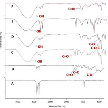
The FTIR spectra of the LDPE (Fig 3A) has bands at 1462cm-1which corresponds to methy-lene and methyl groups respectively. After UV treatment, new bands are formed at 1734 and 1640 cm-1(Fig 3B) indicating the formation of C = O and C = C groups respectively [47]. FTIR spectrum of curcumin coated LDPE (before UV treatment) (Fig 3C) shows bands correspond-ing to OH (3290 cm-1), C = C (1626 cm-1) and C = O (1743 cm-1) groups, which are present in
Fig 1. The chemical changes which occurred on UV treatment of (A) curcumin, (B) LDPE and (C) enzyme.
doi:10.1371/journal.pone.0121665.g001
Fig 2. Step by step reactions which lead to the formation of Papain crosslinked LDPE.
curcumin. These results are in agreement with earlier reports [48,49]. Presence of enol peaks at 1078 cm-1and 1136 cm-1(after UV treatment) indicates that curcumin has crosslinked to the polymer through an oxygen group. The band corresponding to OH group in the non uv treated polymer (3290 cm-1) shifts to 3167 cm-1in the UV treated sample. Bands at 1335 cm-1and 1379 cm-1indicate the CH3bending vibration present in non UV treated polymer which
disap-pears when curcumin is crosslinked to LDPE [49]. Peak at 1050 cm-1, represents C-O-C group which has appeared after the crosslinking of curcumin to LDPE. Appearance of 1272 cm-1 peak indicates C-C group. The carbonyl group of curcumin (C = O) observed at 1743 cm-1in non UV treated sample gets converted to C-O (1150 cm-1) in the UV treated sample. The ap-pearance of the peak at 1150 cm−1(after UV treatment) can be attributed to the crosslinking of
curcumin to LDPE (Fig 3D)
The FTIR spectra of papain coated on CC-LDPE before (Fig 3E) and after UV treatment (Fig 3F) indicates the formation of C = N group (1624 cm-1) in the latter which confirms the covalent immobilization of papain to CC-LDPE [38]. Appearance of peak at 1042 cm-1 repre-sents C-C which is less intense in non UV treated than in the UV treated sample indicating the formation of several carbon-carbon bonds between papain and CC-LDPE. This also indicates the crosslinking of COOH group in papain to CC-LDPE. The band at 1529 cm-1indicates the presence of amide group in PCC-LDPE [50]. Bands at 1042, 1136 and 1285 cm-1indicate the presence of primary amine (CN stretch) and peak at 1624 cm-1indicates the formation of sec-ondary amine (NH) which arises due to the immobilization of papain to LDPE [49]. These changes in the FTIR spectra confirm that the papain is covalently crosslinked to CC-LDPE. The photochemical crosslinking when compared to chemical process is operated at room tem-perature and is easy to control, which is helpful in preserving the 3-dimensional structure of the enzyme after UV treatment [51].
Most of the commonly used photocorsslinkers include aryl azides and diazirines as their reactive groups. These are toxic and hazardous [35]. So, their application in food and medical
Fig 3. FTIR spectra of A) Non UV treated LDPE, B) UV treated LDPE, C) Non UV treated CC-LDPE, D) UV treated CC-LDPE, E) Non UV treated PCC-LDPE and F) UV treated PCC-LDPE.
industry is limited, which necessitates the need for nontoxic photocrosslinker. In this study, curcumin is tried as a novel photocrosslinker. Polyethylene is a polymer widely used for many applications including food packages. Lack of functional group limits it from being used as a base for immobilizing antimicrobials and proteins on its surface. In the present study, UV treatment of curcumin as well as LDPE results in the formation of biradicals, favoring the crosslinking between them and then later to papain. The successful crosslinking of curcumin and further immobilization of papain to LLDPE, HDPE and PCL are confirmed similarly from their respective FTIR spectra (Figs B to D and Table A inS1 File). This is the first report on the use of curcumin as a photocrosslinker. Curcumin could be used to crosslink surfaces that could be used for various applications including food, pharmaceuticals and medicine which would require the use of non-toxic crosslinker.
Elemental composition of the surfaces concentration and morphology of
the polymers
The changes in the elemental composition of the polymers’surfaces after UV crosslinking were investigated by EDAX. These measurements indicated 98.1±0.49 and 1.5±0.18 weight % of ele-mental carbon and oxygen, respectively, on the surface of UV treated LDPE and 92.5±1.80 and 7.5±0.69 weight % of elemental carbon and oxygen, respectively, on the surface of CC-LDPE respectively. The increase in percentage of oxygen in the latter is due to the immobilization of curcumin which possesses several oxygen groups. 67.0±3.70, 13.3±1.60, 18.7±2.20 and 0.1 ±0.06 weight % of elemental carbon, nitrogen, oxygen and sulphur respectively are present on the surface of PCC-LDPE. The appearance of elemental sulphur and nitrogen are due to the immobilization of the enzyme which contains amino acids, once again emphasizing its pres-ence on the polymer surface.
The contact angle of LDPE, CC-LDPE and PCC-LDPE were 128± 2.3°, 80± 1.8° and 71 ±1.3° respectively. The relevant results for HDPE, LLDPE and PCL were 110± 2.7°, 83± 2.6° and 65±1.9°; 100± 3.2°, 85± 2.3° and 70±2.5° and 79± 1.9°, 70± 1.4° and 58±1.3° respectively. It is observed that PCL is the most hydrophilic and LDPE is the most hydrophobic surface. Non treated polymers are the most hydrophobic while the crosslinking successively reduces the hy-drophobicity. Previously, it has been reported that the hydrophilic surfaces generally reduce the adhesion of microorganisms [52].
Stability and activity of immobilized enzyme
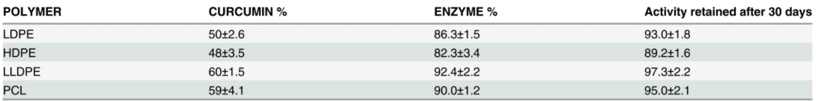
The percentages of curcumin and papain immobilized on the four polymers are listed in Table 1. It can be concluded (based on the one way ANOVA, p<0.05) that the percentage of curcumin and papain crosslinked to LLDPE is the highest, followed by their amount on PCL, LDPE and HDPE. After 30 days of storage, free enzyme retained 87.5 ±2.0% of activity, while PCC-LDPE retained 93.0±1.8% of enzyme activity, PCC-HDPE, PCC-LLDPE and PCC-PCL retained 89.2±1.6, 97.3±2.2 and 95.0±2.1% of enzyme activity respectively (Table 1). These data show that both the enzymatic activity and stability are well maintained after crosslinking.
Table 1. The weight percentages of curcumin and papain immobilized on the four polymers.
POLYMER CURCUMIN % ENZYME % Activity retained after 30 days
LDPE 50±2.6 86.3±1.5 93.0±1.8
HDPE 48±3.5 82.3±3.4 89.2±1.6
LLDPE 60±1.5 92.4±2.2 97.3±2.2
PCL 59±4.1 90.0±1.2 95.0±2.1
Biofilm inhibition
The MIC values of papain, curcumin and the mixture of papain and curcumin needed to inhib-it the growth ofAcinetobactersp. as determined by microdilution broth assay method [53] were 7.80±0.18, 15.60±0.28 and 0.98±0.11μM respectively. ForS.aureus, the corresponding
MIC values of papain, curcumin and a mixture of papain and curcumin were 1.95±0.22, 3.90 ±0.37 and 0.98±0.11μM respectively. The combination of papain and curcumin exhibited
en-hanced activity than the individual compounds when used alone.
Papain immobilized curcumin crosslinked polymers (PCC) showed the least number of Aci-netobatersp. andS.aureusattached cells (Fig4A&4B). Whereas, maximum number of attached cells were observed on the bare polymers. It is observed that PCC-LLDPE showed the best anti-microbial activity against both species (maximum reduction in the number of live biofilm cells) followed by PCL, LDPE and then HDPE (p<0.5 forAcinetobatersp. and p<0.001 forS.aureus). These results negatively correlate with the percentage of enzyme immobilized on the polymers (correlation coefficient between live colony count on the polymer surface and percentage of en-zyme immobilized on the polymer surface = -0.85 forAcinetobatersp. and -0.89 forS.aureus). Highest percentage of enzyme was immobilized on LLDPE and the lowest on HDPE (Table 1). Percentage of enzyme crosslinked to LLDPE was comparatively more, which could probably be due to the presence of more short branches (more atoms/mol) in this polymer when compared to that in LDPE and HDPE. Since HDPE contains no branches, it shows less crosslinking when compared to LLDPE and LDPE. Presence of C = O as well as NH groups in PCL, permits more radical formation leading to increased curcumin and enzyme crosslinking when compared to LDPE and HDPE. These results demonstrate that photo-cross-linking is robust and can dramati-cally improve the structural stability of the enzyme. The conversion of weak ionic bonds to strong covalent bonds prevents the leakage of the enzyme leading to its high activity and stability [54].
Acinetobacteris a contaminant found in many food products and it is a challenge to eradicate it from contaminated food. Papain crosslinked polymer reduces its growth. This enzyme is also effective againstS.aureuswhich is a common food borne pathogen. Veluchamy et al. have re-ported superior performance of subtilisin (a bacterial protease) immobilized on polycaprolactum with glutaraldehyde againstEscherichia coliandS.aureus[41,55]. Orgaz et al. reported the effect of pronase treated chitosan against several foodborne pathogens. They showed 8.0, 7.5, 6.0, 5.0 and 0.5 log reduction againstBacillus cereus,L.monocytogenes,P.fluorescens,S.enterica and S.
aureusrespectively [55]. In another study, Morvay et al. reported that protease fromBacillus licheniformisinhibited the formation of mature biofilms ofB.cereusandPseudomonas aeruginosa
[56]. Lysozyme was covalently attached to polystyrene resin beads by the sole histidine residue (His-15) through peptide spacers of various lengths. Three 6-aminocaproic acid units of spacer length displayed the greatest degree of hydrolytic activity againstMicrococcus lysodeikticus[57].
Fig 4. Population (Log CFU/cm2) of (A)Acinetobactersp. and (B)S.aureusbiofilms formed on bare,
CC and PCC polymers after 24 hours of incubation (*p<0.5,***p<0.001).
Prabhawathi et al. reported 2 and 7 times reduction in carbohydrate and 9 and 5 times reduction in biofilm protein ofS.aureusandE.colirespectively on lipase immobilized polycaprolactam (LIP) when compared to uncoated polycaprolactam (UP) [58]. The immobilization was per-formed using Langmuir Blodgett technique. An arginine—tryptophan-rich peptide (CWR11) immobilized on a silicone surface displayed antimicrobial activity against a broad spectrum of microbes such asS.aureus,E.coliandP.aeruginosavia wall disruption [59].
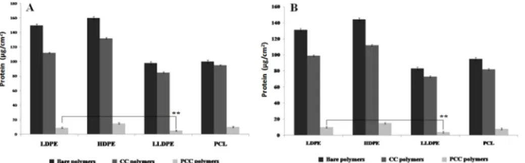
Bacteria enclosed in biofilms are usually more resistant to antimicrobial treatments, when compared to the same bacteria in planktonic form [60]. Since the major components of the bio-film matrices are exopolysaccharides and proteins, the action of the enzyme in reducing their amount is investigated here. Biofilms were developed by the adhesion of microbial cells on the polymer surface. The subsequent colonization of the organisms then is facilitated through the production of exopolysaccharides. Exopolysaccharides generally account for 50–90% of the total organic carbon in the matrix [41]. All the four PCC surfaces had less biofilm protein fol-lowed by the four CC surfaces (Fig 5). The bare polymer surfaces had the highest protein con-tent. The amount of protein in the biofilm formed on PCC-LLDPE was lesser than that on LDPE (p<0.01). Papain increased the membrane permeability as well as acted on the proteins and the peptidoglycan present on the outer membrane of the bacteria [23], leading to the loss of cell contents, and thus, resulting in bacterial destruction. There are literature reports which show that protease [41,61] hydrolyses the protein present on a polymer surface.
All the four types of polymers when crosslinked with papain showed reduced exopolysacchar-ide content in their biofilm and once again this content in the biofilm formed on PCC-LLDPE was lesser than that on LDPE (p<0.01 forAcinetobatersp. and p<0.01 forS.aureus) (Fig 6). Re-duction in exopolysaccharide content could disturb the uniformity and integrity of the biofilm structure which could lead to its breakdown as observed in the present study. A strong correla-tion is observed between the reduccorrela-tion of CFU with reduccorrela-tion in protein content in the biofilm
Fig 5. Protein content of (A)Acinetobactersp. and (B)S.aureusbiofilms formed on bare, CC and PCC polymers after 24 hours of incubation (**p<0.01).
doi:10.1371/journal.pone.0121665.g005
Fig 6. Exopolysaccharide content of (A)Acinetobactersp. and (B)S.aureusbiofilms formed on bare, CC and PCC polymers after 24 hours of incubation (**p<0.01,***p<0.001).
(correlation coefficient>0.95) as well as reduction of CFU with reduction in exopolysaccharide content in the biofilm (correlation coefficient>0.91) for both the organisms with all the four polymers. Maximum reduction of exopolysaccharide and protein were observed on LLDPE sur-face and least on HDPE sursur-face.
SEM images of the biofilm ofAcinetobactersp. (Fig7A,7B&7C) andS.aureuson LDPE, CC-LDPE and PCC-LDPE (Fig7D,7E&7F) support the CFU results. Least number of bacte-ria was observed on PCC-LDPE and maximum on bare LDPE. Cells with compromised and damaged membrane that are considered to be dead are stained red by the Backlight dye, where-as the cells with intact membrane are stained green. Fluorescence images show more live Acine-tobactersp. andS.aureuscells on LDPE surface (Fig8A&8D), a mixture of live and dead cells on CC-LDPE (Fig8B&8E) and more dead cells on PCC-LDPE (Fig8C&8F). These data indi-cate that the enzyme is able to damage the cell membrane and hence reduce the number of live bacterial cells in the biofilm [41].
Also, contact angle indicates that bare polymer is more hydrophobic than the enzyme immo-bilized one and so less bacterial attachment is expected on the latter. It could be observed from CFU data, and SEM and fluorescence microscopic analysis that there is more adhesion of mi-crobes to bare polymer surface than enzyme immobilized surface. So less attachment of bacteria
Fig 7. SEM images ofAcinetobactersp. grown on (A) LDPE (B) Curcumin crosslinked LDPE (C) Papain immobilized CC-LDPE andS.aureusgrown on (D) LDPE (E) Curcumin crosslinked LDPE (F) Papain immobilized CC-LDPE.
doi:10.1371/journal.pone.0121665.g007
Fig 8. Fluorescence microscopic images ofAcinetobactersp. biofilm on (A) LDPE (B) curcumin crosslinked LDPE (C) papain immobilized CC-LDPE (Green-live cells, red- dead cells due to cell membrane damage) andS.aureusbiofilm on (D) LDPE (E) curcumin crosslinked LDPE (F) papain immobilized CC- LDPE.
and biofilm are observed on the enzyme immobilized surface because of the antibacterial activity of papain as well as the relatively hydrophillic nature of this surface
Characterization of bacterial surface properties
Bacterial surface properties, such as the hydrophobicity, are considered important factors influ-encing the adherence of cells to biomaterial surfaces and this property is determined from the BATH assay. As the concentration of hexadecane is increased, the O.D value corresponding to
Acinetobactersp. decreased indicating that more of this organism is partitioning to the hydro-phobic solvent phase while the reverse trend to be observed withS.aureus. (Fig F inS1 File). This indicates that the former organism is relatively more hydrophobic than the latter. Hydro-phobic bacteria tend to adhere more to hydroHydro-phobic biomaterial surfaces. More number of Aci-netobactersp.isattached to the bare surface thanS.aureus, probably because the former is more hydrophobic than the latter. The cell membrane ofAcinetobacterspp. consists of pro-teins, lipopolysaccharides and phospholipids. Its hydrophobicity is due to the presence of pilli, lipopolysaccharides and hydrophobic amino acids in the flagella. The protease enzyme has probably acted on the cell membrane ofAcinetobactersp. thereby altering its surface properties leading to its reduced adhesion on the polymer.
Food packaging experiment
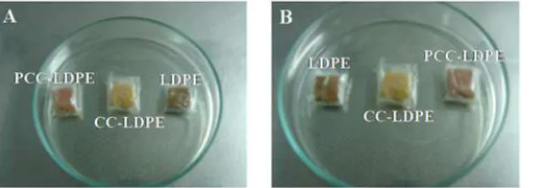
Antimicrobial action of PCC-LDPE was tested on beef samples against both the bacterial strains. The number of liveAcinetobactercells in the meat samples wrapped with LDPE, CC-LDPE and PCC-LDPE at the end of 7thday represented as log(CFU/g of beef) were 11.7±11.1, 7.8±7.2, and 2.5±2.1 respectively indicating the effectiveness of the enzyme (p<0.001) in reducing the num-ber of live bacteria in the food. The numnum-ber of liveS.aureuscells in the meat samples wrapped with the same polymers at the end of 7thday represented as log(CFU/g of beef) were 11.1±10.6, 7.7±6.9 and 1.8±1.5 respectively, once again indicating the effectiveness of the enzyme
well as the Gram positiveS.aureus. It is seen from the images (Fig9A&9B) that, the meat sam-ple wrapped with enzyme immobilized LDPE (PCC-LDPE) appears to be fresh when compared to the one wrapped with bare polymer. Growth of bacteria could be seen on the latter which in-dicates the spoilage of the food.
Conclusion
Curcumin has been successfully used as a novel photo crosslinker to covalently couple papain to four different types of polymers. These papain crosslinked polymers remained stable and ac-tive for a period of 30 days. Moreover, the treated surface turned hydrophilic, thereby becom-ing resistant to adhesion of microbes to it. It also exhibited antibiofilm activity against both a Gram positive, as well as a Gram negative strain. Literature reports generally indicate antibacte-rial coatings that act only on one type of organism but in the approach described here, the en-zyme acts non-specifically on the cell membrane of both types of bacteria. Microorganisms generally develop resistance to antibacterial agents due to the alterations in their genes but the components of the cell membrane remain unaltered and hence the latter could be a good target as demonstrated here. Papain crosslinked polymers show excellent antibacterial activity against
Acinetobacter sps. KC119137.1 andS.aureusonce these have contaminated beef (more than 9 log reduction in the number of live bacteria), due to the combined effect of the crosslinked cur-cumin and the immobilized papain. This strategy provides a promising platform for fabricating robust, nontoxic, renewable and low cost antimicrobial films, which may offer a wide range of applications in food, pharmaceutical and biomedical packaging industries. This strategy can be applied for coating surfaces which do not have any functional groups on them. Both, curcumin and papain, reported here are food-based products and hence can be considered as safe. Fur-ther studies need to be done with oFur-ther food-borne bacteria, as well as aiming to study the ef-fect of this enzyme on food quality (including changes in texture and colour) before it can be taken up for commercial applications.
Supporting Information
S1 File.Fig. A, The phylogenetic tree ofAcinetobactersps. KC119137.1. (FM1). Fig. B, FTIR
spectra of A) Non UV treated PCL, B) UV treated PCL, C) Non UV treated CC-PCL, D) UV treated CC-PCL, E) Non UV treated PCC-PCL and F) UV treated PCC-PCL. Fig. C, FTIR spec-tra of (A) Non UV treated HDPE, (B) UV treated HDPE, (C) Non UV treated CC-HDPE (D), UV treated CC-HDPE, (E) Non UV treated PCC-HDPE and (F) UV treated PCC-HDPE. Fig. D, FTIR spectra of (A) Non UV treated LLDPE, (B) UV treated LLDPE, (C) Non UV ed CC-LLDPE, (D) UV treated CC-LLDPE, (E) Non UV treated PCC-LLDPE and (F) UV treat-ed PCC-LLDPE. Fig. E, Organism hydrophobicity with bath assay. Table A, Table for FTIR. (DOC)
Fig 9. Effect of wrappers formed of LDPE, CC-LDPE and PCC-LDPE on meat samples, inoculated with A)Acinetobactersp. and B)S.aureusafter 7 days of storage at 4°C.
Acknowledgments
We thank Sophisticated Analytical Instrument Faculty (SAIF, IITM) for FTIR and SEM analysis.
Author Contributions
Conceived and designed the experiments: VP MD. Performed the experiments: CMM VP. An-alyzed the data: VP PMS CMM MD. Contributed reagents/materials/analysis tools: MD. Wrote the paper: VP CMM MD.
References
1. Galet VM, Carballo GL, Gavara R, Muñoz PH. Antimicrobial food packaging film based on the release
of LAE from EVOH. Int J Food Microbiol. 2012; 157: 239–244. doi:10.1016/j.ijfoodmicro.2012.05.009 PMID:22640726
2. Fuciños C, Guerra NP, Teijón JM, Pastrana LM, Rúa ML. Use of poly (N-isopropyl acrylamide)
nanohy-drogels for the controlled release of pimaricin in active packaging. J Food Sci. 2012; 77: 21–28. 3. Cerqueira MA, Souza BWS, Teixeira JA, Vicente AA. Effect of glycerol and corn oil on physicochemical
properties of polysaccharide films—A comparative study. Food Hydrocolloid. 2012; 27: 175–184. 4. Ouattara B, Simard RE, Piette G, Bégin A, Holley RA. Diffusion of acetic and propionic acids from
chito-san-based antimicrobial packaging films. J Food Sci. 2000; 65: 768–773.
5. Choi JH, Choi WY, Cha DS, Chinnan MJ, Park HJ. Diffusivity of potassium sorbate inκ-carrageenan
based antimicrobial film. LWT—Food Sci Technol. 2005; 38: 417–423.
6. Fajardo P, Martins JT, Fuciños C, Pastrana L, Teixeira JA. Evaluation of a chitosan-based edible film
as carrier of natamycin to improve the storability of saloio cheese. J Food Eng. 2010; 101: 349–356. 7. Calzolai L, Gilliland D, Rossi F. Measuring nanoparticles size distribution in food and consumer
prod-ucts: a review. Food Addit Contam Part A. 2012; 29: 1183–1193. doi:10.1080/19440049.2012.689777 PMID:22725833
8. Chau CF, Wu SH, Yen GC. The development of regulations for food nanotechnology. Trends Food Sci Tech. 2007; 18: 269–280.
9. Tang W, Zhang H, Wang L, Qian H. Antimicrobial peptide isolated from ovalbumin hydrolysate by im-mobilized liposome-binding extraction. Euro Food Res Technol. 2013; 237: 591–600.
10. Manso S, Nerin FC, Becerril R, Nerín C. Combined analytical and microbiological tools to study the ef-fect onAspergillusflavusof cinnamon essential oil contained in food packaging. Food Control. 2013; 30: 370–378.
11. Han C, Wang J, Li Y, Lu F, Cui Y. Antimicrobial-coated polypropylene films with polyvinyl alcohol in packaging of fresh beef. Meat Sci. 2013; 157: 239–244.
12. Hauser C, Wunderlich J. Antimicrobial packaging films with a sorbic acid based coating. Food Sci. 2011; 1: 197–202.
13. Limjaroen P, Ryser E, Lockhart H, Harte B. Development of a Food Packaging Coating Material with Antimicrobial Properties. J Plast Film Sheet. 2003; 19: 95–109
14. Gemili S, Yemenicioglu A, Altınkaya SA. Development of cellulose acetate based antimicrobial food packaging materialsfor controlled release of lysozyme. J Food Eng. 2008; 90: 453–462.
15. Hanusova K, Vapenka L, Dobias J, Miskova L. Development of antimicrobial packaging materials with immobilized glucose oxidase and lysozyme. Cent Eur J Chem. 2013; 11: 1066–1078.
16. Ramos M, Jimenez A, Peltzer M, Garrigos MC. Characterization and antimicrobial activity studies of polypropylene films with carvacrol and thymol for active packaging. J Food Eng. 2012; 109: 513–519. 17. Suppakul P, Sonneveld K, Bigger SW, Miltz J. Diffusion of linalool and ethylchavicol from
polyethylene-based antimicrobial packaging films. J Food Sci Technol. 2011; 44: 1888–1893.
18. new.dhh.louisiana.gov website.http://new.dhh.louisiana.gov/assets/oph/Center-PHCH/Center-CH/ infectious-epi/EpiManual/AcinetobacterManual.pdf. Accessed 2015 Feb 26.
19. Trouillet JL, Chastre J, Vuagnat A, Joly GML, Combaux D, Dombret MC, et al. Ventilator-associated Pneumonia Caused by Potentially Drug-resistant Bacteria. Am J Respir Crit Care Med. 1951; 157: 531–539.
21. Loir YL, Baron F, Gautier M.Staphylococcus aureusand food poisoning. Genet Mol Res. 2003; 2:63–76. PMID:12917803
22. Stevenson DE, Storer AC. Papain in organic solvents: Determination of conditions suitable for biocatal-ysis and the effect on substrate specificity and inhibition. Biotechnol Bioeng. 1991; 37: 519–527. PMID:18600639
23. Amri E, Mamboya F. Papain, A plant enzyme of Biological importance a review. Am J Biochem Biotech-nol. 2012; 8: 99–104.
24. Chukwuemeka NO, Anthonia AB. Antifungal effects of pawpaw seed extracts and papain on post
har-vestCarica papayaL. fruit rot. Afri J Agri Res. 2010; 5: 1531–1535.
25. Beeley JA, Yip HK, Stevenson AG. Chemochemical caries removal: A review of the techniques and lat-est developments. Br Den J. 2000; 188: 427–430.
26. Lapidot A, Romling U, Yaron S. Biofilm formation and thesurvival ofSalmonella typhimuriumon pars-ley. Int J Food Microbiol. 2006; 109: 229–233. PMID:16616389
27. Quintavalla S, Vicini L. Antimicrobial food packaging in meat industry. Meat Sci. 2002; 62: 373–380. PMID:22061613
28. Mateo C, Abian O, Fernandez-Lorente G, Pedroche J, Fernandez-Lafuente R, Guisan JM. Epoxy sepa-beads: A novel epoxy support for stabilization of industrial enzymes via very intense multipoint covalent attachment. Biotechnol Progr. 2002; 18: 629–634. PMID:12052083
29. Siracusa V, Rocculi P, Romani S, Rosa MD. Biodegradable polymers for food packaging: A review. Trends Food Sci Tech. 2008; 19: 634–643.
30. Robertson GL. Food packaging: Principles and practice. 3rd ed., Vol. 3. CRC press, USA; 2012. 31. Rooney ML. Active packaging in polymer films. In: Active food packaging. Blackie Academic &
Profes-sional, London; 1995.
32. Iseppi R, Pilati F, Marini M, Toselli M, de Niederhäusern S, Guerrieri E, et al. Anti-listerial activity of a polymeric film coated with hybrid coatings doped with Enterocin 416K1 for use as bioactive food pack-aging. Int J Food Microbiol. 2008; 123: 281–287. doi:10.1016/j.ijfoodmicro.2007.12.015PMID:
18262299
33. essentialchemicalindsutry.org website.http://www.essentialchemicalindustry.org/polymers/ polyethene.html. Accessed 2015 Feb 26
34. Nguyen KT, West JL. Photopolymerizable hydrogels for tissue engineering applications. Biomaterials. 2002; 23: 4307–4314. PMID:12219820
35. Bräse S, Gil C, Knepper K, Zimmermann V. Organic Azides: An Exploding Diversity of a Unique Class of Compounds. Angew Chemie. 2005; 44: 5188–5240.
36. Beevers CS, Huang S. Pharmacological and clinical properties of curcumin. Botanics: Targets and Therapy. 2011; 1: 5–18.
37. Liggieri C, Obregon W, Trejo S, Priolo N. Biochemical analysis of a papain-like protease isolated from the latex ofAsclepiascurassavicaL. Acta Biochim Biophys Sinica. 2009; 41: 154–162. PMID:
19204833
38. Sharma K, Agrawal SS, Gupta M. Development and Validation of UV spectrophotometric method for the estimation of Curcumin in Bulk Drug and Pharmaceutical Dosage Forms. Int J Drug Devel Res. 2012; 4: 375–380.
39. Bueno C, Villegas ML, Bertolotti SG, Previtali CM, Neumann MG, Encinas M. The Excited-State Inter-action of Resazurin and Resorufin with Aminesin Aqueous Solutions. Photophysics and Photochemical Reaction. J Photochem Photobiol. 2002; 76: 385–390. PMID:12405144
40. Sarker SD, Nahar L, Kumarasamy Y. Microtitre plate-based antibacterial assay incorporating resazurin as an indicator of cell growth, and its application in the in vitro antibacterial screening of phytochemi-cals. Methods. 2007; 43: 321–324.
41. Veluchamy P, Sivakumar PM, Doble M. Immobilization of Subtilsin on polycaprolactam for antimicrobial food packaging applications. J Agri Food Chem. 2011; 59: 10869–10878. doi:10.1021/jf201124v PMID:21910484
42. Lowry OH, Rosenbrough NJ, Farr AL, Randall RJ. Protein measurement with the folin phenol reagent. J Biol Chem. 1951; 193: 265–275. PMID:14907713
43. Sivakumar PM, Veluchamy P, Doble M. 2-methoxy-2', 4'- dichlorochalcone as an antimicrofoulant against marine bacterial biofilm. Colloids Surf B Biointerfaces. 2010; 81: 439–446. doi:10.1016/j.
colsurfb.2010.07.037PMID:20708908
45. Besse NG, Audinet N, Beaufort A, Colin P, Cornu M, Lombard B. A contribution to the improvement
ofListeria monocytogenesenumeration in cold-smoked salmon. Int J Food Microbiol. 2004; 91:
119–127. PMID:14996455
46. Dankbar DM, Gauglitz G. A study on photolinkers used for biomolecule attachment to polymer sur-faces. Anal Bioanal Chem. 2006; 386: 1967–1974. PMID:17089100
47. Kondyurin AV, Naseri P, Tilley JMR, Nosworthy NJ, Bilek MMM, McKenzie DR. Mechanisms for Cova-lent Immobilization of HorseradishPeroxidase on Ion-Beam-Treated Polyethylene. Scientifica. 2012; 1: 1–28.
48. Rajandas H, Parimannan S, Sathasivam K, Ravichandran M, Yin LS. A novel FTIR-ATR spectroscopy based technique for the estimation of low-density polyethylene biodegradation. Polym Test. 2012; 31: 1094–1099.
49. Mohan PRK, Sreelakshmi G, Muraleedharan CV, Joseph R. Water soluble complexes of curcumin with cyclodextrins: Characterization by FT-Raman spectroscopy. Vib Spec. 2012; 62:77–84
50. Li B, Shan CL, Zhou Q, Fang Y, Wang YL, Xu F, et al. Synthesis, Characterization, and Antibacterial Activity of Cross-Linked Chitosan-Glutaraldehyde. Mar Drugs. 2013; 11: 1534–1552. doi:10.3390/
md11051534PMID:23670533
51. Tripathi S, Mehrotra GK, Dutta PK. Physicochemical and bioactivity of cross-linked chitosan-PVA film for food packaging applications. Int J Biol Macromol. 2009; 45: 372–376. doi:10.1016/j.ijbiomac.2009.
07.006PMID:19643129
52. Erdogan X, Song LL, Wilson JN, Park JO, Srinivasarao M, Bunz UHF. Permanent bubble arrays from a cross-linked poly(para-phenyleneethynylene): picoliter holes without microfabrication. J Am Chem Soc. 2004; 126: 3678–3679. PMID:15038697
53. Doyle RJ. Contribution of the hydrophobic effect to microbial infection. Microb Infect. 2000; 2: 391–400. 54. Candan F, Unlu M, Tepe B, Daferera D, Polissiou M, Sokmen A, et al. Antioxidant and antimicrobial
ac-tivity of the essential oil and methanol extracts ofAchilleamillefoliumsubsp.MillefoliumAfan (Astera-ceae). J Ethnopharmacol. 2003; 87: 215–220. PMID:12860311
55. Fu G, Dai Z. Efficient immobilization of glucose oxidase by in situ photo-cross-linking for glucose bio-sensing. Talanta. 2012; 97: 438–444. doi:10.1016/j.talanta.2012.04.059PMID:22841105 56. Orgaz B, Lobete MM, Puga CH, Jose CS. Effectiveness of Chitosan against Mature Biofilms Formed
by Food Related Bacteria. Int J Mol Sci. 2011; 12: 817–828. doi:10.3390/ijms12010817PMID:
21340015
57. Morvay AA, Decun M, Sala C, Morar A, Morvay PLG. The Ability ofBacillus licheniformisProtease to RemoveBacillus cereusandPseudomonas aeruginosabiofilm. Vet Med-czech. 2011; 68: 245–250. 58. Wu Y, Daeschel MA. Lytic Antimicrobial Activity of Hen Egg White Lysozyme Immobilized to
Polysty-rene Beads. J Food Sci. 2007; 72: 369–374.
59. Prabhawathi V, Boobalan T, Sivakumar PM, Doble M. Antibiofilm Properties of Interfacially Active Li-pase Immobilized Porous Polycaprolactam Prepared by LB Technique. Plos One. 2014; 5: 1–11. 60. Lim K, Chua RRYC, Saravanan R, Basu A, Mishra B, Tambyah PA, et al. Immobilization studies of an
engineered arginine-tryptophan-rich peptide on a silicone surface with antimicrobial and antibiofilm ac-tivity. ACS Appl Mater Interfaces. 2013; 5: 6412–6422. doi:10.1021/am401629pPMID:23758173 61. Simoes M, Simoes LC, Vieira MJ. A review of current and emergent biofilm control strategies.
LWT-Food Sci Technol. 2010; 43: 573–583.
62. Eshamah H, Han I, Naas H, Acton J, Dawson P. Antibacterial effects of natural tenderizing enzymes on different strains ofEscherichia coliO157:H7 andListeria onocytogeneson beef. Meat Sci. 2013; 96: 1494–1500. doi:10.1016/j.meatsci.2013.12.010PMID:24447905
63. Sung SY, Sin LT, Tee TT, Bee ST, Rahmat AR, Rahman WA. Control of bacteria growth on ready to eat beef loaves by antimicrobial plastic packaging incorporated with garlic oil. Food Control. 2013; 39: 214–221.
64. Gamage GR, Park HJ, Kim KM. Effectiveness of antimicrobial coated oriented polypropylene/polyethyl-ene films in sprout packaging. Food Res Int. 2009; 42: 832–839.
65. Ercolini D, Ferrocino I, Storia AL, Mauriello G, Gigli S, Masi P, et al. Development of spoilage microbiota in beef stored in nisin activated packaging. Food Microbiol. 2009; 27: 137–143. doi:10.1016/j.fm.2009.