Mohtasham et al. JDMT, Volume 1, Number 1, September 2012
35
Case Report
Calcifying Odontogenic Cyst with Complex Odontoma: Histological
and Immunohistochemical Features
Nooshin Mohtasham
1, Amin Rahpeyma
2, Saeedeh Khajeh Ahmadi
3,
Mohsen Merati
41
Oral and Maxillofacial Diseases Research Center, Department of Oral and Maxillofacial
Pathology, Faculty of Dentistry, Mashhad University of Medical Sciences, Mashhad, Iran
2
Dental Research Center, Department of Oral and Maxillofacial Surgery, Faculty of Dentistry,
Mashhad University of Medical Sciences, Mashhad, Iran
3
Dental Research Center, Department of Oral and Maxillofacial Pathology, Faculty of Dentistry,
Mashhad University of Medical Sciences, Mashhad, Iran
4
Department of Orthodontics and Dental Research Center, Faculty of Dentistry, Mashhad
University of Medical Sciences, Mashhad, Iran
Received 27 September 2011 and Accepted 10 January 2012
Abstract
The calcifying odontogenic cyst (COC) is a rare odontogenic cyst. Only 2% of all odontogenic cysts and tumors are COC. COC associated with odontoma (COCaO) reported in 24% of COCs. COCaO presents a greater incidence in female, with a ratio of 2 to 1. The highest incidence of COCaO occurs during the second decade with a mean age of 16 years, most frequently occurring in the maxilla (61.5%). Here, we describe a classic case of COCaO of the maxillary incisor-canine region in 17-year-old girl, and discuss the clinicopathological features and immunohistochemical finding of this tumor.
Key Words: Calcifying odontogenic cyst, histopathologic feature, immunohistochemical straining, odontoma.
--- Mohtasham N, Rahpeyma A, Khajeh Ahmadi S, Merati M. Calcifying Odontogenic Cyst with Complex Odontoma: Histological and Immunohistochemical Features. J Dent Mater Tech 2012; 1(1): 35-9.
Introduction
The calcifying odontogenic cyst (COC) was first described as a distinct entity by Gorlin et al. (1962) and Gold (1963) (1). They reported 15 cases of this entity that were called as an intraoral marlherbe's calcifying epithelium (pilomatricoma). Several variants of the cyst may be seen are cystic and neoplastic variants; peripheral and central types. Rare cases of malignant transformation have been reported (2). The basic features of COC consist of 1) cystic, nonproliferative 2) cystic, proliferative/ameloblastomatous 3) odontoma-associated 4) epithelial odontogenic ghost cell tumor (3).
Various terms have been used for description of this lesion such as COC (4), keratinizing calcifying odontogenic cyst (KCOC) (1), calcifying ghost cell odontogenic tumor (CGCOT) (5), calcifying cystic odontogenic tumor (CCOT) (6,7), dentinogenic ghost cell tumor (DGCT) (8), epithelial odontogenic ghost cell tumor (EOGCT) (9), odontogenic ghost cell tumor (OGCT) (10), odontocalcifying odontogenic cyst (11).
Cystic variant composed of 85% of the cases (12). In addition, occasionally dysplastic dentine and an area of dental hard tissue formation and resembling odontoma can be found.
association impacted teeth is approxima Multiple impacted teeth are a well kn COC (4). Radicular resorption is uncom is usually treated by enucleation Recurrence is uncommon.
Case Report
A 17-year-old girl without any rem history, with two-month history of swe maxilla region was referred to Departm Maxillofacial surgery for definitive dia examination showed a painless buccal e left maxilla from medial line to the can associated teeth were vital without path A well-defined unilocular radiolucentFigure 1.
Panoramic radiograp
mate 10-32% (15). known feature of ommon (2). COC n and curettage.
markable medical welling in the left tment of Oral and iagnosis. Intraoral l expansion on the canine region. The thologic mobility. nt area extending
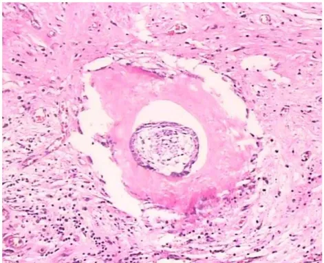
from medial line to the canin mass, was observed in the pan Root divergence of lateral and resorption was present (Fig. removed under local anesthes of biopsy tissue showed a proliferation of odontogenic e were present within epithelial like structure were present in Immunohistochemical stainin (Fig. 4). Histopathologic d odontogenic cyst with comple 2-year follow-up after cy recurrence.
aph: COC associated with odontoma (COCaO) wit
ine region, with radiopaque anoramic radiograph (OPG). nd canine teeth without root . 1). A thick wall cyst was esia. Histologic examination a cystic cavity lined by epithelial cells. Ghost cells al layer (Fig. 2). Odontoma-in the wall of cyst (Fig. 3).
ng for CK7 was negative diagnosis was calcifying plex odontoma (COCaO). A yst excision showed no
Mohtasham et al. JDMT, Volume 1, Number 1, September 2012
37
Figure 3.
Histopathology feature and detail of the complex odontoma (H&E staining, original
magnification ×400)
Figure 4.
Immunohistochemical staining is negative for CK7 (IHC staining, original
Discussion
COC may be arising from odontogenic epithelial remnants within the jaw or gingival. There is no gender predilection. It occurs in the maxilla and mandible with equal frequency (4). Rare peripheral variant of this lesion have been described.
COC may occur at any age with the prevalence peaks in the second and third decades (mean age, 33 years) but COC associated with odontoma (COCaO) occurs in younger persons, with a mean age of 17 years. In pathological feature, COC composed of a fibrous capsule that is lined with a proliferation of odontogenic epithelial cells and the ghost cell change that characterizes in these lesions (13).
COC rarely can occur in conjunction with other odontogenic tumors such as ameloblastoma, ameloblastic fibroma, amelobastic fibro-odontoma and adenomatoid odontogenic tumor (16). Radioluscency accompanying odontoma or presence of soft tissue with odontoma during biopsy or operation, guided clinician toward four differential diagnosis including: cystic odontoma, COCaO, amelobelastic fibro-odontoma, odontoameloblastoma. Unlike other lesions, surgical resection with safe bony margin is recommended for odontoameloblastoma. However, conservative treatment (enucleation) is required for another lesion. COCaO is similar to the cystic COC, it has tooth like structures in the connective tissue of the cyst (11).
COC may occur in association with odontoma; Buchner (2) shows this association in 35% of his cases, Nagao et al. (17) in 22% and Shamaskin et al. (18) in 47%. Radiographically COCaO appears as a mixed radiolucent-radiopaque lesion (80%) occasionally calcifications cannot be observed on OPG but can be visualized in CT scan (19). Treatment of COCaO consists of conservative enucleation and the prognosis is excellent. Recurrence after rather conservative therapy is uncommon (2).
Roudrigues-Fregnani et al. (20) showed that the epithelial cells of COC express antibodies directed against cytokeratins 7, 8, 14, and 19 but staining for CK7 was negative in present case. Finally, this finding denotes more research in this field.
Conclusion
Microscopic evaluation of soft tissue associated with odontoma is important; because this tissue can be dentigerous cyst, COC or ameloblastic fibroma. In this situation conservation complete removal of tissue recommended, but if soft tissue component to be
References
1. Gorlin RJ, Pindborg JJ, Clausen FP, Vickers RA.
The calcifying odontogenic cyst-a possible
analogue of the cutaneous calcifying epithelioma of
Malherbe. An analysis of fifteen cases. Oral Surg
Oral Med Oral Pathol 1962; 15: 1235-43.
2. Buchner A. The central (intraosseous) calcifying
odontogenic cyst: an analysis of 215 cases. J Oral
Maxillofac Surg 1991; 49: 330-9.
3. Gnepp DR. Diagnostic surgical pathology of the
head and neck. Philadelphia: Elsevier Inc, 2009.
4. McIntosh JJ, Campbell JH, Aguirre A, Reddy LV,
Elhadi H. Expansible mass of the maxilla. J Oral
Maxillofac Surg 2008; 66: 1253-8.
5. Fejerskov O, Krogh J. The calcifying ghost cell
odontogenic tumor - or the calcifying odontogenic
cyst. J Oral Pathol 1972; 1: 273-87.
6. Freedman PD, Lumerman H, Gee JK. Calcifying
odontogenic cyst. A review and analysis of seventy
cases. Oral Surg Oral Med Oral Pathol 1975; 40:
93-106.
7. Lucchese A, Petruzzi M, Scivetti M, et al.
Calcifying odontogenic cysts associated with
odontomas: confocal laser scanning microscopy
analysis of 13 cases. Ultrastruct Pathol 2011; 35:
146-50.
8. Praetorius F, Hjørting-Hansen E, Gorlin RJ,
Vickers RA. Calcifying odontogenic cyst. Range,
variations and neoplastic potential. Acta Odontol
Scand 1981; 39: 227-40.
9. Ellis GL, Shmookler BM. Aggressive epithelial
odontogenic ghost cell tumor. Oral Surg Oral Med
Oral Pathol 1986; 61: 471-8.
10. Colmenero C, Patron M, Colmenero B.
Odontogenic ghost cell tumors. The neoplastic form
of calcifying odontogenic cyst. J Craniomaxillofac
Surg 1990; 18: 215-8.
11. Hirshberg A, Kaplan I, Buchner A. Calcifying
Mohtasham et al. JDMT, Volume 1, Number 1, September 2012
39
12. Hong SP, Ellis GL, Hartman KS. Calcifying
odontogenic cyst. A review of ninety-two cases
with reevaluation of their nature as cysts or
neoplasms, the nature of ghost cells, and
subclassification. Oral Surg Oral Med Oral Pathol
1991; 72: 56-64.
13. Gallana-Alvarez S, Mayorga-Jimenez F,
Torres-Gómez FJ, Avellá-Vecino FJ, Salazar-Fernandez C.
Calcifying odontogenic cyst associated with
complex odontoma: case report and review of the
literature. Med Oral Patol Oral Cir Bucal 2005; 10:
243-7.
14. Devlin H, Horner K. The radiological features of
calcifying odontogenic cyst. Br J Radiol 1993; 66:
403-7.
15. Praetorius F, Hjorting-Hansen E, Gorlin RJ,
Vickers RA. Calcifying odontogenic cyst. Acta
Odont Scand 1981; 39: 227-40.
16. Zeitoun IM, Dhanrajani PJ, Mosadomi HA.
Adenomatoid odontogenic tumor arising in a
calcifying odontogenic cyst. J Oral Maxillofac Surg
1996; 54: 634-7.
17. Nagao T, Nakajima T, Fukushima M, Ishiki T.
Calcifying odontogenic cyst: a survey of 23 cases
in the Japanese literature. J Maxillofac Surg 1983;
11: 174-9.
18. Shamaskin RG, Svirsky JA, Kaugars GE.
Intraosseous and extraosseous calcifying
odontogenic cyst (Gorlin cyst). J Oral Maxillofac
Surg 1989; 47: 562-5.
19. Iida S, Fukuda Y, Ueda T, Aikawa T, Arizpe JE,
Okura M. Calcifying odontogenic cyst: radiologic
findings in 11 cases. Oral Surg Oral Med Oral
Pathol Oral Radiol Endod 2006; 101: 356-62.
20. Rodrigues-Fregnani E, Ramona-Pires F,
Rivera-Quezada D, et al. Calcifying odontogenic cyst:
clinicopathological features and
immunohistochemical profile of 10 cases. Oral
Pathol Med 2003; 32: 163-70.
Corresponding Author: Saeedeh Khajeh Ahmadi
Dental Research Center
Mashhad University of Medical Sciences Vakilabad Blvd, Mashhad, Iran
P.O. Box: 91735-984 Tel: +98-511-8829501 Fax: +98-511-8829500